| Rabbit Anti-Cyclin D1 antibody |
| 反应物种(预测) |
Dog |
| 产品应用(已验证) |
WB,IHC,ICC,FCM |
| 产品应用(可尝试) |
IF,ELISA |
| 推荐稀释比例 |
WB=1:500-2000,Elisa=1:5000-10000,IHC-P=1:100-500,IHC-F=1:100-500,Flow Cyt=1μg/Test,IF=1:100-500,ICC=1:100, |
| 研究领域 |
肿瘤,细胞生物,染色质和核信号,细胞周期蛋白,表观遗传学, |
| 标签 |
Array |
-
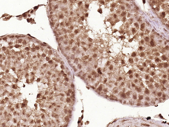
Paraformaldehyde-fixed, paraffin embedded (Rat testis); Antigen retrieval by boiling in sodium citrate buffer (pH6.0) for 15min; Block endogenous peroxidase by 3% hydrogen peroxide for 20 minutes; Blocking buffer (normal goat serum) at 37°C for 30min; Antibody incubation with (Cyclin D1) Polyclonal Antibody, Unconjugated (bs-0623R) at 1:400 overnight at 4°C, followed by operating according to SP Kit(Rabbit) (sp-0023) instructionsand DAB staining.
-
Paraformaldehyde-fixed, paraffin embedded (Rat esophageal); Antigen retrieval by boiling in sodium citrate buffer (pH6.0) for 15min; Block endogenous peroxidase by 3% hydrogen peroxide for 20 minutes; Blocking buffer (normal goat serum) at 37°C for 30min; Antibody incubation with (Cyclin D1) Polyclonal Antibody, Unconjugated (bs-0623R) at 1:400 overnight at 4°C, followed by operating according to SP Kit(Rabbit) (sp-0023) instructionsand DAB staining.
-
Paraformaldehyde-fixed, paraffin embedded (Human cervical cancer); Antigen retrieval by boiling in sodium citrate buffer (pH6.0) for 15min; Block endogenous peroxidase by 3% hydrogen peroxide for 20 minutes; Blocking buffer (normal goat serum) at 37°C for 30min; Antibody incubation with (Cyclin D1) Polyclonal Antibody, Unconjugated (bs-0623R) at 1:400 overnight at 4°C, followed by operating according to SP Kit(Rabbit) (sp-0023) instructionsand DAB staining.
-
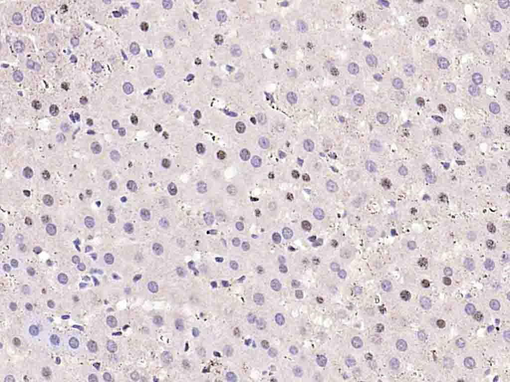
Paraformaldehyde-fixed, paraffin embedded (Rat liver); Antigen retrieval by boiling in sodium citrate buffer (pH6.0) for 15min; Block endogenous peroxidase by 3% hydrogen peroxide for 20 minutes; Blocking buffer (normal goat serum) at 37°C for 30min; Antibody incubation with (Cyclin D1) Polyclonal Antibody, Unconjugated (bs-0623R) at 1:400 overnight at 4°C, followed by operating according to SP Kit(Rabbit) (sp-0023) instructionsand DAB staining.
-
Sample:
A549 Cell (Human) Lysate at 30 ug
Primary: Anti-Cyclin D1 (bs-0623R) at 1/300 dilution
Secondary: IRDye800CW Goat Anti-Rabbit IgG at 1/20000 dilution
Predicted band size: 32 kD
Observed band size: 35 kD
-
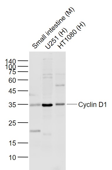
Sample:
Lane 1: Small intestine (Mouse) Lysate at 40 ug
Lane 2: U251 (Human) Cell Lysate at 30 ug
Lane 3: HT1080 (Human) Cell Lysate at 30 ug
Primary: Anti-Cyclin D1 (bs-0623R) at 1/1000 dilution
Secondary: IRDye800CW Goat Anti-Rabbit IgG at 1/20000 dilution
Predicted band size: 34 kD
Observed band size: 34 kD
-
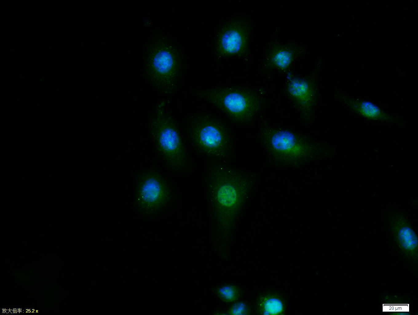
Tissue/cell:MCF7 cell; 4% Paraformaldehyde-fixed; Triton X-100 at room temperature for 20 min; Blocking buffer (normal goat serum, C-0005) at 37°C for 20 min; Antibody incubation with (Cyclin D1) polyclonal Antibody, Unconjugated (bs-0623R) 1:100, 90 minutes at 37°C; followed by a FITC conjugated Goat Anti-Rabbit IgG antibody at 37°C for 90 minutes, DAPI (blue, C02-04002) was used to stain the cell nuclei.
-
Paraformaldehyde-fixed, paraffin embedded (rat liver); Antigen retrieval by boiling in sodium citrate buffer (pH6.0) for 15min; Block endogenous peroxidase by 3% hydrogen peroxide for 20 minutes; Blocking buffer (normal goat serum) at 37°C for 30min; Antibody incubation with (Cyclin D1) Polyclonal Antibody, Unconjugated (bs-0623R) at 1:200 overnight at 4°C, followed by operating according to SP Kit(Rabbit) (sp-0023) instructionsand DAB staining.
-
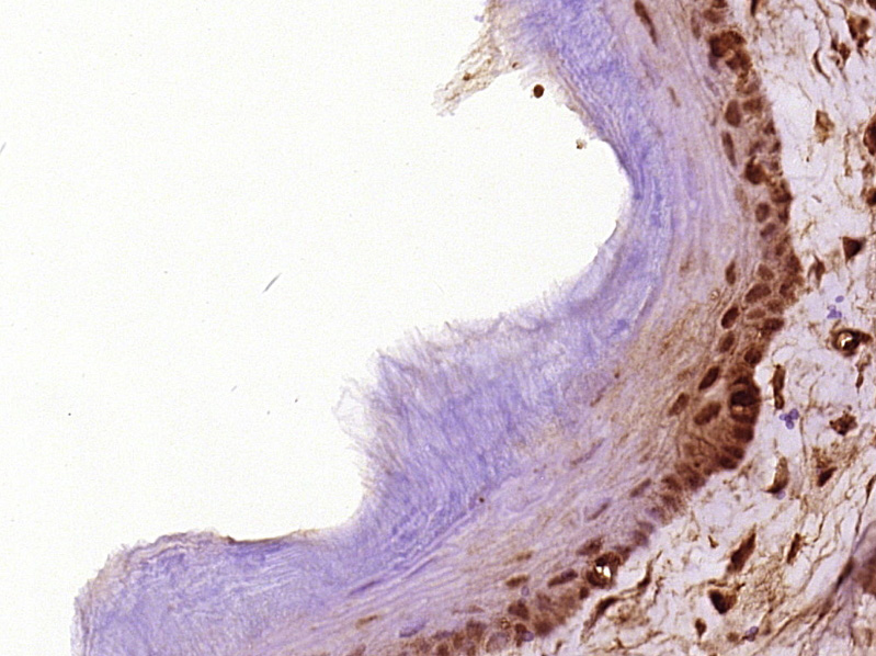
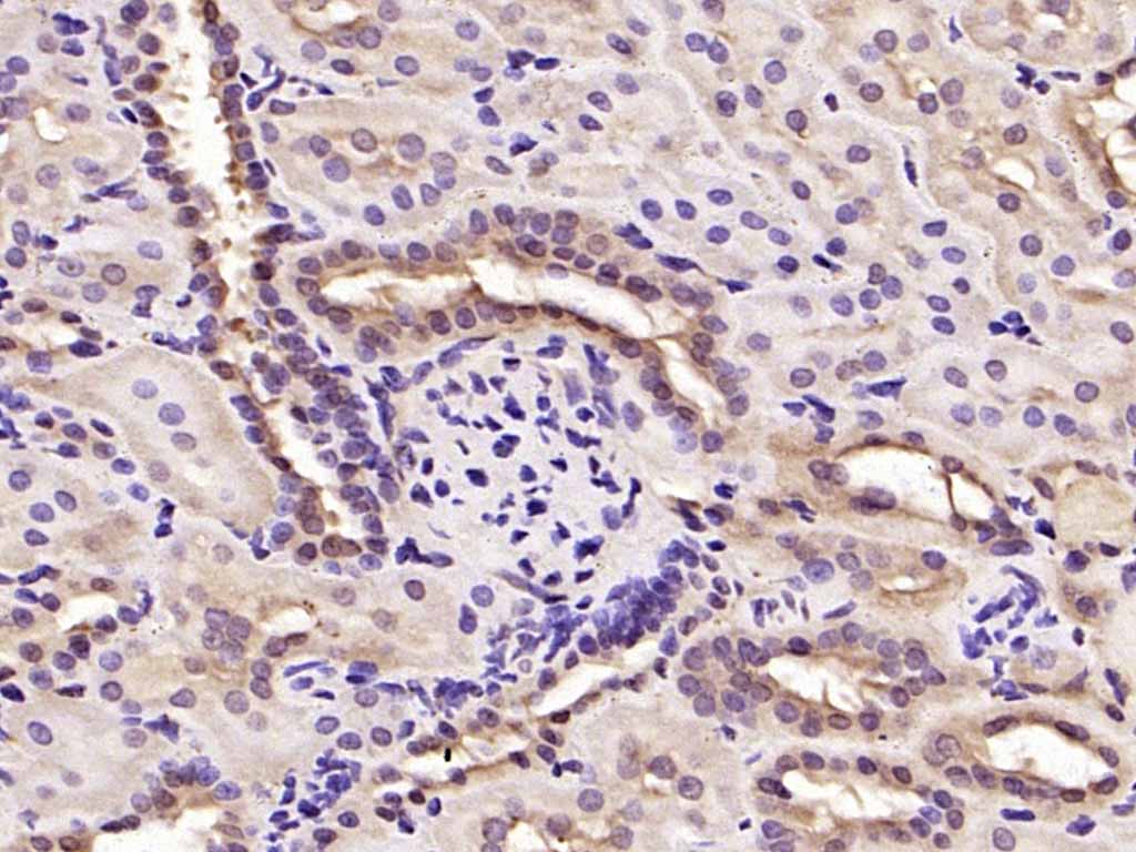
Paraformaldehyde-fixed, paraffin embedded (rat kidney); Antigen retrieval by boiling in sodium citrate buffer (pH6.0) for 15min; Block endogenous peroxidase by 3% hydrogen peroxide for 20 minutes; Blocking buffer (normal goat serum) at 37°C for 30min; Antibody incubation with (Cyclin D1) Polyclonal Antibody, Unconjugated (bs-0623R) at 1:200 overnight at 4°C, followed by operating according to SP Kit(Rabbit) (sp-0023) instructionsand DAB staining.
-
Tissue/cell: human placenta tissue; 4% Paraformaldehyde-fixed and paraffin-embedded;
Antigen retrieval: citrate buffer ( 0.01M, pH 6.0 ), Boiling bathing for 15min; Block endogenous peroxidase by 3% Hydrogen peroxide for 30min; Blocking buffer (normal goat serum,C-0005) at 37℃ for 20 min;
Incubation: Anti-Cyclin D1 Polyclonal Antibody, Unconjugated(bs-0623R) 1:200, overnight at 4°C, followed by conjugation to the secondary antibody(SP-0023) and DAB(C-0010) staining
-
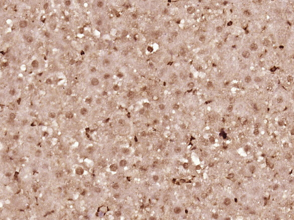
Tissue/cell: human endometrium carcinoma; 4% Paraformaldehyde-fixed and paraffin-embedded;
Antigen retrieval: citrate buffer ( 0.01M, pH 6.0 ), Boiling bathing for 15min; Block endogenous peroxidase by 3% Hydrogen peroxide for 30min; Blocking buffer (normal goat serum,C-0005) at 37℃ for 20 min;
Incubation: Anti-Cyclin D1 Polyclonal Antibody, Unconjugated(bs-0623R) 1:200, overnight at 4°C, followed by conjugation to the secondary antibody(SP-0023) and DAB(C-0010) staining
-
Sample: A549 Lysate at 40 ug
Primary: Anti-CyclinD1 (bs-0623R) at 1/300 dilution
Secondary: IRDye800CW Goat Anti-Rabbit IgG at 1/10000 dilution
Predicted band size: 32 kD
Observed band size: 35 kD
-
Blank control: RSC96(blue), the cells were fixed with 2% paraformaldehyde (10 min) and then permeabilized with ice-cold 90% methanol for 30 min on ice.
Isotype Control Antibody: Rabbit IgG(orange) ;
Secondary Antibody: Goat anti-rabbit IgG-PE(white blue),
Dilution: 1:200 in 1 X PBS containing 0.5% BSA ;
Primary Antibody Dilution: 1μg in 100 μL1X PBS containing 0.5% BSA(green).
-
Sample:
Mcf-7(Human) Cell Lysate at 30 ug
Primary: Anti-Cyclin D1 (bs-0623R) at 1/300 dilution
Secondary: IRDye800CW Goat Anti-Rabbit IgG at 1/20000 dilution
Predicted band size: 32 kD
Observed band size: 32 kD
RRID:AB_10856925
产品名称:Rabbit Anti-Cyclin D1 antibody
别名: CyclinD1; Cyclin-D1; B cell ccl/lymphoma 1; B cell leukemia 1; B-cell CLL/lymphoma 1; B-cell leukemia 1; B-cell lymphoma 1 protein; BCL-1; BCL1; BCL1 oncogene; CCND 1; CCND1; CCND1 protein; CCND1/FSTL3 fusion gene, included; CCND1/IGHG1 fusion gene; CCND1
中文名称:周期素D1抗体
英文名称:Rabbit Anti-Cyclin D1 antibody
抗体来源: Rabbit
克隆类型:多克隆
细胞定位:细胞核,细胞浆,细胞膜
性 状:Liquid
亚 型:IgG
纯化方法:affinity purified by Protein A
保存条件:Shipped at 4℃. Store at -20 °C for one year. Avoid repeated freeze/thaw cycles.
免 疫 原:KLH conjugated synthetic peptide derived from human Cyclin D1
抗原表位:61-110/295
SWISS:P25322
Gene ID :595
Human Gene ID:595
The protein encoded by this gene belongs to the highly conserved cyclin family, whose members are characterized by a dramatic periodicity in protein abundance throughout the cell cycle. Cyclins function as regulators of CDK kinases. Different cyclins exhibit distinct expression and degradation patterns which contribute to the temporal coordination of each mitotic event. This cyclin forms a complex with and functions as a regulatory subunit of CDK4 or CDK6, whose activity is required for cell cycle G1/S transition. This protein has been shown to interact with tumor suppressor protein Rb and the expression of this gene is regulated positively by Rb. Mutations, amplification and overexpression of this gene, which alters cell cycle progression, are observed frequently in a variety of tumors and may contribute to tumorigenesis. [provided by RefSeq, Jul 2008].
Function:Regulatory component of the cyclin D1-CDK4 (DC) complexthat phosphorylates and inhibits members of the retinoblastoma (RB)protein family including RB1 and regulates the cell-cycle duringG(1)/S transition. Phosphorylation of RB1 allows dissociation ofthe t
Subunit:Interacts with FBXO4. Interacts witheither CDK4 or CDK6 protein kinase to form a serine/threoninekinase holoenzyme complex. The cyclin subunit imparts substratespecificity to the complex. Component of the ternary complexCCND1/CDK4/CDKN1B required for nucl
Subcellular Location:Nucleus. Cytoplasm. Membrane. Note=CyclinD-CDK4 complexes accumulate at the nuclear membrane and are thentranslocated to the nucleus through interaction with KIP/CIP familymembers.
Post-translational modifications:Phosphorylation at Thr-286 by MAP kinases is required forubiquitination and degradation following DNA damage. It probablyplays an essential role for recognition by the FBXO31 component ofSCF (SKP1-cullin-F-box) protein ligase complex.
Ubiquitinated, p
DISEASE:Note=A chromosomal aberration involving CCND1 may be acause of B-lymphocytic malignancy, particularly mantle-celllymphoma (MCL). Translocation t(11;14)(q13;q32) with immunoglobulingene regions. Activation of CCND1 may be oncogenic by directlyaltering prog
Similarity:Belongs to the cyclin family. Cyclin D subfamily.
Important Note:This product as supplied is intended for research use only, not for use in human, therapeutic or diagnostic applications.
 400-901-9800


 说明书
说明书 联系我们
联系我们 打印此页面
打印此页面 收藏
收藏