| Rabbit Anti-E cadherin antibody |
| 反应物种(预测) |
Mouse,Rat,Chicken,Dog,Pig,Cow,Horse |
| 产品应用(已验证) |
WB,IHC,ICC,FCM |
| 产品应用(可尝试) |
IF,ELISA |
| 推荐稀释比例 |
WB=1:500-2000,Elisa=1:5000-10000,IHC-P=1:100-500,IHC-F=1:100-500,Flow Cyt=1μg/Test,IF=1:100-500,ICC=1:100, |
| 研究领域 |
肿瘤,细胞生物,免疫学,细胞粘附分子,细胞表面分子,上皮细胞, |
| 标签 |
Array |
-
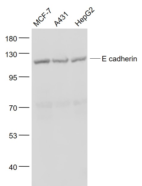
Sample:
MCF-7(Human) Cell Lysate at 30 ug
A431(Human) Cell Lysate at 30 ug
HepG2(Human) Cell Lysate at 30 ug
Primary: Anti- E cadherin (bs-1519R) at 1/1000 dilution
Secondary: IRDye800CW Goat Anti-Rabbit IgG at 1/20000 dilution
Predicted band size: 90/97 kD
Observed band size: 125 kD
-
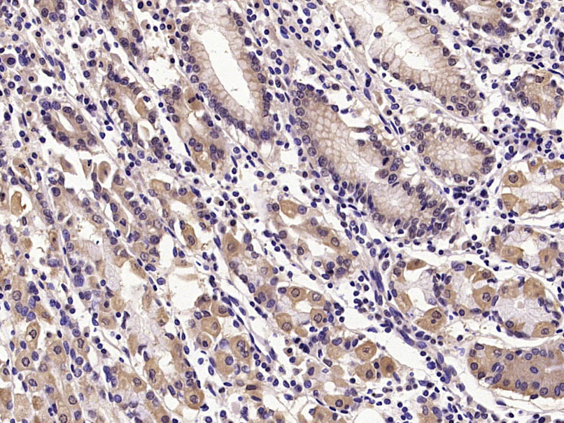
Paraformaldehyde-fixed, paraffin embedded (Human stomach); Antigen retrieval by microwave in sodium citrate buffer (pH6.0) ; Block endogenous peroxidase by 3% hydrogen peroxide for 30 minutes; Blocking buffer (3% BSA) at RT for 30min; Antibody incubation with (E cadherin) Polyclonal Antibody, Unconjugated (bs-1519R) at 1:400 overnight at 4℃, followed by conjugation to the secondary antibody (labeled with HRP)and DAB staining.
-
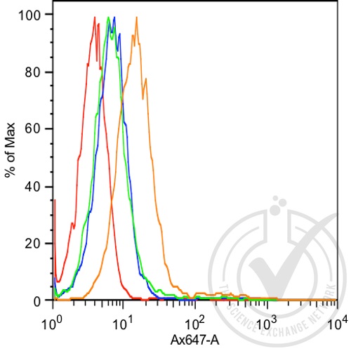
Histogram of MCF7 cells stained with anti-E-cadherin (orange), isotype control antibody (green), secondary antibody only (blue) and unstained (red).
-
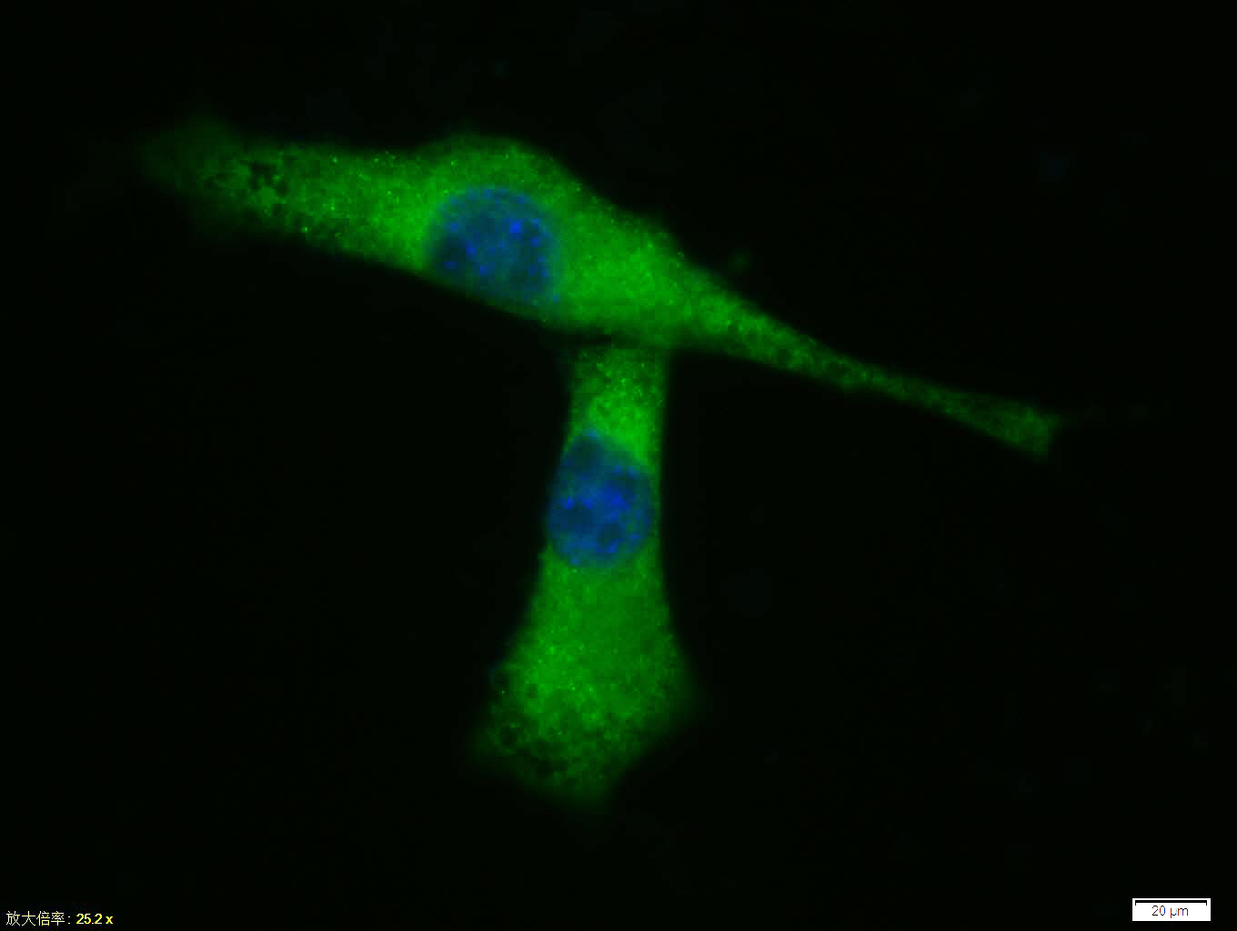
Tissue/cell: A431 cell; 4% Paraformaldehyde-fixed; Triton X-100 at room temperature for 20 min; Blocking buffer (normal goat serum, C-0005) at 37°C for 20 min; Antibody incubation with (E cadherin) polyclonal Antibody, Unconjugated (bs-1519R) 1:100, 90 minutes at 37°C; followed by a FITC conjugated Goat Anti-Rabbit IgG antibody at 37°C for 90 minutes, DAPI (blue, C02-04002) was used to stain the cell nuclei.
-
Sample:
Lane 1: MDA-MB-231 (Human) Cell Lysate at 30 ug
Lane 2: A431 (Human) Cell Lysate at 30 ug
Lane 3: A549 (Human) Cell Lysate at 30 ug
Lane 4: HepG2 (Human) Cell Lysate at 30 ug
Primary:
Anti- E cadherin (bs-1519R) at 1/500 dilution
Secondary: IRDye800CW Goat Anti-Rabbit IgG at 1/20000 dilution
Predicted band size: 125 kD
Observed band size: 120 kD
RRID:AB_10855050
产品名称:Rabbit Anti-E cadherin antibody
别名: E-cadherin; anion exchanger protein 3; Arc 1; Cadherin 1; cadherin 1 type 1 E-cadherin; Cadherin1; CAM 120/80; CD 234; CD324; CD324 antigen; CDH1; CDHE; ECAD; Epithelial cadherin; epithelial calcium dependant adhesion protein; LCAM; Liver cell adhesion mo
中文名称:上皮钙粘附分子抗体
英文名称:Rabbit Anti-E cadherin antibody
抗体来源: Rabbit
克隆类型:多克隆
细胞定位:细胞膜
性 状:Liquid
亚 型:IgG
纯化方法:affinity purified by Protein A
保存条件:Shipped at 4℃. Store at -20 °C for one year. Avoid repeated freeze/thaw cycles.
免 疫 原:KLH conjugated synthetic peptide derived from human E-cadherin
抗原表位:841-882/882
抗原细胞定位:Cytoplasmic
SWISS:P12830
Gene ID :999
Human Gene ID:999
This gene encodes a classical cadherin of the cadherin superfamily. Alternative splicing results in multiple transcript variants, at least one of which encodes a preproprotein that is proteolytically processed to generate the mature glycoprotein. This calcium-dependent cell-cell adhesion protein is comprised of five extracellular cadherin repeats, a transmembrane region and a highly conserved cytoplasmic tail. Mutations in this gene are correlated with gastric, breast, colorectal, thyroid and ovarian cancer. Loss of function of this gene is thought to contribute to cancer progression by increasing proliferation, invasion, and/or metastasis. The ectodomain of this protein mediates bacterial adhesion to mammalian cells and the cytoplasmic domain is required for internalization. This gene is present in a gene cluster with other members of the cadherin family on chromosome 16. [provided by RefSeq, Nov 2015]
Function:Cadherins are calcium-dependent cell adhesion proteins. They preferentially interact with themselves in a homophilic manner in connecting cells; cadherins may thus contribute to the sorting of heterogeneous cell types. CDH1 is involved in mechanisms regul
Subunit:Homodimer.
Subcellular Location:Cell junction. Cell membrane; Single-pass type I membrane protein.
Tissue Specificity:Non-neural epithelial tissues.
Post-translational modifications:During apoptosis or with calcium influx, cleaved by a membrane-bound metalloproteinase (ADAM10), PS1/gamma-secretase and caspase-3 to produce fragments of about 38 kDa (E-CAD/CTF1), 33 kDa (E-CAD/CTF2) and 29 kDa (E-CAD/CTF3), respectively. Processing by
DISEASE:Defects in CDH1 are involved in dysfunction of the cell-cell adhesion system, triggering cancer invasion (gastric, breast, ovary, endometrium and thyroid) and metastasis.
Defects in CDH1 are a cause of gastric cancer [MIM:137215]; also known as heredi
Similarity:Contains 5 cadherin domains.
Important Note:This product as supplied is intended for research use only, not for use in human, therapeutic or diagnostic applications.
 400-901-9800


 说明书
说明书 联系我们
联系我们 打印此页面
打印此页面 收藏
收藏