| Rabbit Anti-CD163 antibody |
| 反应物种(预测) |
Rat,Dog,Pig,Horse |
| 产品应用(已验证) |
WB,IHC,ICC,IF,FCM |
| 产品应用(可尝试) |
ELISA |
| 推荐稀释比例 |
WB=1:500-2000,Elisa=1:5000-10000,IHC-P=1:100-500,IHC-F=1:100-500,Flow Cyt=1ug/Test,IF=1:100-500,ICC=1:100-500, |
| 研究领域 |
心血管,免疫学,细胞膜受体 |
| 标签 |
Array |
-
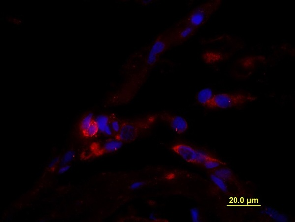
Formalin-fixed and paraffin embedded Human testis tissue labeled with unconjugated Anti-CD163/M130 Polyclonal Antibody, unconjugated (bs-2527R) at 1:100 for 40 minutes at 37°C followed by labeling Donkey Anti-Rabbit, Cy3 conjugated 1:300, 60 minutes at 37°C. DAPI nuclear stain employed. Image shows membrane staining of testicular macrophages in the interstitial compartment of the testis, while cells in the seminiferous tubules are negative.
-
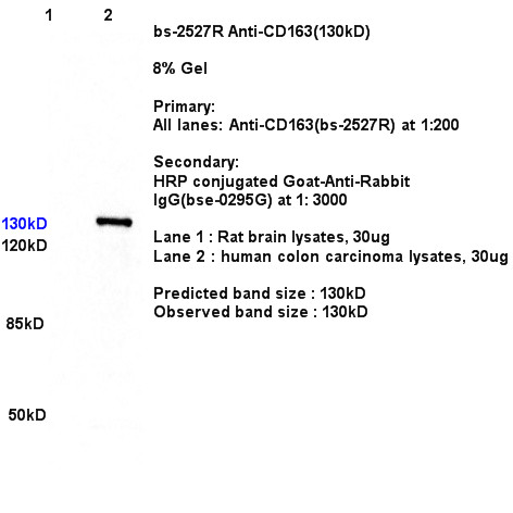
Sample:
Lane1: Brain (Rat) Lysate at 30 ug
Lane2: Colon carcinoma(Human) Lysate at 30 ug
Primary: Anti- CD163 (bs-2527R) at 1/300 dilution
Secondary: IRDye800CW Goat Anti-Rabbit IgG at 1/20000 dilution
Predicted band size: 130 kD
Observed band size: 130kD
-
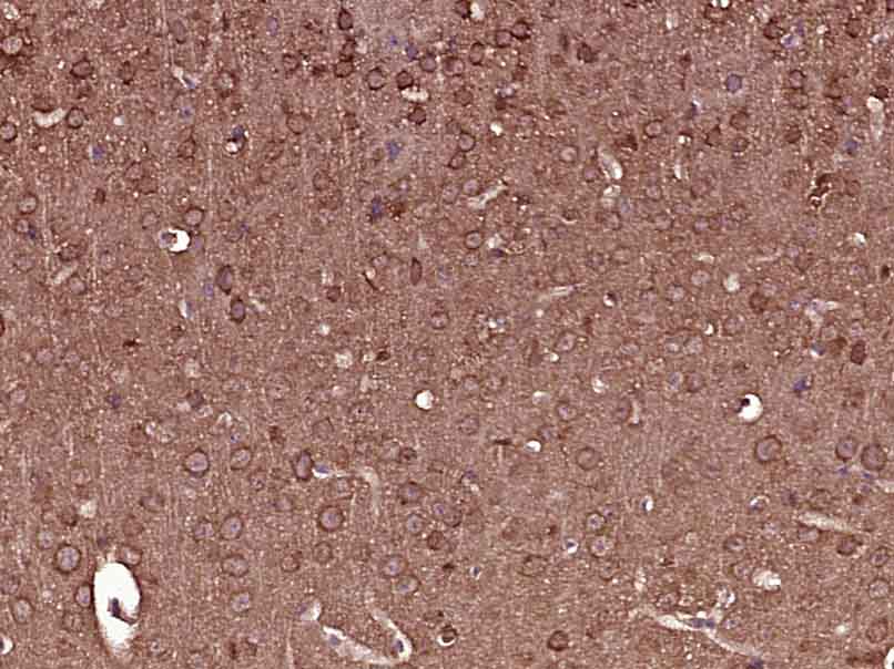
Paraformaldehyde-fixed, paraffin embedded (Mouse brain); Antigen retrieval by boiling in sodium citrate buffer (pH6.0) for 15min; Block endogenous peroxidase by 3% hydrogen peroxide for 20 minutes; Blocking buffer (normal goat serum) at 37°C for 30min; Antibody incubation with (CD163) Polyclonal Antibody, Unconjugated (bs-2527R) at 1:400 overnight at 4°C, followed by operating according to SP Kit(Rabbit) (sp-0023) instructionsand DAB staining.
-
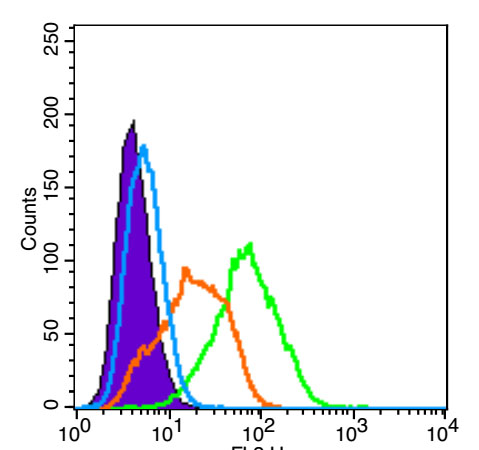
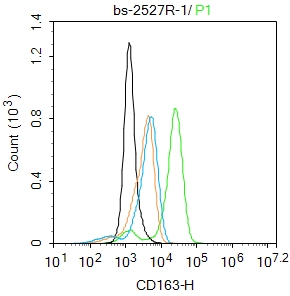
Blank control (Black line): U2OS.
Primary Antibody (green line): Rabbit Anti-CD163 antibody (bs-2527R)
Dilution: 3μg /10^6 cells;
Isotype Control Antibody (orange line): Rabbit IgG .
Secondary Antibody (white blue line): Goat anti-rabbit IgG-PE
Dilution: 1μg /test.
Protocol
The cells were fixed with 4% paraformaldehyde for 10 min at room temperature. Cells incubated in 5%BSA to block non-specific protein-protein interactions for 30 min at room temperature. The cells were then stained with Primary Antibody for 30 min at room temperature. The secondary antibody used for 40 min at room temperature. Acquisition of 20,000 events was performed.
-
Blank control: THP-1.
Primary Antibody (green line): Rabbit Anti-CD163 antibody (bs-2527R)
Dilution: 1μg /10^6 cells;
Isotype Control Antibody (orange line): Rabbit IgG .
Secondary Antibody : Goat anti-rabbit IgG-FITC
Dilution: 1μg /test.
Protocol
The cells were fixed with 4% PFA (10min at room temperature)and then permeabilized with 0.1% PBST for 20 min at room temperature. The cells were then incubated in 5%BSA to block non-specific protein-protein interactions for 30 min at room temperature .Cells stained with Primary Antibody for 30 min at room temperature. The secondary antibody used for 40 min at room temperature. Acquisition of 20,000 events was performed.
RRID:AB_10856166
产品名称:Rabbit Anti-CD163 antibody
别名: Scavenger receptor cysteine-rich type 1 protein M130; CD 163; CD163 antigen; CD163 molecule; Hemoglobin Scavenger Receptor; M130; M130 antigen precursor; Macrophage associated antigen; MM130; C163A_HUMAN.
中文名称:CD163抗体
英文名称:Rabbit Anti-CD163 antibody
抗体来源: Rabbit
克隆类型:多克隆
细胞定位:细胞膜,分泌型蛋白
性 状:Liquid
亚 型:IgG
纯化方法:affinity purified by Protein A
保存条件:Shipped at 4℃. Store at -20 °C for one year. Avoid repeated freeze/thaw cycles.
免 疫 原:KLH conjugated synthetic peptide derived from human CD163
抗原表位:1001-1121/1156
抗原细胞定位:Extracellular
SWISS:Q86VB7
Gene ID :9332
Human Gene ID:9332
The protein encoded by this gene is a member of the scavenger receptor cysteine-rich (SRCR) superfamily, and is exclusively expressed in monocytes and macrophages. It functions as an acute phase-regulated receptor involved in the clearance and endocytosis of hemoglobin/haptoglobin complexes by macrophages, and may thereby protect tissues from free hemoglobin-mediated oxidative damage. This protein may also function as an innate immune sensor for bacteria and inducer of local inflammation. Alternatively spliced transcript variants encoding different isoforms have been described for this gene. [provided by RefSeq, Aug 2011]
Function:Acute phase-regulated receptor involved in clearance and endocytosis of hemoglobin/haptoglobin complexes by macrophages and may thereby protect tissues from free hemoglobin-mediated oxidative damage. May play a role in the uptake and recycling of iron, vi
Subcellular Location:Secreted and Cell membrane. Isoform 1 and isoform 2 show a lower surface expression when expressed in cells.
Tissue Specificity:Expressed in monocytes and mature macrophages such as Kupffer cells in the liver, red pulp macrophages in the spleen, cortical macrophages in the thymus, resident bone marrow macrophages and meningeal macrophages of the central nervous system. Expressed a
Post-translational modifications:A soluble form (sCD163) is produced by proteolytic shedding which can be induced by lipopolysaccharide, phorbol ester and Fc region of immunoglobulin gamma. This cleavage is dependent on protein kinase C and tyrosine kinases and can be blocked by protease
Similarity:Contains 9 SRCR domains.
Important Note:This product as supplied is intended for research use only, not for use in human, therapeutic or diagnostic applications.
 400-901-9800


 说明书
说明书 联系我们
联系我们 打印此页面
打印此页面 收藏
收藏