| Rabbit Anti-Elastin antibody |
| 产品应用(已验证) |
WB,IHC,ICC,FCM |
| 产品应用(可尝试) |
IF,ELISA |
| 推荐稀释比例 |
WB=1:500-2000,Elisa=1:5000-10000,IHC-P=1:100-500,IHC-F=1:100-500,Flow Cyt=1μg /test,IF=1:100-500,ICC=1:100, |
| 研究领域 |
肿瘤,免疫学,神经生物学,转录调节因子 |
| 标签 |
Array |
-
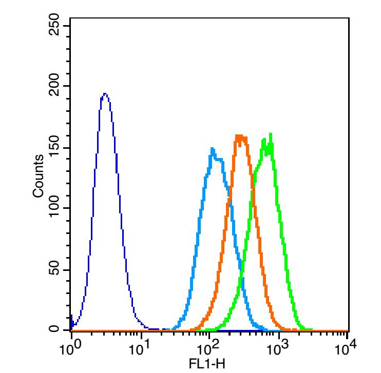
The figure annotation: The blue histogram is unstained cells.The green histogram is cells stained with Rabbit Anti-alpha Elastin/Tropoelastin antibody (bs-1756R)plus secondary antibody.
Controls: Positive control: A549 cells .Isotype control: Cell lines treated with rabbit IgG (bs-0295P)instead of the primary antibody to confirm that primary antibody binding is specific.Secondary only control: Both cell lines treated with Goat Anti-rabbit IgG/FITC antibody (bs-0295G-FITC) to confirm no background signal produced from secondary antibody alone. 1ug in 100uL 1 X PBS containing 0.5% BSA.
-
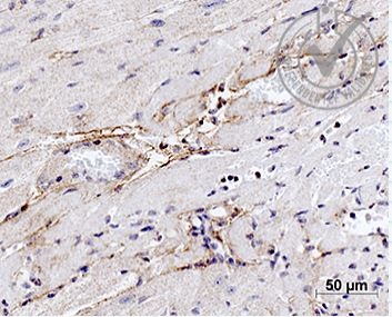
Images provided the Independent Validation Program (badge number 28751): Formalin-fixed and paraffin embedded mouse heart labeled with Rabbit Anti-alpha Elastin Polyclonal Antibody (bs-1756R) at 1:1000 4℃ temperature overnight 4℃followed by conjugation to secondary antibody.
-
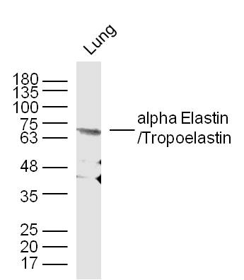
Sample: Lung (Mouse) Lysate at 30 ug
Primary: Anti- alpha Elastin/Tropoelastin (bs-1756R) at 1/300 dilution
Secondary: IRDye800CW Goat Anti-Mouse IgG at 1/10000 dilution
Predicted band size: 70 kD
Observed band size: 68 kD
-
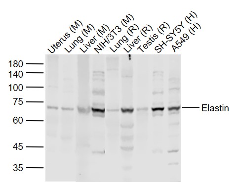
Sample:
Lane 1: Uterus (Mouse) Lysate at 40 ug
Lane 2: Lung (Mouse) Lysate at 40 ug
Lane 3: Liver (Mouse) Lysate at 40 ug
Lane 4: NIH/3T3 (Mouse) Cell Lysate at 30 ug
Lane 5: Lung (Rat) Lysate at 40 ug
Lane 6: Liver (Rat) Lysate at 40 ug
Lane 7: Testis (Rat) Lysate at 40 ug
Lane 8: SH-SY5Y (Human) Cell Lysate at 30 ug
Lane 9: A549 (Human) Cell Lysate at 30 ug
Primary: Anti-Elastin (bs-1756R) at 1/1000 dilution
Secondary: IRDye800CW Goat Anti-Rabbit IgG at 1/20000 dilution
Predicted band size: 66 kD
Observed band size: 68 kD
RRID:AB_10856940
产品名称:Rabbit Anti-Elastin antibody
别名: alpha Elastin/Tropoelastin; Elastin isoform a; ELN; ELN_HUMAN; elastin isoform m precursor; FLJ38671; FLJ43523; Supravalvular aortic stenosis; Tropoelastin; Williams Beuren syndrome; Williams syndrome region; ADCL1; SVAS; WBS; WS.
中文名称:弹性蛋白抗体
英文名称:Rabbit Anti-Elastin antibody
抗体来源: Rabbit
克隆类型:多克隆
细胞定位:细胞外基质,分泌型蛋白
性 状:Liquid
亚 型:IgG
纯化方法:affinity purified by Protein A
保存条件:Shipped at 4℃. Store at -20 °C for one year. Avoid repeated freeze/thaw cycles.
免 疫 原:KLH conjugated synthetic peptide derived from human Elastin
抗原表位:681-786/786
SWISS:P15502
Gene ID :2006
Human Gene ID:2006
This gene encodes a protein that is one of the two components of elastic fibers. Elastic fibers comprise part of the extracellular matrix and confer elasticity to organs and tissues including the heart, skin, lungs, ligaments, and blood vessels. The encoded protein is rich in hydrophobic amino acids such as glycine and proline, which form mobile hydrophobic regions bounded by crosslinks between lysine residues. Degradation products of the encoded protein, known as elastin-derived peptides or elastokines, bind the elastin receptor complex and other receptors and stimulate migration and proliferation of monocytes and skin fibroblasts. Elastokines can also contribute to cancer progression. Deletions and mutations in this gene are associated with supravalvular aortic stenosis (SVAS) and autosomal dominant cutis laxa. [provided by RefSeq, Aug 2017].
Function:Major structural protein of tissues such as aorta and nuchal ligament, which must expand rapidly and recover completely. Molecular determinant of the late arterial morphogenesis, stabilizing arterial structure by regulating proliferation and organization
Subunit:The polymeric elastin chains are cross-linked together into an extensible 3D network. Forms a ternary complex with BGN and MFAP2. Interacts with MFAP2 via divalent cations (calcium > magnesium > manganese) in a dose-dependent and saturating manner.
Subcellular Location:Secreted, extracellular space, extracellular matrix. Note=Extracellular matrix of elastic fibers.
Tissue Specificity:Expressed within the outer myometrial smooth muscle and throughout the arteriolar tree of uterus (at protein level). Also expressed in the large arteries, lung and skin.
DISEASE:Defects in ELN are the cause of cutis laxa, autosomal dominant, type 1 (ADCL1) . A connective tissue disorder characterized by loose, hyperextensible skin with decreased resilience and elasticity leading to a premature aged appearance. Face, hands, feet,
Similarity:Belongs to the elastin family.
Important Note:This product as supplied is intended for research use only, not for use in human, therapeutic or diagnostic applications.
 400-901-9800


 说明书
说明书 联系我们
联系我们 打印此页面
打印此页面 收藏
收藏