| Rabbit Anti-phospho-ERK1 + 2 (Thr183/Tyr185) antibody |
| 反应物种(预测) |
Rat,Chicken,Dog,Cow,Horse,Rabbit,GuineaPig |
| 产品应用(已验证) |
WB,IHC,ICC,IF |
| 产品应用(可尝试) |
ELISA |
| 推荐稀释比例 |
WB=1:500-2000,Elisa=1:5000-10000,IHC-P=1:100-500,IHC-F=1:100-500,IF=1:100-500,ICC=1:100, |
| 研究领域 |
免疫学,神经生物学,信号转导,干细胞,激酶和磷酸酶 |
| 标签 |
Array |
-
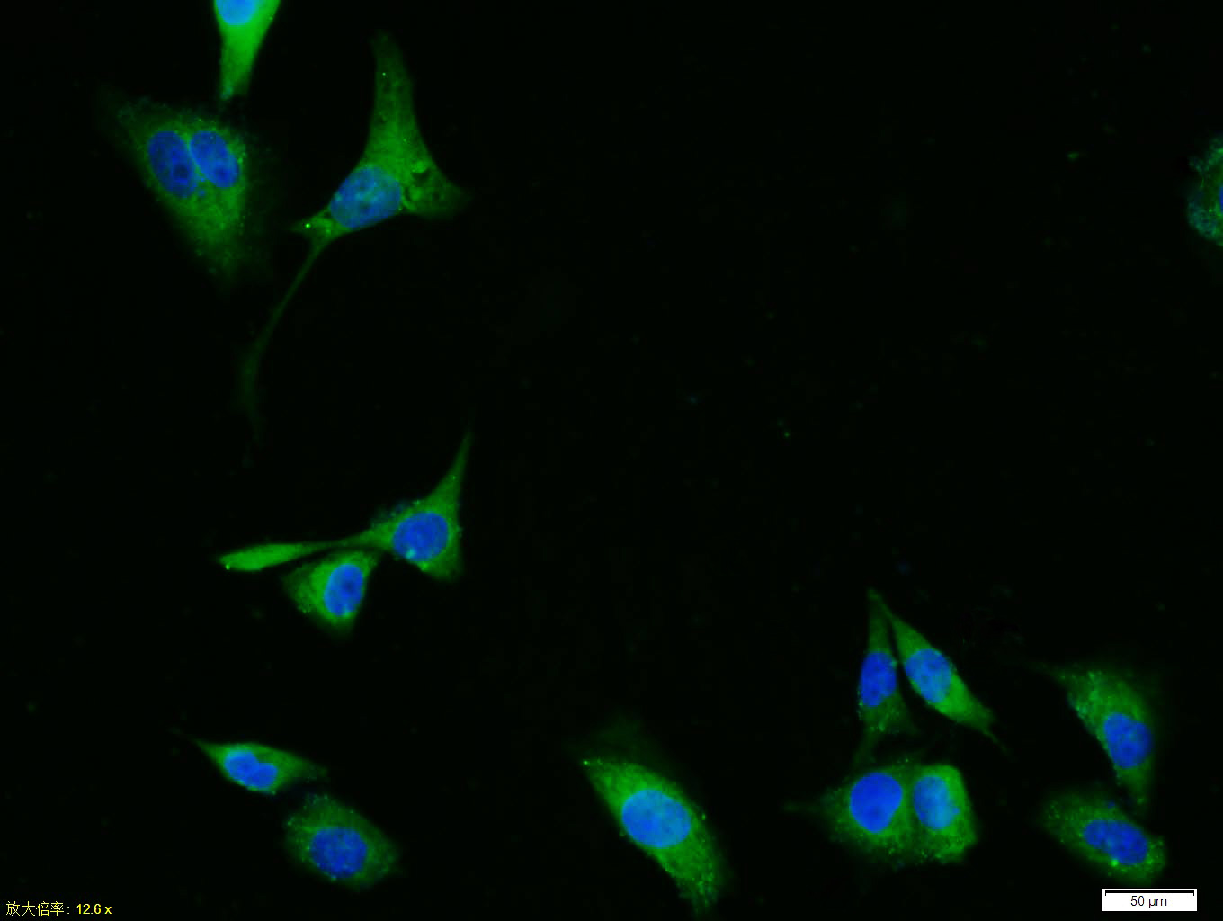
Tissue/cell: HUVEC cell; 4% Paraformaldehyde-fixed; Triton X-100 at room temperature for 20 min; Blocking buffer (normal goat serum, C-0005) at 37°C for 20 min; Antibody incubation with (phospho-ERK1 + 2 (Thr183/Tyr185)) Polyclonal Antibody, Unconjugated (bs-1522R) 1:100, 90 minutes at 37°C; followed by a conjugated Goat Anti-Rabbit IgG antibody (bs-0295G-FITC) at 37°C for 90 minutes, DAPI (blue, C02-04002) was used to stain the cell nuclei.
-
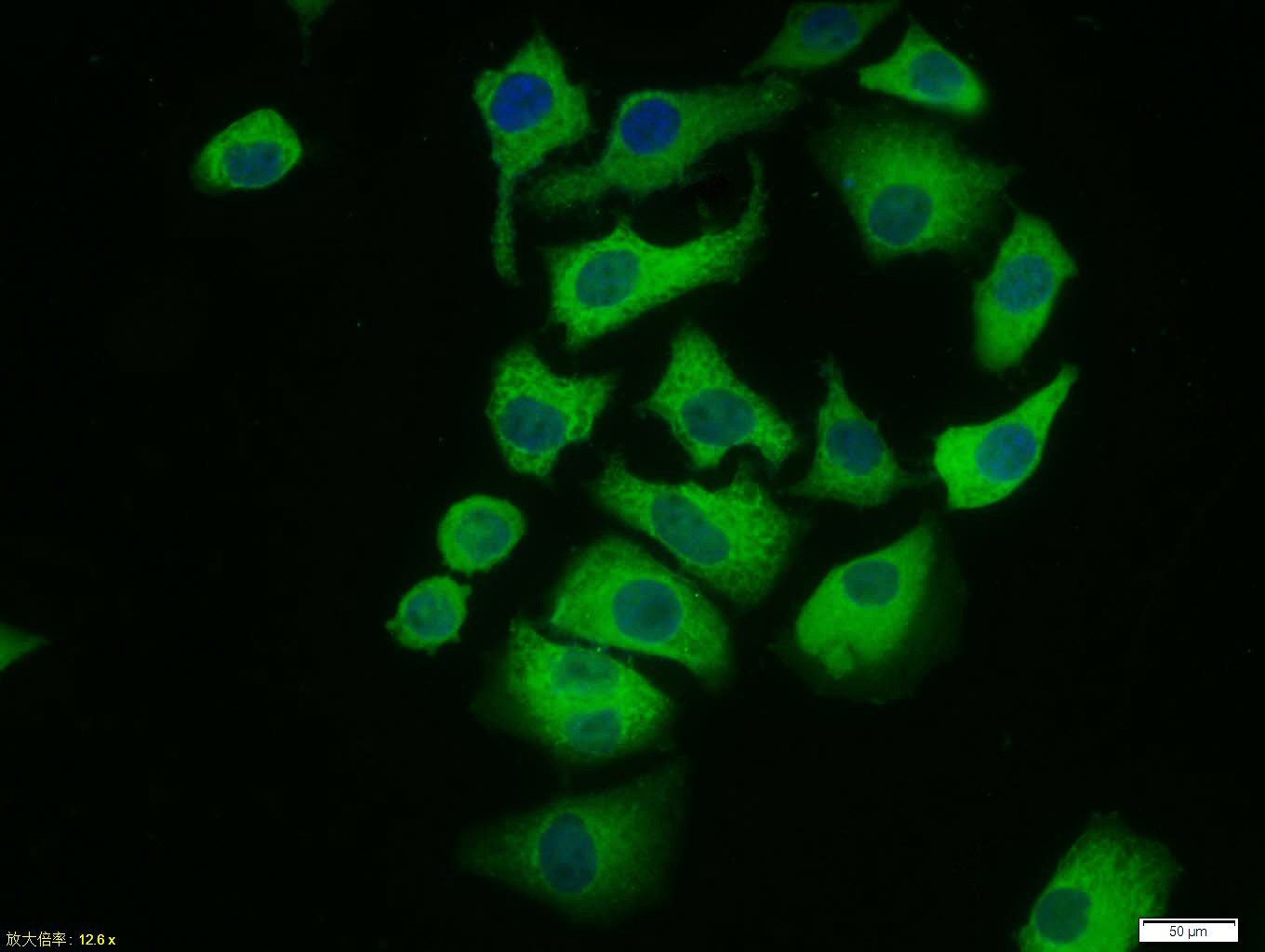
Tissue/cell: Hela cell; 4% Paraformaldehyde-fixed; Triton X-100 at room temperature for 20 min; Blocking buffer (normal goat serum, C-0005) at 37°C for 20 min; Antibody incubation with (phospho-ERK1 + 2 (Thr183/Tyr185)) polyclonal Antibody, Unconjugated (bs-1522R) 1:100, 90 minutes at 37°C; followed by a FITC conjugated Goat Anti-Rabbit IgG antibody at 37°C for 90 minutes, DAPI (blue, C02-04002) was used to stain the cell nuclei.
-
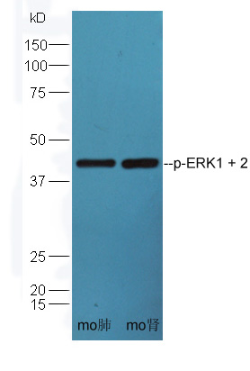
Sample:
Lane1: Lung (Mouse) Lysate at 30 ug
Lane2: Kidney (Mouse) Lysate at 30 ug
Primary:Anti-phospho-ERK1+2 (bs-1522R) at 1:300 dilution;
Secondary:HRP conjugated Goat-Anti-Rabbit IgG(bs-0295G-HRP) at 1: 5000 dilution;
Predicted band size:42/44 kD
Observed band size:42 kD
-
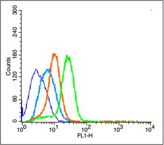
Blank control (blue line): U251 (blue).
Primary Antibody (green line): Rabbit Anti-phospho-ERK1 + 2 (Thr183185) antibody (bs-1522R)
Dilution: 3μg /10^6 cells;
Isotype Control Antibody (orange line): Rabbit IgG .
Secondary Antibody (white blue line): Goat anti-rabbit IgG-PE
Dilution: 1μg /test.
Protocol
The cells were fixed with 2% paraformaldehyde (10 min)and then permeabilized with 0.1% PBS-Tween for 20 min at room temperature. Cells stained with Primary Antibody for 30 min at room temperature. The cells were then incubated in 1 X PBS/2%BSA/10% goat serum to block non-specific protein-protein interactions followed by the antibody for 15 min at room temperature. The secondary antibody used for 40 min at room temperature. Acquisition of 20,000 events was performed.
RRID:AB_10885923
产品名称:Rabbit Anti-phospho-ERK1 + 2 (Thr183/Tyr185) antibody
别名: ERK1 + ERK2 (phospho Thr183/Tyr185); phospho-ERK1/MAPK-1/2(Thr183/Tyr185); ERK 1; ERK 2; ERK-2; ERK1; ERK2; ERT1; ERT2; Extracellular signal regulated kinase 1; Extracellular signal regulated kinase 1; Extracellular signal regulated kinase 2; Extracellula
中文名称:磷酸化丝裂原活化蛋白激酶1/2抗体
英文名称:Rabbit Anti-phospho-ERK1 + 2 (Thr183/Tyr185) antibody
抗体来源: Rabbit
克隆类型:多克隆
细胞定位:细胞核,细胞浆
性 状:Liquid
亚 型:IgG
纯化方法:affinity purified by Protein A
保存条件:Shipped at 4℃. Store at -20 °C for one year. Avoid repeated freeze/thaw cycles.
免 疫 原:KLH conjugated Synthesised phosphopeptide derived from mouse ERK1 around the phosphorylation site of Thr183/Tyr185
抗原表位:FL(p-T)E(p-Y)V
SWISS:P63085
Gene ID :5595
Human Gene ID:5595
Mitogen-activated protein kinase (MAPK) signaling cascades include MAPK or extracellular signal-regulated kinase (ERK), MAPK kinase (MKK or MEK), and MAPK kinase kinase (MAPKKK or MEKK). MAPKK kinase/MEKK phosphorylates and activates its downstream protein kinase, MAPK kinase/MEK, which in turn activates MAPK. The kinases of these signaling cascades are highly conserved, and homologs exist in yeast, Drosophila, and mammalian cells. MAPKKK5 contains 1,374 amino acids with all 11 kinase subdomains. Northern blot analysis shows that MAPKKK5 transcript is abundantly expressed in human heart and pancreas. The MAPKKK5 protein phosphorylates and activates MKK4 (aliases SERK1, MAPKK4) in vitro, and activates c-Jun N-terminal kinase (JNK)/stress-activated protein kinase (SAPK) during transient expression in COS and 293 cells; MAPKKK5 does not activate MAPK/ERK. [provided by RefSeq, Jul 2008]
Function:Serine/threonine kinase which acts as an essentialcomponent of the MAP kinase signal transduction pathway. MAPK1/ERK2and MAPK3/ERK1 are the 2 MAPKs which play an important role in theMAPK/ERK cascade. They participate also in a signaling cascadeinitiated
Subunit:Binds both upstream activators and downstream substratesin multimolecular complexes. Interacts with ADAM15, ARHGEF2, ARRB2,DAPK1 (via death domain), HSF4, IER3, IPO7, DUSP6, NISCH, SGK1, andisoform 1 of NEK2. Interacts (via phosphorylated form) with TPR(v
Subcellular Location:Cytoplasm, cytoskeleton, spindle (Bysimilarity). Nucleus. Cytoplasm, cytoskeleton, centrosome (Bysimilarity). Cytoplasm. Note=Associated with the spindle duringprometaphase and metaphase (By similarity). PEA15-binding andphosphorylated DAPK1 promote its c
Tissue Specificity:Widely expressed.
Post-translational modifications:Dually phosphorylated on Thr-183 and Tyr-185, which activatesthe enzyme. Ligand-activated ALK induces tyrosine phosphorylation(By similarity). Dephosphorylated by PTPRJ at Tyr-185 (Bysimilarity). Phosphorylated upon FLT3 and KIT signaling (Bysimilarity).
Similarity:Belongs to the protein kinase superfamily. CMGCSer/Thr protein kinase family. MAP kinase subfamily.
Contains 1 protein kinase domain.
Important Note:This product as supplied is intended for research use only, not for use in human, therapeutic or diagnostic applications.
 400-901-9800


 说明书
说明书 联系我们
联系我们 打印此页面
打印此页面 收藏
收藏