| Rabbit Anti-FOXP3 antibody |
| 反应物种(预测) |
Dog,Pig,Cow,Horse,Rabbit,Sheep,GuineaPig |
| 产品应用(已验证) |
WB,IHC,ICC,FCM |
| 产品应用(可尝试) |
IF,ELISA |
| 推荐稀释比例 |
WB=1:500-2000,Elisa=1:5000-10000,IHC-P=1:100-500,IHC-F=1:100-500,Flow Cyt=0.2ug/Test,IF=1:100-500,ICC=1:100, |
| 研究领域 |
转录调节因子 |
| 标签 |
Array |
-
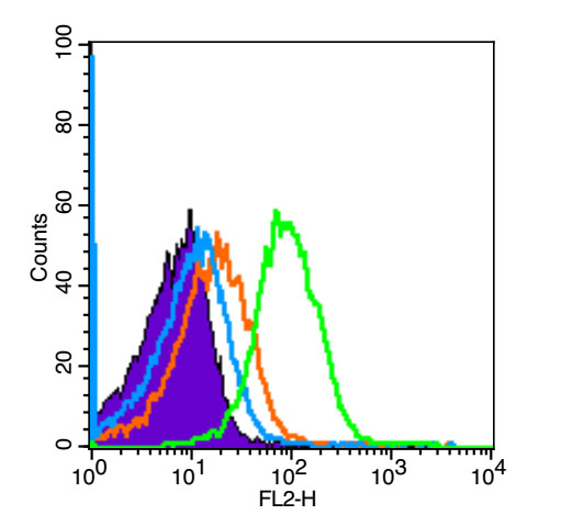
Blank control (Black line):Mouse spleen (Black).
Primary Antibody (green line): Rabbit Anti-FoxP3 antibody (bs-10211R)
Dilution: 3μg /10^6 cells;
Isotype Control Antibody (orange line): Rabbit IgG .
Secondary Antibody (white blue line): Goat anti-rabbit IgG-PE
Dilution: 1μg /test.
Protocol
The cells were fixed with 4% PFA (10min at room temperature)and then permeabilized with 90% ice-cold methanol for 20 min at room temperature. The cells were then incubated in 5%BSA goat serum to block non-specific protein-protein interactions for 15 min at room temperature .Cells stained with Primary Antibody for 30 min at room temperature. The secondary antibody used for 40 min at room temperature. Acquisition of 20,000 events was performed.
-
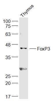
Sample:
Thymus (Mouse) Lysate at 40 ug
Primary: Anti-FoxP3 (bs-10211R) at 1/300 dilution
Secondary: IRDye800CW Goat Anti-Rabbit IgG at 1/20000 dilution
Predicted band size: 47 kD
Observed band size: 47 kD
-
Sample:
Spleen (Rat) Lysate at 40 ug
Primary: Anti-FoxP3 (bs-10211R) at 1/300 dilution
Secondary: IRDye800CW Goat Anti-Rabbit IgG at 1/20000 dilution
Predicted band size: 47 kD
Observed band size: 47 kD
-
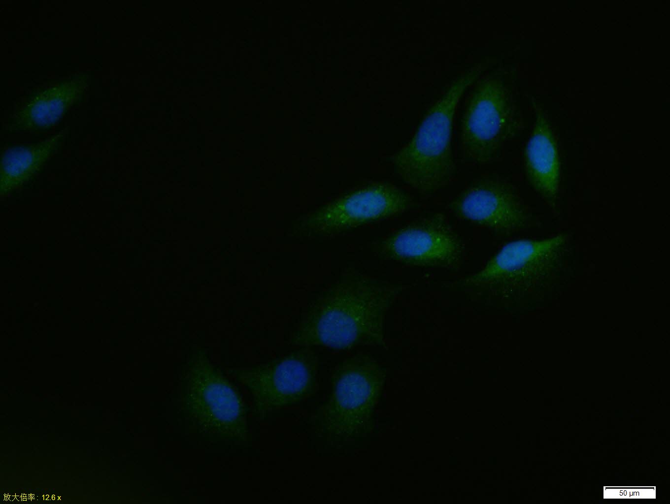
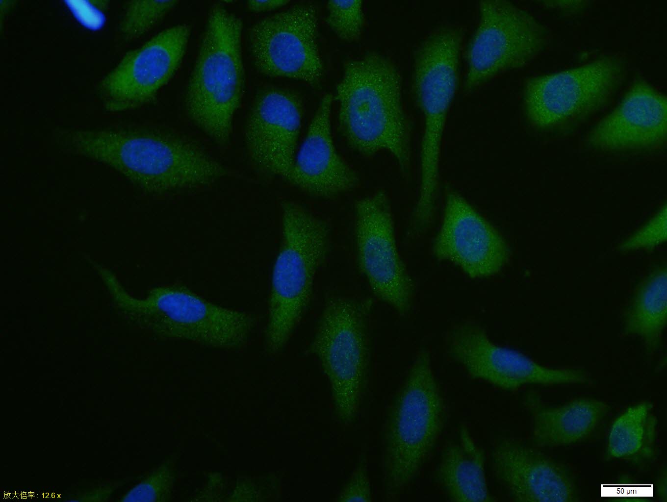
U2OS cell; 4% Paraformaldehyde-fixed; Triton X-100 at room temperature for 20 min; Blocking buffer (normal goat serum, C-0005) at 37°C for 20 min; Antibody incubation with (FoxP3) polyclonal Antibody, Unconjugated (bs-10211R) 1:100, 90 minutes at 37°C; followed by a conjugated Goat Anti-Rabbit IgG antibody at 37°C for 90 minutes, DAPI (blue, C02-04002) was used to stain the cell nuclei.
-
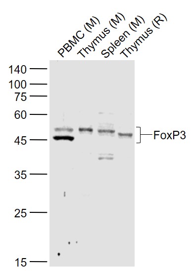
Sample:
Lane 1: PBMC (Mouse) Lysate at 40 ug
Lane 2: Thymus (Mouse) Lysate at 40 ug
Lane 3: Spleen (Mouse) Lysate at 40 ug
Lane 4: Thymus (Rat) Lysate at 40 ug
Primary: Anti-FoxP3 (bs-10211R) at 1/1000 dilution
Secondary: IRDye800CW Goat Anti-Rabbit IgG at 1/20000 dilution
Predicted band size: 43/45 kD
Observed band size: 43/45 kD
-
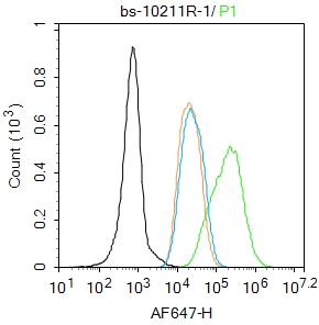
Blank control: MCF7.
Primary Antibody (green line): Rabbit Anti-FoxP3 antibody (bs-10211R)
Dilution: 2μg /10^6 cells;
Isotype Control Antibody (orange line): Rabbit IgG .
Secondary Antibody : Goat anti-rabbit IgG-AF647
Dilution: 1μg /test.
Protocol
The cells were fixed with 4% PFA (10min at room temperature)and then permeabilized with 90% ice-cold methanol for 20 min at-20℃.The cells were then incubated in 5%BSA to block non-specific protein-protein interactions for 30 min at room temperature .Cells stained with Primary Antibody for 30 min at room temperature. The secondary antibody used for 40 min at room temperature. Acquisition of 20,000 events was performed.
-
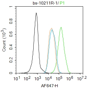
Blank control: MCF7.
Primary Antibody (green line): Rabbit Anti-FoxP3 antibody (bs-10211R)
Dilution: 2μg /10^6 cells;
Isotype Control Antibody (orange line): Rabbit IgG .
Secondary Antibody : Goat anti-rabbit IgG-AF647
Dilution: 1μg /test.
Protocol
The cells were fixed with 4% PFA (10min at room temperature)and then permeabilized with 90% ice-cold methanol for 20 min at-20℃.The cells were then incubated in 5%BSA to block non-specific protein-protein interactions for 30 min at room temperature .Cells stained with Primary Antibody for 30 min at room temperature. The secondary antibody used for 40 min at room temperature. Acquisition of 20,000 events was performed.
-
U2OS cell; 4% Paraformaldehyde-fixed; Triton X-100 at room temperature for 20 min; Blocking buffer (normal goat serum, C-0005) at 37°C for 20 min; Antibody incubation with (FoxP3) polyclonal Antibody, Unconjugated (bs-10211R) 1:100, 90 minutes at 37°C; followed by a conjugated Goat Anti-Rabbit IgG antibody at 37°C for 90 minutes, DAPI (blue, C02-04002) was used to stain the cell nuclei.
-
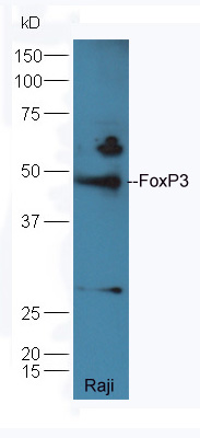
Protein: Raji(human) lysates at 40ug;
Primary: rabbit Anti-FoxP3 (bs-10211R) at 1:300;
Secondary: HRP conjugated Goat-Anti-rabbit IgG(bs-0295G-HRP) at 1: 5000;
Predicted band size:47 kD
Observed band size:47 kD
-
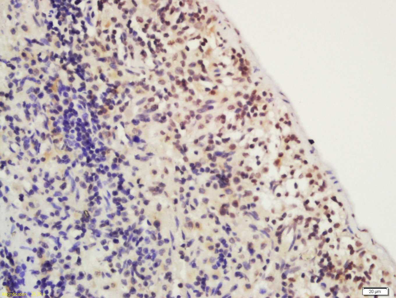
Tissue/cell: rat spleen; 4% Paraformaldehyde-fixed and paraffin-embedded;
Antigen retrieval: citrate buffer ( 0.01M, pH 6.0 ), Boiling bathing for 15min; Block endogenous peroxidase by 3% Hydrogen peroxide for 30min; Blocking buffer (normal goat serum,C-0005) at 37℃ for 20 min;
Incubation: Anti-FoxP3 Polyclonal Antibody, Unconjugated(bs-10211R) 1:200, overnight at 4°C, followed by conjugation to the secondary antibody(SP-0023) and DAB(C-0010) staining
-
Tissue/cell: Rat brain tissue; 4% Paraformaldehyde-fixed and paraffin-embedded;
Antigen retrieval: citrate buffer ( 0.01M, pH 6.0 ), Boiling bathing for 15min; Block endogenous peroxidase by 3% Hydrogen peroxide for 30min; Blocking buffer (normal goat serum,C-0005) at 37℃ for 20 min;
Incubation: Anti-FoxP3 Polyclonal Antibody, Unconjugated(bs-10211R) 1:500, overnight at 4°C, followed by conjugation to the secondary antibody(SP-0023) and DAB(C-0010) staining
-
Blank control: Mouse spleen.
Primary Antibody (green line): Rabbit Anti-FoxP3/PE Conjugated antibody (bs-10211R-PE)
Dilution: 0.2μg /10^6 cells;
Isotype Control Antibody (orange line): Rabbit IgG-PE .
Protocol
The cells were fixed with 4% PFA (10min at room temperature)and then permeabilized with 90% ice-cold methanol for 20 min at-20℃. The cells were then incubated in 5% BSA to block non-specific protein-protein interactions for 30 min at room temperature. The cells were stained with Primary Antibody for 30 min at room temperature. Acquisition of 20,000 events was performed.
-
Sample:
U937 Cell (Human) Lysate at 30 ug
Primary: Anti-FoxP3 (Bs-10211R) at 1/300 dilution
Secondary: IRDye800CW Goat Anti-Rabbit IgG at 1/20000 dilution
Predicted band size: 47 kD
Observed band size: 48 kD
RRID:RRID
产品名称:Rabbit Anti-FOXP3 antibody
别名: Forkhead box protein P3; forkhead box P3; Scurfin; Forkhead box protein P3, C-terminally processed; Forkhead box protein P3 41 kDa form; JM2; AIID; IPEX; PIDX; XPID; DIETER; FOXP3_HUMAN;
中文名称:叉头蛋白P3抗体
英文名称:Rabbit Anti-FOXP3 antibody
抗体来源: Rabbit
克隆类型:多克隆
细胞定位:细胞核
性 状:Liquid
亚 型:IgG
纯化方法:affinity purified by Protein A
保存条件:Shipped at 4℃. Store at -20 °C for one year. Avoid repeated freeze/thaw cycles.
免 疫 原:KLH conjugated synthetic peptide derived from human FoxP3
抗原表位:331-431/431
SWISS:Q9BZS1
Gene ID :50943
Human Gene ID:50943
The protein encoded by this gene is a member of the forkhead/winged-helix family of transcriptional regulators. Defects in this gene are the cause of immunodeficiency polyendocrinopathy, enteropathy, X-linked syndrome (IPEX), also known as X-linked autoimmunity-immunodeficiency syndrome. Alternatively spliced transcript variants encoding different isoforms have been identified. [provided by RefSeq, Jul 2008].
Function:Probable transcription factor. Plays a critical role in the control of immune response.
Subunit:Interacts with IKZF3.
Subcellular Location:Nucleus (Potential).
Post-translational modifications:Acetylation on lysine residues stabilizes FOXP3 and promotes differentiation of T-cells into induced regulatory T-cells (iTregs) associated with suppressive functions. Deacetylated by SIRT1.
DISEASE:Defects in FOXP3 are the cause of immunodeficiency polyendocrinopathy, enteropathy, X-linked syndrome (IPEX) [MIM:304790]; also known as X-linked autoimmunity-immunodeficiency syndrome. IPEX is characterized by neonatal onset insulin-dependent diabetes me
Similarity:Contains 1 C2H2-type zinc finger.
Contains 1 fork-head DNA-binding domain.
Important Note:This product as supplied is intended for research use only, not for use in human, therapeutic or diagnostic applications.
 400-901-9800


 说明书
说明书 联系我们
联系我们 打印此页面
打印此页面 收藏
收藏