| Rabbit Anti-VEGFR2 antibody |
| 反应物种(预测) |
Mouse,Rat,Dog,Pig,Cow,Horse,Sheep |
| 产品应用(已验证) |
IHC,FCM |
| 产品应用(可尝试) |
ELISA |
| 推荐稀释比例 |
Elisa=1:5000-10000,IHC-P=1:100-500,IHC-F=1:100-500,Flow Cyt=1μg/Test, |
| 研究领域 |
肿瘤,细胞生物,免疫学,神经生物学,信号转导,生长因子和激素,激酶和磷酸酶,细胞膜受体,细胞类型标志物(Hemangioblast Marker),内皮细胞, |
| 标签 |
Array |
-
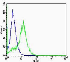
Cell: Hela
Concentration:1:100
Host/Isotype:Rabbit/IgG
Flow cytometric analysis of Rabbit IgG isotype control (Cat#: bs-10412R) on Hela(green) compared with control in the absence of primary antibody (blue) followed by Alexa Fluor 488-conjugated goat anti-rabbit IgG(H+L) secondary antibody .
-
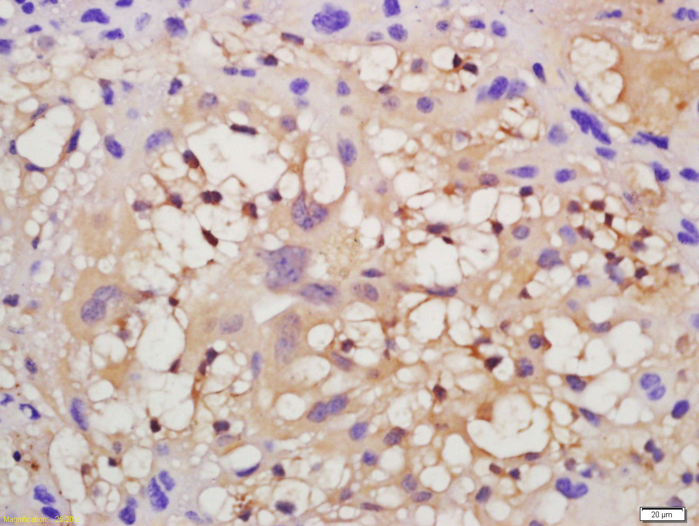
Tissue/cell: human lung carcinoma; 4% Paraformaldehyde-fixed and paraffin-embedded;
Antigen retrieval: citrate buffer ( 0.01M, pH 6.0 ), Boiling bathing for 15min; Block endogenous peroxidase by 3% Hydrogen peroxide for 30min; Blocking buffer (normal goat serum,C-0005) at 37℃ for 20 min;
Incubation: Anti-VEGFR2 Polyclonal Antibody, Unconjugated(bs-10412R) 1:200, overnight at 4°C, followed by conjugation to the secondary antibody(SP-0023) and DAB(C-0010) staining
-
Tissue/cell: mouse placenta tissue; 4% Paraformaldehyde-fixed and paraffin-embedded;
Antigen retrieval: citrate buffer ( 0.01M, pH 6.0 ), Boiling bathing for 15min; Block endogenous peroxidase by 3% Hydrogen peroxide for 30min; Blocking buffer (normal goat serum,C-0005) at 37℃ for 20 min;
Incubation: Anti-VEGFR2 Polyclonal Antibody, Unconjugated(bs-10412R) 1:200, overnight at 4°C, followed by conjugation to the secondary antibody(SP-0023) and DAB(C-0010) staining
-
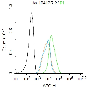
Blank control: MCF7.
Primary Antibody (green line): Rabbit Anti-VEGFR2 antibody (bs-10412R)
Dilution: 2μg /10^6 cells;
Isotype Control Antibody (orange line): Rabbit IgG .
Secondary Antibody : Goat anti-rabbit IgG-AF647
Dilution: 1μg /test.
Protocol
The cells were fixed with 4% PFA (10min at room temperature)and then permeabilized with 90% ice-cold methanol for 20 min at-20℃.The cells were then incubated in 5%BSA to block non-specific protein-protein interactions for 30 min at at room temperature .Cells stained with Primary Antibody for 30 min at room temperature. The secondary antibody used for 40 min at room temperature. Acquisition of 20,000 events was performed.
RRID:RRID
产品名称:Rabbit Anti-VEGFR2 antibody
别名: VEGF Receptor 2; CD309; CD309 antigen; Fetal liver kinase 1; FLK-1; FLK1; KDR; Kinase insert domain receptor (a type III receptor tyrosine kinase); Kinase insert domain receptor; KRD1; Ly73; Protein tyrosine kinase receptor FLK1; Protein-tyrosine kinase r
中文名称:血管内皮生长因子受体2抗体
英文名称:Rabbit Anti-VEGFR2 antibody
抗体来源: Rabbit
克隆类型:多克隆
细胞定位:细胞核,细胞浆,细胞膜,分泌型蛋白
性 状:Liquid
亚 型:IgG
纯化方法:affinity purified by Protein A
保存条件:Shipped at 4℃. Store at -20 °C for one year. Avoid repeated freeze/thaw cycles.
免 疫 原:KLH conjugated synthetic peptide derived from human VEGFR2
抗原表位:101-200/1356
抗原细胞定位:Extracellular
SWISS:P35968
Gene ID :3791
Human Gene ID:3791
Vascular endothelial growth factor (VEGF) is a major growth factor for endothelial cells. This gene encodes one of the two receptors of the VEGF. This receptor, known as kinase insert domain receptor, is a type III receptor tyrosine kinase. It functions as the main mediator of VEGF-induced endothelial proliferation, survival, migration, tubular morphogenesis and sprouting. The signalling and trafficking of this receptor are regulated by multiple factors, including Rab GTPase, P2Y purine nucleotide receptor, integrin alphaVbeta3, T-cell protein tyrosine phosphatase, etc.. Mutations of this gene are implicated in infantile capillary hemangiomas. [provided by RefSeq, May 2009].
Function:Tyrosine-protein kinase that acts as a cell-surface receptor for VEGFA, VEGFC and VEGFD. Plays an essential role in the regulation of angiogenesis, vascular development, vascular permeability, and embryonic hematopoiesis. Promotes proliferation, survival
Subunit:Interacts with MYOF. Interacts with VEGFA, VEGFC and VEGFD. Monomer in the absence of bound VEGFA, VEGFC or VEGFD. Homodimer in the presence of bound dimeric VEGFA, VEGFC or VEGFD. Can also form heterodimers with FLT1 and FLT4. Interacts (tyrosine phospho
Subcellular Location:Isoform 1: Cell membrane; Single-pass type I membrane protein. Cytoplasm. Nucleus. Cytoplasmic vesicle. Early endosome. Note=Detected on caveolae-enriched lipid rafts at the cell surface. Is recycled from the plasma membrane to endosomes and back again. P
Tissue Specificity:Detected in cornea (at protein level). Widely expressed.
Post-translational modifications:N-glycosylated.
Ubiquitinated. Tyrosine phosphorylation of the receptor promotes its poly-ubiquitination, leading to its degradation via the proteasome or lysosomal proteases.
Autophosphorylated on tyrosine residues upon ligand binding. Autophosph
DISEASE:Defects in KDR are associated with susceptibility to hemangioma capillary infantile (HCI) [MIM:602089]. HCI are benign, highly proliferative lesions involving aberrant localized growth of capillary endothelium. They are the most common tumor of infancy, o
Similarity:Belongs to the protein kinase superfamily. Tyr protein kinase family. CSF-1/PDGF receptor subfamily.
Contains 7 Ig-like C2-type (immunoglobulin-like) domains.
Contains 1 protein kinase domain.
Important Note:This product as supplied is intended for research use only, not for use in human, therapeutic or diagnostic applications.
 400-901-9800


 说明书
说明书 联系我们
联系我们 打印此页面
打印此页面 收藏
收藏