| Rabbit Anti-CD146 antibody |
| 反应物种(预测) |
Mouse,Dog,Cow,Horse |
| 产品应用(已验证) |
WB |
| 产品应用(可尝试) |
ELISA |
| 推荐稀释比例 |
WB=1:500-2000,Elisa=1:5000-10000, |
| 研究领域 |
肿瘤,免疫学,细胞粘附分子,细胞表面分子, |
| 标签 |
Array |
-
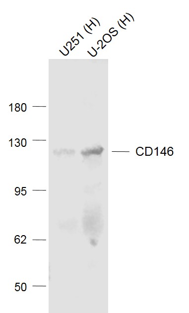
Sample:
Lane 1: U251 (Human) Cell Lysate at 30 ug
Lane 2: U-2OS (Human) Cell Lysate at 30 ug
Primary: Anti-CD146 (bs-1618R) at 1/1000 dilution
Secondary: IRDye800CW Goat Anti-Rabbit IgG at 1/20000 dilution
Predicted band size: 120 kD
Observed band size: 120 kD
RRID:AB_10855068
产品名称:Rabbit Anti-CD146 antibody
别名: A32 antigen; CD 146; CD146 antigen; Cell surface glycoprotein MUC18; Cell surface glycoprotein P1H12; MCAM; Melanoma adhesion molecule; Melanoma associated antigen A32; Melanoma associated antigen MUC18; Melanoma associated glycoprotein MUC18; Melanoma ce
中文名称:黑色素瘤细胞粘附分子CD146抗体
英文名称:Rabbit Anti-CD146 antibody
抗体来源: Rabbit
克隆类型:多克隆
细胞定位:细胞膜
性 状:Liquid
亚 型:IgG
纯化方法:affinity purified by Protein A
保存条件:Shipped at 4℃. Store at -20 °C for one year. Avoid repeated freeze/thaw cycles.
免 疫 原:KLH conjugated synthetic peptide derived from human CD146
抗原表位:201-300/646
抗原细胞定位:Extracellular
SWISS:P43121
Gene ID :4162
Human Gene ID:4162
MCAM (MUC18 antigen, CD146), a member of the immuoglobulin superfamily, is an intrinsic membrane glycoprotein of 110-120 kDa found on the surface of endothelial cells, bone marrow fibroblasts and various melanomas. MCAM (Melanoma adhesion molecule) has been used as a marker of tumor progression in human melanoma because expression in those tumors correlates strongly with poor prognosis and the development of metastic disease. In addition, a number of human T, B and myeloid leukemic cell lines seem to express MCAM. The close structural relationship with N-CAM and related molecules suggests that MCAM may be also a developmentally regulated cell adhesion.
Function:Plays a role in cell adhesion, and in cohesion of the endothelial monolayer at intercellular junctions in vascular tissue. Its expression may allow melanoma cells to interact with cellular elements of the vascular system, thereby enhancing hematogeneous t
Subcellular Location:Membrane; Single-pass type I membrane protein.
Tissue Specificity:Detected in endothelial cells in vascular tissue throughout the body. May appear at the surface of neural crest cells during their embryonic migration. Appears to be limited to vascular smooth muscle in normal adult tissues. Associated with tumor progress
Similarity:Contains 3 Ig-like C2-type (immunoglobulin-like) domains.
Contains 2 Ig-like V-type (immunoglobulin-like) domains.
Important Note:This product as supplied is intended for research use only, not for use in human, therapeutic or diagnostic applications.
 400-901-9800


 说明书
说明书 联系我们
联系我们 打印此页面
打印此页面 收藏
收藏