| Rabbit Anti-Profilin 1 antibody |
| 反应物种(预测) |
Dog,Cow,Horse |
| 产品应用(已验证) |
IHC,FCM |
| 产品应用(可尝试) |
WB,IF,ELISA |
| 推荐稀释比例 |
WB=1:500-2000,Elisa=1:5000-10000,IHC-P=1:100-500,IHC-F=1:100-500,Flow Cyt=3ug/Test,IF=1:100-500, |
| 研究领域 |
肿瘤,细胞生物,神经生物学,信号转导,干细胞,细胞周期蛋白,细胞分化,细胞类型标志物, |
| 标签 |
Array |
-
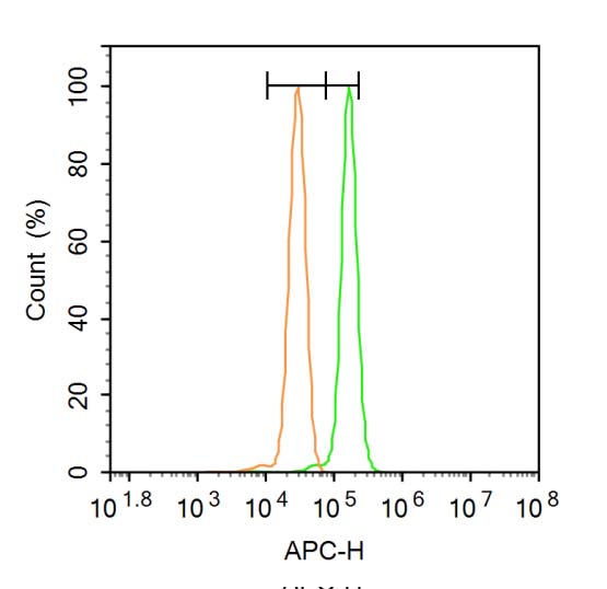
Blank control: A431.
Primary Antibody (green line): Rabbit Anti-Profilin 1 antibody (bs-1556R)
Dilution: 1μg /10^6 cells;
Isotype Control Antibody (orange line): Rabbit IgG .
Secondary Antibody: Goat anti-rabbit IgG-AF647
Dilution: 1μg /test.
Protocol
The cells were fixed with 4% PFA (10min at room temperature)and then permeabilized with 0.1% PBST for 20 min at room temperature. The cells were then incubated in 5%BSA to block non-specific protein-protein interactions for 30 min at room temperature .Cells stained with Primary Antibody for 30 min at room temperature. The secondary antibody used for 40 min at room temperature. Acquisition of 20,000 events was performed.
-
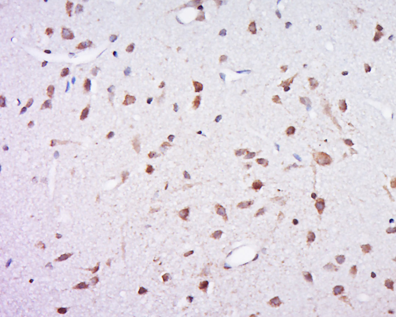
Tissue/cell: Rat brain tissue; 4% Paraformaldehyde-fixed and paraffin-embedded;
Antigen retrieval: citrate buffer ( 0.01M, pH 6.0 ), Boiling bathing for 15min; Block endogenous peroxidase by 3% Hydrogen peroxide for 30min; Blocking buffer (normal goat serum,C-0005) at 37∩ for 20 min;
Incubation: Anti-PFN1 Polyclonal Antibody, Unconjugated(bs-1556R) 1:500, overnight at 4∑C, followed by conjugation to the secondary antibody(SP-0023) and DAB(C-0010) staining
-
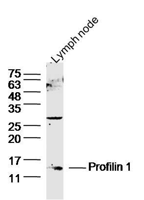
Sample: Lymph node (Mouse) Lysate at 30 ug
Primary: Anti- Profilin 1 (bs-1556R) at 1/300 dilution
Secondary: IRDye800CW Goat Anti-Mouse IgG at 1/10000 dilution
Predicted band size: 15 kD
Observed band size: 15 kD
RRID:AB_10855686
产品名称:Rabbit Anti-Profilin 1 antibody
别名: Actin binding protein; PFN 1; Pfn; PFN1; Profilin I; Profilin1; ProfilinI; PROF1_HUMAN.
中文名称:前纤维蛋白1抗体
英文名称:Rabbit Anti-Profilin 1 antibody
抗体来源: Rabbit
克隆类型:多克隆
细胞定位:细胞浆
性 状:Liquid
亚 型:IgG
纯化方法:affinity purified by Protein A
保存条件:Shipped at 4℃. Store at -20 °C for one year. Avoid repeated freeze/thaw cycles.
免 疫 原:KLH conjugated synthetic peptide derived from human Profilin 1
抗原表位:41-140/140
SWISS:P07737
Gene ID :5216
Human Gene ID:5216
Profilin 1 is a ubiquitous actin monomer-binding protein involved in actin polymerization in response to extracellular signals. Three profilin genes have been identified: profilin 1, 2, and 3. Profilin 1 is the most ubiquitous and abundant, and is highly expressed throughout development and adulthood inmost tissues including brain. Profilin 2 is the neuronal specific isoform and profilin 3 is a testis specific isoform. Profilins of eukaryotic cells are small (12-15 kDa) cytoplasmic proteins that bind to actin monomers, polyphosphoinositides and polymers of L-proline. Profilins were shown to be important for normal cell proliferation, differentiation and motility. Deletion of profilin 1 gene leads to an embryonic lethal phenotype.
Function:Binds to actin and affects the structure of the cytoskeleton. At high concentrations, profilin prevents the polymerization of actin, whereas it enhances it at low concentrations. By binding to PIP2, it inhibits the formation of IP3 and DG. Inhibits androg
Subunit:Occurs in many kinds of cells as a complex with monomeric actin in a 1:1 ratio. Found in a complex with XPO6, Ran, ACTB and PFN1. Interacts with VASP. Interacts with HTT.
Subcellular Location:Cytoplasm, cytoskeleton.
Tissue Specificity:Expressed in epididymis (at protein level).
Post-translational modifications:Phosphorylation at Ser-138 reduces its affinity for G-actin and blocks its interaction with HTT, reducing its ability to inhibit androgen receptor (AR) and HTT aggregation.
DISEASE:Amyotrophic lateral sclerosis 18 (ALS18) [MIM:614808]: A neurodegenerative disorder affecting upper motor neurons in the brain and lower motor neurons in the brain stem and spinal cord, resulting in fatal paralysis. Sensory abnormalities are absent. The p
Similarity:Belongs to the profilin family.
Important Note:This product as supplied is intended for research use only, not for use in human, therapeutic or diagnostic applications.
 400-901-9800


 说明书
说明书 联系我们
联系我们 打印此页面
打印此页面 收藏
收藏