| Rabbit Anti-MAP1LC3A antibody |
| 反应物种(预测) |
Rat,Chicken,Pig,Cow |
| 产品应用(已验证) |
WB,FCM |
| 产品应用(可尝试) |
IHC,IF,ELISA |
| 推荐稀释比例 |
WB=1:500-2000,Elisa=1:5000-10000,IHC-P=1:100-500,IHC-F=1:100-500,Flow Cyt=2μg/Test,IF=1:100-500, |
| 研究领域 |
肿瘤,细胞生物,免疫学,神经生物学,信号转导,细胞凋亡,激酶和磷酸酶,细胞自噬, |
| 标签 |
Array |
-
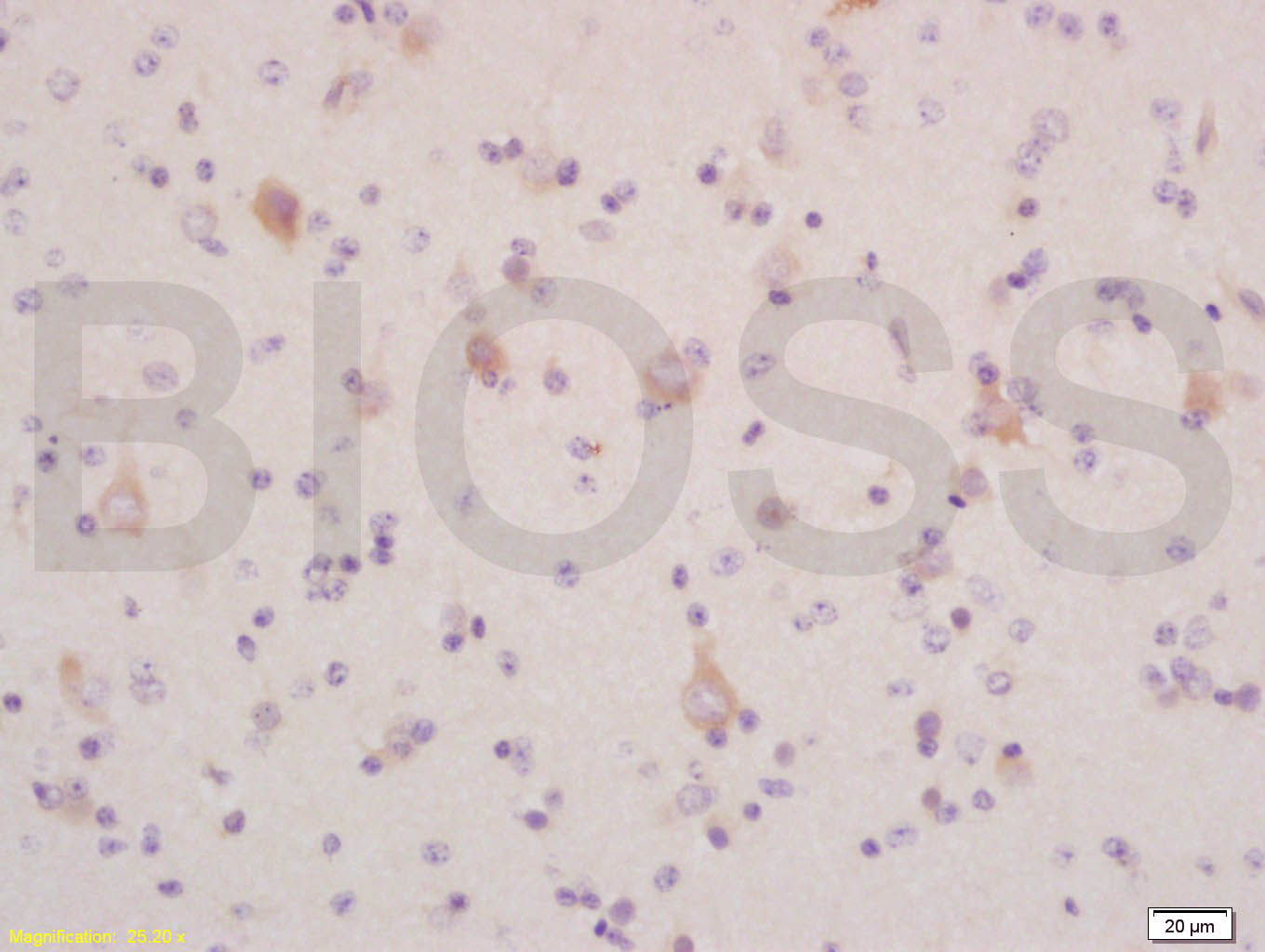
Tissue/cell: human glioma tissue; 4% Paraformaldehyde-fixed and paraffin-embedded;
Antigen retrieval: citrate buffer ( 0.01M, pH 6.0 ), Boiling bathing for 15min; Block endogenous peroxidase by 3% Hydrogen peroxide for 30min; Blocking buffer (normal goat serum,C-0005) at 37℃ for 20 min;
Incubation: Anti-LC3 α/MAP1A/MAP LC3 Alpha/Beta Polyclonal Antibody, Unconjugated (bs-1534R) 1:200, overnight at 4°C, followed by conjugation to the secondary antibody(SP-0023) and DAB(C-0010) staining
-
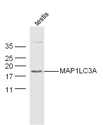
Sample:Testis (Mouse) Lysate at 30 ug
Primary: Anti-MAP1LC3A (bs-1534R) at 1/300 dilution
Secondary: IRDye800CW Goat Anti-Rabbit IgG at 1/20000 dilution
Predicted band size: 14 kD
Observed band size: 18 kD
-
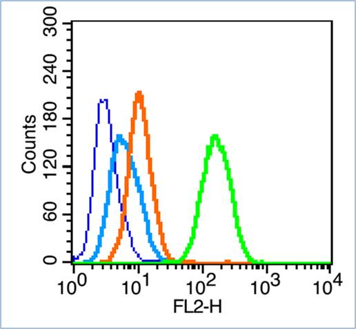
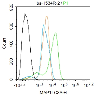
Blank control (blue line): Hela (blue).
Primary Antibody (green line): Rabbit Anti-MAP1LC3A antibody (bs-1534R)
Dilution: 0.2μg /10^6 cells;
Isotype Control Antibody (orange line): Rabbit IgG .
Secondary Antibody (white blue line): Goat anti-rabbit IgG-PE
Dilution: 1μg /test.
Protocol
The cells were fixed with 70% ethanol (Overmight at 4℃) and then permeabilized with 90% ice-cold methanol for 30 min at -20℃.Cells stained with Primary Antibody for 30 min at room temperature. The cells were then incubated in 1 X PBS/2%BSA/10% goat serum to block non-specific protein-protein interactions followed by the antibody for 15 min at room temperature. The secondary antibody used for 40 min at room temperature. Acquisition of 20,000 events was performed.
-
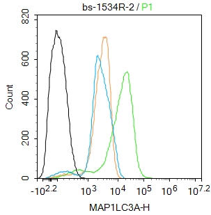
Blank control: RAW264.7.
Primary Antibody (green line): Rabbit Anti-MAP1LC3A antibody (bs-1534R)
Dilution: 2ug/Test;
Secondary Antibody : Goat anti-rabbit IgG-FITC
Dilution: 0.5ug/Test.
Protocol
The cells were fixed with 4% PFA (10min at room temperature)and then permeabilized with 0.1% PBST for 20 min at room temperature.The cells were then incubated in 5%BSA to block non-specific protein-protein interactions for 30 min at room temperature .Cells stained with Primary Antibody for 30 min at room temperature. The secondary antibody used for 40 min at room temperature. Acquisition of 20,000 events was performed.
-
Blank control: RAW264.7.
Primary Antibody (green line): Rabbit Anti-MAP1LC3A antibody (bs-1534R)
Dilution: 2ug/Test;
Secondary Antibody : Goat anti-rabbit IgG-FITC
Dilution: 0.5ug/Test.
Protocol
The cells were fixed with 4% PFA (10min at room temperature)and then permeabilized with 0.1% PBST for 20 min at room temperature.The cells were then incubated in 5%BSA to block non-specific protein-protein interactions for 30 min at room temperature .Cells stained with Primary Antibody for 30 min at room temperature. The secondary antibody used for 40 min at room temperature. Acquisition of 20,000 events was performed.
RRID:AB_10852738
产品名称:Rabbit Anti-MAP1LC3A antibody
别名: ATG8E;
Autophagy-related protein LC3 A;
Autophagy-related ubiquitin-like modifier LC3 A;
LC3;
LC3A;
MAP1 light chain 3 like protein 1;
MAP1 light chain 3-like protein 1;
MAP1A/1B light chain 3 A;
MAP1A/MAP1B LC3 A;
MAP1A/MAP1B light chain 3 A;
M
中文名称:自噬微管相关蛋白轻链3抗体
英文名称:Rabbit Anti-MAP1LC3A antibody
抗体来源: Rabbit
克隆类型:多克隆
细胞定位:细胞浆,细胞膜
性 状:Liquid
亚 型:IgG
纯化方法:affinity purified by Protein A
保存条件:Shipped at 4℃. Store at -20 °C for one year. Avoid repeated freeze/thaw cycles.
免 疫 原:KLH conjugated synthetic peptide derived from human MAP1LC3A
抗原表位:75-121/121
SWISS:Q9H492
Gene ID :84557
Human Gene ID:84557
Microtubule-associated MAPILC3A constitutes nearly half of the mass of all the microtubule associated proteins that copurify with brain microtubules. MAP1LC3A is one of three human orthologs of the rat Map1LC3, (named MAP1LC3A, MAP1LC3B, and MAP1LC3C). The three isoforms of human MAP1LC3 exhibit distinct expression patterns in different human tissues and also differ in their post-translation modifications. MAP1LC3A and MAP1LC3C are produced by the proteolytic cleavage after the conserved C-terminal Gly residue; MAP1LC3B does not undergo C-terminal cleavage and exists in a single modified form.
Function:Probably involved in formation of autophagosomal vacuoles (autophagosomes).
Subunit:3 different light chains, LC1, LC2 and LC3, can associate with MAP1A and MAP1B proteins. Interacts with SQSTM1. Interacts with TP53INP1 and TP53INP2.
Subcellular Location:Cytoplasm, cytoskeleton. Endomembrane system; Lipid-anchor. Cytoplasmic vesicle, autophagosome membrane; Lipid-anchor. Cytoplasmic vesicle, autophagosome. Note=LC3-II binds to the autophagic membranes.
Tissue Specificity:Most abundant in heart, brain, liver, skeletal muscle and testis but absent in thymus and peripheral blood leukocytes.
Post-translational modifications:The precursor molecule is cleaved by APG4B/ATG4B to form the cytosolic form, LC3-I. This is activated by APG7L/ATG7, transferred to ATG3 and conjugated to phospholipid to form the membrane-bound form, LC3-II.
Similarity:Detects a band of approximately 16 kDa (predicted molecular weight: 14 kDa).
Important Note:This product as supplied is intended for research use only, not for use in human, therapeutic or diagnostic applications.
 400-901-9800


 说明书
说明书 联系我们
联系我们 打印此页面
打印此页面 收藏
收藏