| Rabbit Anti-HSP70 antibody |
| 反应物种(预测) |
Chicken,Cow,Rabbit,Sheep |
| 产品应用(已验证) |
WB,IHC,ICC,FCM |
| 产品应用(可尝试) |
ELISA |
| 推荐稀释比例 |
WB=1:500-2000,Elisa=1:5000-10000,IHC-P=1:100-500,Flow Cyt=1ug/Test,ICC=1:100, |
| 研究领域 |
肿瘤,细胞生物,免疫学,信号转导 |
| 标签 |
Array |
-
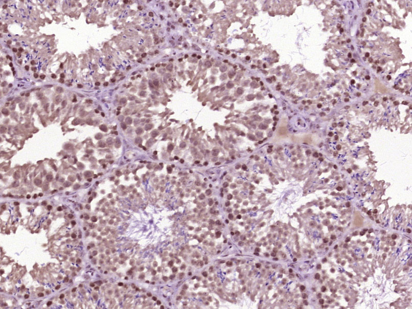
Paraformaldehyde-fixed, paraffin embedded (Mouse testis); Antigen retrieval by boiling in sodium citrate buffer (pH6.0) for 15min; Block endogenous peroxidase by 3% hydrogen peroxide for 20 minutes; Blocking buffer (normal goat serum) at 37°C for 30min; Antibody incubation with (HSP70) Polyclonal Antibody, Unconjugated (bs-0244R) at 1:400 overnight at 4°C, followed by operating according to SP Kit(Rabbit) (sp-0023) instructionsand DAB staining.
-
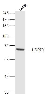
Sample:
Lung (Mouse) Lysate at 40 ug
Primary: Anti-HSP70 (bs-0244R) at 1/300 dilution
Secondary: IRDye800CW Goat Anti-Rabbit IgG at 1/20000 dilution
Predicted band size: 70 kD
Observed band size: 70 kD
-
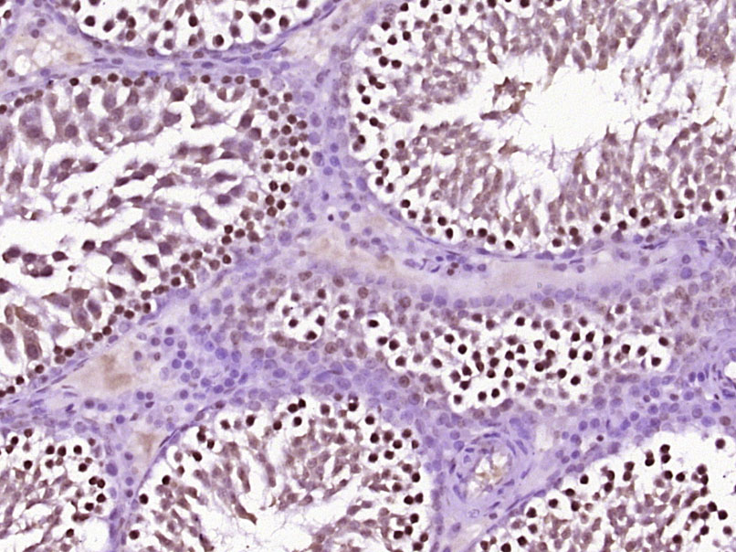
Paraformaldehyde-fixed, paraffin embedded (Rat testis); Antigen retrieval by boiling in sodium citrate buffer (pH6.0) for 15min; Block endogenous peroxidase by 3% hydrogen peroxide for 20 minutes; Blocking buffer (normal goat serum) at 37°C for 30min; Antibody incubation with (HSP70) Polyclonal Antibody, Unconjugated (bs-0244R) at 1:400 overnight at 4°C, followed by operating according to SP Kit(Rabbit) (sp-0023) instructionsand DAB staining.
-
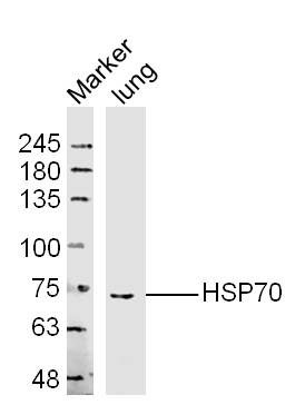
Sample: Lung (Mouse) Lysate at 40 ug
Primary: Anti-HSP70 (bs-0244R) at 1/300 dilution
Secondary: IRDye800CW Goat Anti-Rabbit IgG at 1/20000 dilution
Predicted band size: 70 kD
Observed band size: 70 kD
-
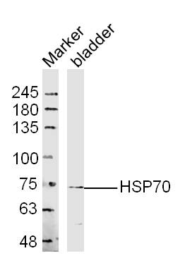
Sample: Bladder (Mouse) Lysate at 40 ug
Primary: Anti-HSP70 (bs-0244R) at 1/300 dilution
Secondary: IRDye800CW Goat Anti-Rabbit IgG at 1/20000 dilution
Predicted band size: 70 kD
Observed band size: 70 kD
-
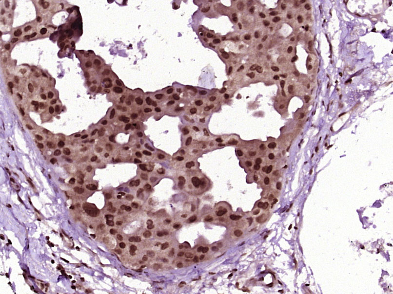
Paraformaldehyde-fixed, paraffin embedded (Human breast carcinoma); Antigen retrieval by boiling in sodium citrate buffer (pH6.0) for 15min; Block endogenous peroxidase by 3% hydrogen peroxide for 20 minutes; Blocking buffer (normal goat serum) at 37°C for 30min; Antibody incubation with (HSP70) Polyclonal Antibody, Unconjugated (bs-0244R) at 1:400 overnight at 4°C, followed by operating according to SP Kit(Rabbit) (sp-0023) instructionsand DAB staining.
-
Tissue/cell: U-87MG cell; 4% Paraformaldehyde-fixed; Triton X-100 at room temperature for 20 min; Blocking buffer (normal goat serum, C-0005) at 37°C for 20 min; Antibody incubation with (HSP70) polyclonal Antibody, Unconjugated (bs-0244R) 1:100, 90 minutes at 37°C; followed by a FITC conjugated Goat Anti-Rabbit IgG antibody at 37°C for 90 minutes, DAPI (blue, C02-04002) was used to stain the cell nuclei.
-
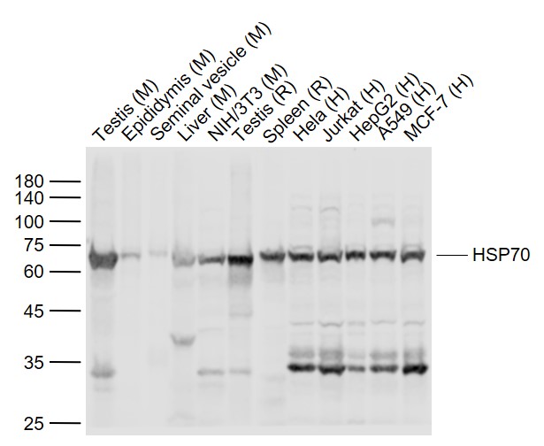
Sample:
Lane 1: Testis (Mouse) Lysate at 40 ug
Lane 2: Epididymis (Mouse) Lysate at 40 ug
Lane 3: Seminal vesicle (Mouse) Lysate at 40 ug
Lane 4: Liver (Mouse) Lysate at 40 ug
Lane 5: NIH/3T3 (Mouse) Cell Lysate at 30 ug
Lane 6: Testis (Rat) Lysate at 40 ug
Lane 7: Spleen (Rat) Lysate at 40 ug
Lane 8: Hela (Human) Cell Lysate at 30 ug
Lane 9: Jurkat (Human) Cell Lysate at 30 ug
Lane 10: HepG2 (Human) Cell Lysate at 30 ug
Lane 11: A549 (Human) Cell Lysate at 30 ug
Lane 12: MCF-7 (Human) Cell Lysate at 30 ug
Primary:
Anti-HSP70 (bs-0244R) at 1/1000 dilution
Secondary: IRDye800CW Goat Anti-Rabbit IgG at 1/20000 dilution
Predicted band size: 70 kD
Observed band size: 68 kD
-
Blank control:A549.
Primary Antibody (green line): Rabbit Anti-HSP70 antibody (bs-0244R)
Dilution: 1μg /10^6 cells;
Isotype Control Antibody (orange line): Rabbit IgG .
Secondary Antibody : Goat anti-rabbit IgG-AF488
Dilution: 1μg /test.
Protocol
The cells were fixed with 4% PFA (10min at room temperature)and then permeabilized with 90% ice-cold methanol for 20 min at -20℃. The cells were then incubated in 5%BSA to block non-specific protein-protein interactions for 30 min at room temperature .Cells stained with Primary Antibody for 30 min at room temperature. The secondary antibody used for 40 min at room temperature. Acquisition of 20,000 events was performed.
-
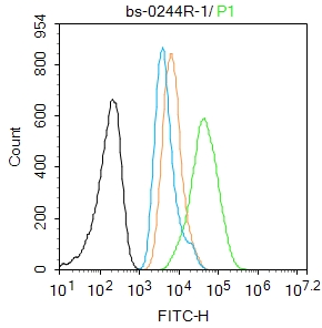
Blank control:293T.
Primary Antibody (green line): Rabbit Anti-HSP70 antibody (bs-0244R)
Dilution: 2μg /10^6 cells;
Isotype Control Antibody (orange line): Rabbit IgG .
Secondary Antibody : Goat anti-rabbit IgG-FITC
Dilution: 1μg /test.
Protocol
The cells were fixed with 4% PFA (10min at room temperature)and then permeabilized with 0.1% PBST for 20 min at room temperature. The cells were then incubated in 5%BSA to block non-specific protein-protein interactions for 30 min at room temperature .Cells stained with Primary Antibody for 30 min at room temperature. The secondary antibody used for 40 min at room temperature. Acquisition of 20,000 events was performed.
RRID:RRID
产品名称:Rabbit Anti-HSP70 antibody
别名: HSP70; HSP-70; HSP 70; Heat shock 70 kDa protein 1; heat shock 70kDa protein 1A; Heat shock 70kDa protein 1B; Heat shock induced protein; heat shock protein 70; HSP70 1; HSP70 2; HSP70.1; HSP72; HSPA1; HSPA1A; HSPA1B; XXbac BCX40G17.3 001; Heat shock 70 k
中文名称:热休克蛋白70抗体
英文名称:Rabbit Anti-HSP70 antibody
抗体来源: Rabbit
克隆类型:多克隆
细胞定位:细胞浆
性 状:Liquid
亚 型:IgG
纯化方法:affinity purified by Protein A
保存条件:Shipped at 4℃. Store at -20 °C for one year. Avoid repeated freeze/thaw cycles.
免 疫 原:KLH conjugated synthetic peptide derived from human HSP70
抗原表位:500-600/641
SWISS:P0DMV8
Gene ID :3303
Human Gene ID:3303
This intronless gene encodes a 70kDa heat shock protein which is a member of the heat shock protein 70 family. In conjuction with other heat shock proteins, this protein stabilizes existing proteins against aggregation and mediates the folding of newly translated proteins in the cytosol and in organelles. It is also involved in the ubiquitin-proteasome pathway through interaction with the AU-rich element RNA-binding protein 1. The gene is located in the major histocompatibility complex class III region, in a cluster with two closely related genes which encode similar proteins. [provided by RefSeq, Jul 2008].
Function:In cooperation with other chaperones, Hsp70s stabilize preexistent proteins against aggregation and mediate the folding of newly translated polypeptides in the cytosol as well as within organelles. These chaperones participate in all these processes throu
Subunit:Component of the CatSper complex. Identified in a mRNP granule complex, at least composed of ACTB, ACTN4, DHX9, ERG, HNRNPA1, HNRNPA2B1, HNRNPAB, HNRNPD, HNRNPL, HNRNPR, HNRNPU, HSPA1, HSPA8, IGF2BP1, ILF2, ILF3, NCBP1, NCL, PABPC1, PABPC4, PABPN1, RPLP0,
Subcellular Location:Cytoplasm. Note=Localized in cytoplasmic mRNP granules containing untranslated mRNAs.
Tissue Specificity:HSPA1B is testis-specific.
Similarity:Belongs to the heat shock protein 70 family.
Important Note:This product as supplied is intended for research use only, not for use in human, therapeutic or diagnostic applications.
 400-901-9800


 说明书
说明书 联系我们
联系我们 打印此页面
打印此页面 收藏
收藏