| Rabbit Anti-PD-1 antibody |
| 反应物种(预测) |
Cow,Horse,Rabbit |
| 产品应用(已验证) |
WB,FCM |
| 产品应用(可尝试) |
IHC,IF,ELISA |
| 推荐稀释比例 |
WB=1:500-2000,Elisa=1:5000-10000,IHC-P=1:100-500,IHC-F=1:100-500,Flow Cyt=1μg /test,IF=1:100-500, |
| 研究领域 |
肿瘤,细胞生物,免疫学,细胞凋亡 |
| 标签 |
Array |
-
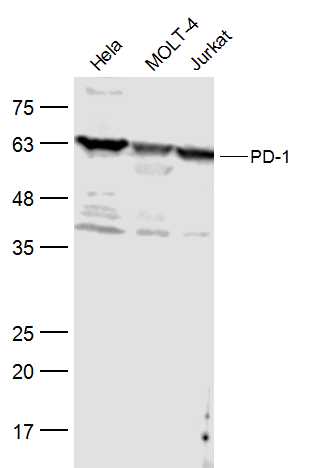
Sample:
Hela(Human) Cell Lysate at 30 ug
MOLT-4(Human) Cell Lysate at 30 ug
Jurkat(Human) Cell Lysate at 30 ug
Primary: Anti-PD-1 (bs-1867R) at 1/500 dilution
Secondary: IRDye800CW Goat Anti-Rabbit IgG at 1/20000 dilution
Predicted band size: 32 kD
Observed band size: 55 kD
-
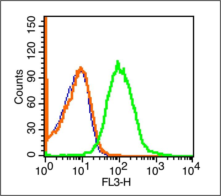
Blank control (blue line): Mouse spleen cells(blue).
Primary Antibody (green line): Rabbit Anti-PD-1/PE-CY7 Conjugated antibody (bs-1867R-PE-CY7)
Dilution: 1μg /10^6 cells;
Isotype Control Antibody (orange line): Rabbit IgG-PE-CY7 .
Protocol
The cells were fixed with 70% ice-cold methanol overnight at 4℃ . The cells were then incubated in 1 X PBS/2%BSA/10% goat serum to block non-specific protein-protein interactions followed by the antibody for 15 min at room temperature. Cells stained with Primary Antibody for 30 min at room temperature.Acquisition of 20,000 events was performed.
-
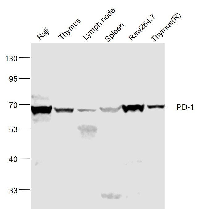
Sample:
Raji(Human) Cell Lysate at 30 ug
Thymus(Mouse) Lysate at 40 ug
Lymph node(Mouse) Lysate at 40 ug
Spleen(Mouse) Lysate at 40 ug
Raw264.7(Mouse) Cell Lysate at 40 ug
Thymus (Rat) Lysate at 40 ug
Primary: Anti-PD-1 (bs-1867R) at 1/1000 dilution
Secondary: IRDye800CW Goat Anti-Rabbit IgG at 1/20000 dilution
Predicted band size: 55 kD
Observed band size: 58 kD
RRID:AB_10856073
产品名称:Rabbit Anti-PD-1 antibody
别名: Programmed cell death protein 1; CD279; CD279 antigen; hPD 1; hPD-1; hSLE1; PD 1; PD1; PDCD 1; PDCD1; PDCD1_HUMAN; Programmed cell death 1; Protein PD 1; Protein PD-1; SLEB2; Systemic lupus erythematosus susceptibility 2.
中文名称:程序性死亡1(CD279)抗体
英文名称:Rabbit Anti-PD-1 antibody
抗体来源: Rabbit
克隆类型:多克隆
细胞定位:细胞膜
性 状:Liquid
亚 型:IgG
纯化方法:affinity purified by Protein A
保存条件:Shipped at 4℃. Store at -20 °C for one year. Avoid repeated freeze/thaw cycles.
免 疫 原:KLH conjugated synthetic peptide derived from human PD-1
抗原表位:201-288/288
SWISS:Q15116
Gene ID :5133
Human Gene ID:5133
Programmed cell death protein 1 (PDCD1) is an immune-inhibitory receptor expressed in activated T cells; it is involved in the regulation of T-cell functions, including those of effector CD8+ T cells. In addition, this protein can also promote the differentiation of CD4+ T cells into T regulatory cells. PDCD1 is expressed in many types of tumors including melanomas, and has demonstrated to play a role in anti-tumor immunity. Moreover, this protein has been shown to be involved in safeguarding against autoimmunity, however, it can also contribute to the inhibition of effective anti-tumor and anti-microbial immunity. [provided by RefSeq, Aug 2020]
Function:Inhibitory cell surface receptor involved in the regulation of T-cell function during immunity and tolerance. Upon ligand binding, inhibits T-cell effector functions in an antigen-specific manner. Possible cell death inducer, in association with other fac
Subunit:Monomer.
Subcellular Location:Membrane; Single-pass type I membrane protein.
Tissue Specificity:Ta,Ba,Ma,Thy
DISEASE:Systemic lupus erythematosus 2 (SLEB2) [MIM:605218]: A chronic, relapsing, inflammatory, and often febrile multisystemic disorder of connective tissue, characterized principally by involvement of the skin, joints, kidneys and serosal membranes. It is of u
Similarity:Contains 1 Ig-like V-type (immunoglobulin-like) domain.
Important Note:This product as supplied is intended for research use only, not for use in human, therapeutic or diagnostic applications.
 400-901-9800


 说明书
说明书 联系我们
联系我们 打印此页面
打印此页面 收藏
收藏