| Rabbit Anti-CD27 antibody |
| 反应物种(预测) |
Human,Dog,Pig,Cow |
| 产品应用(已验证) |
WB |
| 产品应用(可尝试) |
ELISA |
| 推荐稀释比例 |
WB=1:500-2000,Elisa=1:5000-10000, |
| 研究领域 |
肿瘤,免疫学,干细胞,细胞因子,淋巴细胞,T-淋巴细胞, |
| 标签 |
Array |
-
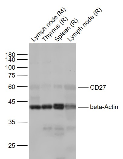
Sample:
Lane 1: Lymph node (Mouse) Lysate at 30 ug
Lane 2: Thymus (Rat) Lysate at 40 ug
Lane 3: Spleen (Rat) Lysate at 40 ug
Lane 4: Lymph node (Rat) Lysate at 30 ug
Primary:
Anti-CD27 (bs-2491R) at 1/1000 dilution
Anti-beta-Actin (bs-0061R) at 1/2000 dilution
Secondary: IRDye800CW Goat Anti-Rabbit IgG at 1/20000 dilution
Predicted band size: 50-55 kD
Observed band size: 60 kD
RRID:AB_10856440
产品名称:Rabbit Anti-CD27 antibody
别名: CD 27; CD27 antigen; CD27 molecule; CD27_HUMAN; CD27L receptor; LPFS2; MGC20393; OTTHUMP00000238557; S152; T cell activation antigen CD27; T cell antivation antigen S152; T-cell activation antigen CD27; T14; TNFRSF 7; TNFRSF7; TNFSF7; Tp 55; Tp55; Tumor n
中文名称:CD27抗体
英文名称:Rabbit Anti-CD27 antibody
抗体来源: Rabbit
克隆类型:多克隆
细胞定位:细胞膜
性 状:Liquid
亚 型:IgG
纯化方法:affinity purified by Protein A
保存条件:Shipped at 4℃. Store at -20 °C for one year. Avoid repeated freeze/thaw cycles.
免 疫 原:KLH conjugated synthetic peptide derived from human CD27
抗原表位:201-260/260
SWISS:P26842
Gene ID :939
Human Gene ID:939
The protein encoded by this gene is a member of the TNF-receptor superfamily. This receptor is required for generation and long-term maintenance of T cell immunity. It binds to ligand CD70, and plays a key role in regulating B-cell activation and immunoglobulin synthesis. This receptor transduces signals that lead to the activation of NF-kappaB and MAPK8/JNK. Adaptor proteins TRAF2 and TRAF5 have been shown to mediate the signaling process of this receptor. CD27-binding protein (SIVA), a proapoptotic protein, can bind to this receptor and is thought to play an important role in the apoptosis induced by this receptor. [provided by RefSeq, Jul 2008]
Function:Receptor for CD70/CD27L. May play a role in survival of activated T-cells. May play a role in apoptosis through association with SIVA1.
Subunit:Homodimer. Interacts with SIVA1 and TRAF2.
Subcellular Location:Membrane; Single-pass type I membrane protein.
Tissue Specificity:Found in most T-lymphocytes.
Post-translational modifications:Phosphorylated.
O-glycosylated with core 1 or possibly core 8 glycans.
Similarity:Contains 3 TNFR-Cys repeats.
Important Note:This product as supplied is intended for research use only, not for use in human, therapeutic or diagnostic applications.
 400-901-9800


 说明书
说明书 联系我们
联系我们 打印此页面
打印此页面 收藏
收藏