| Rabbit Anti-WDR26 antibody |
| 反应物种(预测) |
Chicken,Dog,Cow,Horse |
| 产品应用(已验证) |
WB,IHC,ICC,FCM |
| 产品应用(可尝试) |
IF,ELISA |
| 推荐稀释比例 |
WB=1:500-2000,Elisa=1:5000-10000,IHC-P=1:100-500,IHC-F=1:100-500,Flow Cyt=1µg/Test,IF=1:100-500,ICC=1:100, |
| 研究领域 |
心血管,细胞生物,信号转导,细胞周期蛋白,转录调节因子 |
| 标签 |
Array |
-
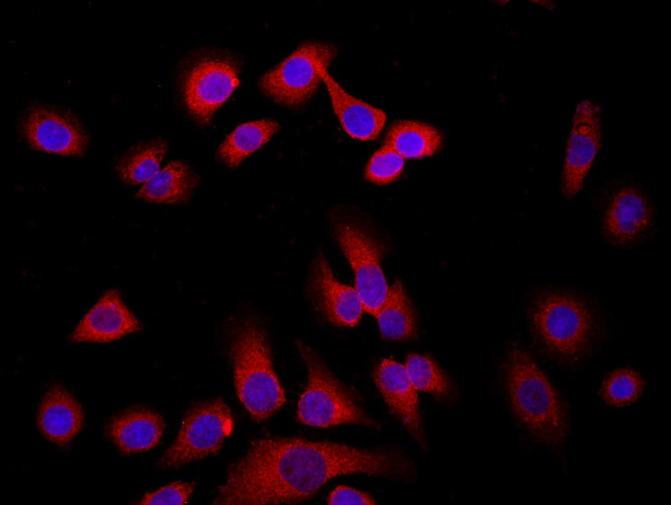
Tissue/cell: MCF7; 4% Paraformaldehyde-fixed; Triton X-100 at room temperature for 20 min; Blocking buffer (normal goat serum, C-0005) at 37°C for 20 min; Antibody incubation with (WDR26) Polyclonal Antibody, Unconjugated (bs-0932R) 1:200, 90 minutes at 37°C; followed by a conjugated Goat Anti-Rabbit IgG antibody (bs-0295G-FITC) at 37°C for 90 minutes, DAPI (blue, C02-04002) was used to stain the cell nuclei.
-
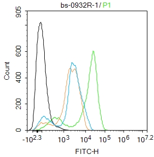
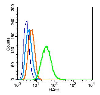
Blank control:THP-1.
Primary Antibody (green line): Rabbit Anti-WDR26 antibody (bs-0932R)
Dilution: 1ug/Test;
Secondary Antibody : Goat anti-rabbit IgG-FITC
Dilution: 0.5ug/Test.
Protocol
The cells were fixed with 4% PFA (10min at room temperature)and then permeabilized with 90% ice-cold methanol for 20 min at -20℃.The cells were then incubated in 5%BSA to block non-specific protein-protein interactions for 30 min at room temperature .Cells stained with Primary Antibody for 30 min at room temperature. The secondary antibody used for 40 min at room temperature. Acquisition of 20,000 events was performed.
-
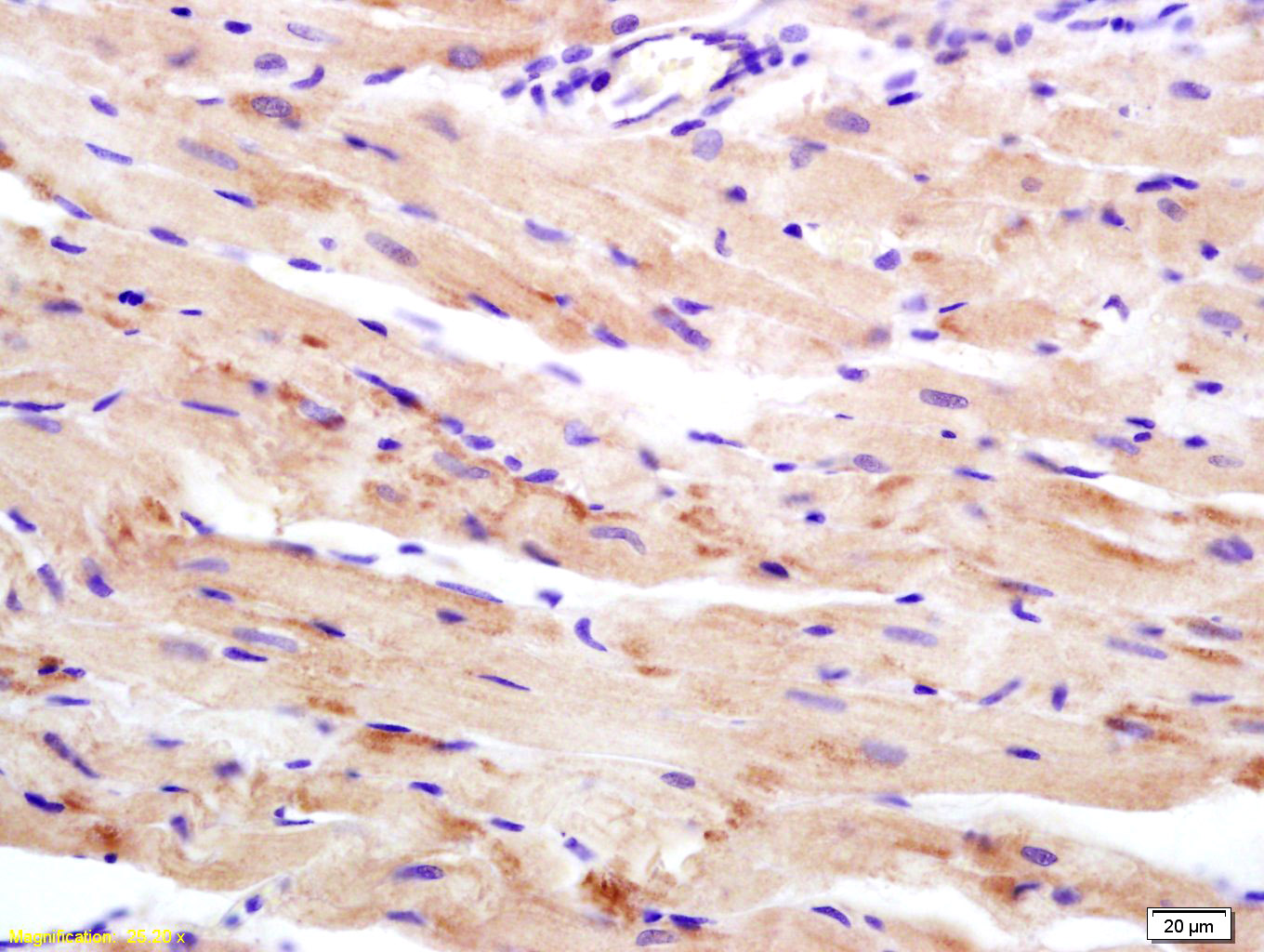
Tissue/cell: rat heart tissue; 4% Paraformaldehyde-fixed and paraffin-embedded;
Antigen retrieval: citrate buffer ( 0.01M, pH 6.0 ), Boiling bathing for 15min; Block endogenous peroxidase by 3% Hydrogen peroxide for 30min; Blocking buffer (normal goat serum,C-0005) at 37℃ for 20 min;
Incubation: Anti-WDR26 Polyclonal Antibody, Unconjugated(bs-0932R) 1:200, overnight at 4°C, followed by conjugation to the secondary antibody(SP-0023) and DAB(C-0010) staining
-
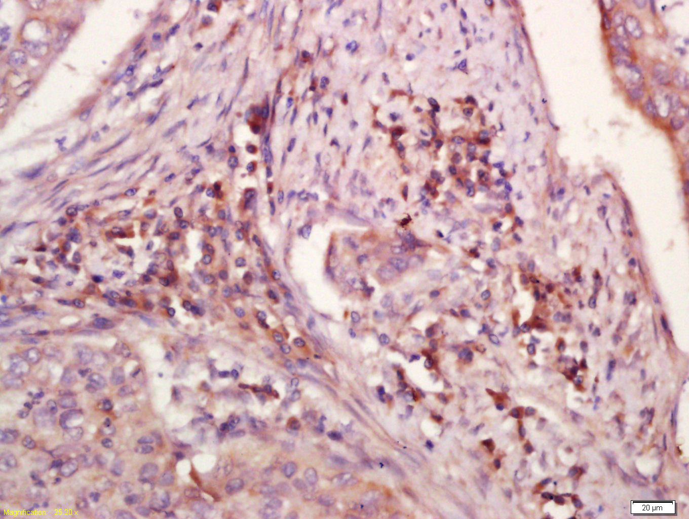
Tissue/cell: human laryngocarcinoma; 4% Paraformaldehyde-fixed and paraffin-embedded;
Antigen retrieval: citrate buffer ( 0.01M, pH 6.0 ), Boiling bathing for 15min; Block endogenous peroxidase by 3% Hydrogen peroxide for 30min; Blocking buffer (normal goat serum,C-0005) at 37℃ for 20 min;
Incubation: Anti-WDR26 Polyclonal Antibody, Unconjugated(bs-0932R) 1:200, overnight at 4°C, followed by conjugation to the secondary antibody(SP-0023) and DAB(C-0010) staining
-
Blank control: RSC96(blue), the cells were fixed with 2% paraformaldehyde (10 min) and then permeabilized with ice-cold 90% methanol for 30 min on ice.
Isotype Control Antibody: Rabbit IgG(orange) ;
Secondary Antibody: Goat anti-rabbit IgG-PE(white blue),
Dilution: 1:200 in 1 X PBS containing 0.5% BSA ;
Primary Antibody Dilution: 1μg in 100 μL1X PBS containing 0.5% BSA(green).
-
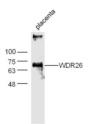
Sample:
Placenta (Mouse) Lysate at 40 ug
Primary: Anti-WDR26 (bs-0932R) at 1/300 dilution
Secondary: IRDye800CW Goat Anti-Rabbit IgG at 1/20000 dilution
Predicted band size: 72 kD
Observed band size: 70 kD
RRID:AB_10857073
产品名称:Rabbit Anti-WDR26 antibody
别名: myocardial ischemic preconditioning upregulated protein 2; WDR26; PRO0852; CDW2; MIP2; CUL4 and DDB1 associated WDR protein 2; Myocardial ischemic preconditioning up regulated protein 2; myocardial ischemic preconditioning upregulated protein 2; WD repeat
中文名称:心肌缺血预处理正调节蛋白2抗体
英文名称:Rabbit Anti-WDR26 antibody
抗体来源: Rabbit
克隆类型:多克隆
细胞定位:细胞浆
性 状:Liquid
亚 型:IgG
纯化方法:affinity purified by Protein A
保存条件:Shipped at 4℃. Store at -20 °C for one year. Avoid repeated freeze/thaw cycles.
免 疫 原:KLH conjugated synthetic peptide derived from human WDR26
抗原表位:101-200/514
SWISS:Q9H7D7
Gene ID :80232
Human Gene ID:80232
This gene encodes a member of the WD repeat protein family. WD repeats are minimally conserved regions of approximately 40 amino acids typically bracketed by gly-his and trp-asp (GH-WD), which may facilitate formation of heterotrimeric or multiprotein complexes. Members of this family are involved in a variety of cellular processes, including cell cycle progression, signal transduction, apoptosis, and gene regulation. Two transcript variants encoding two different isoforms have been found for this gene. [provided by RefSeq].
Function:May be involved in MAPK pathways.
Subunit:Interacts with DDB1-CUL4A/B E3 ligase complexes.
Subcellular Location:Cytoplasm.
Tissue Specificity:Broadly expressed, with highest levels in heart and skeletal muscle.
Similarity:Contains 1 CTLH domain.
Contains 1 LisH domain.
Contains 6 WD repeats.
Important Note:This product as supplied is intended for research use only, not for use in human, therapeutic or diagnostic applications.
 400-901-9800


 说明书
说明书 联系我们
联系我们 打印此页面
打印此页面 收藏
收藏