| Rabbit Anti-CK7 antibody |
| 产品应用(已验证) |
WB,IHC,ICC,FCM |
| 产品应用(可尝试) |
IF,ELISA |
| 推荐稀释比例 |
WB=1:500-2000,Elisa=1:5000-10000,IHC-P=1:100-500,IHC-F=1:100-500,Flow Cyt=1ug/Test,IF=1:100-500,ICC=1:100, |
| 研究领域 |
肿瘤,细胞生物,信号转导 |
| 标签 |
Array |
-
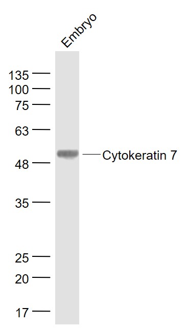
Sample:
Embryo (Mouse) Lysate at 40 ug
Primary: Anti- Cytokeratin 7 (bs-1744R) at 1/1000 dilution
Secondary: IRDye800CW Goat Anti-Rabbit IgG at 1/20000 dilution
Predicted band size: 54 kD
Observed band size: 54 kD
-
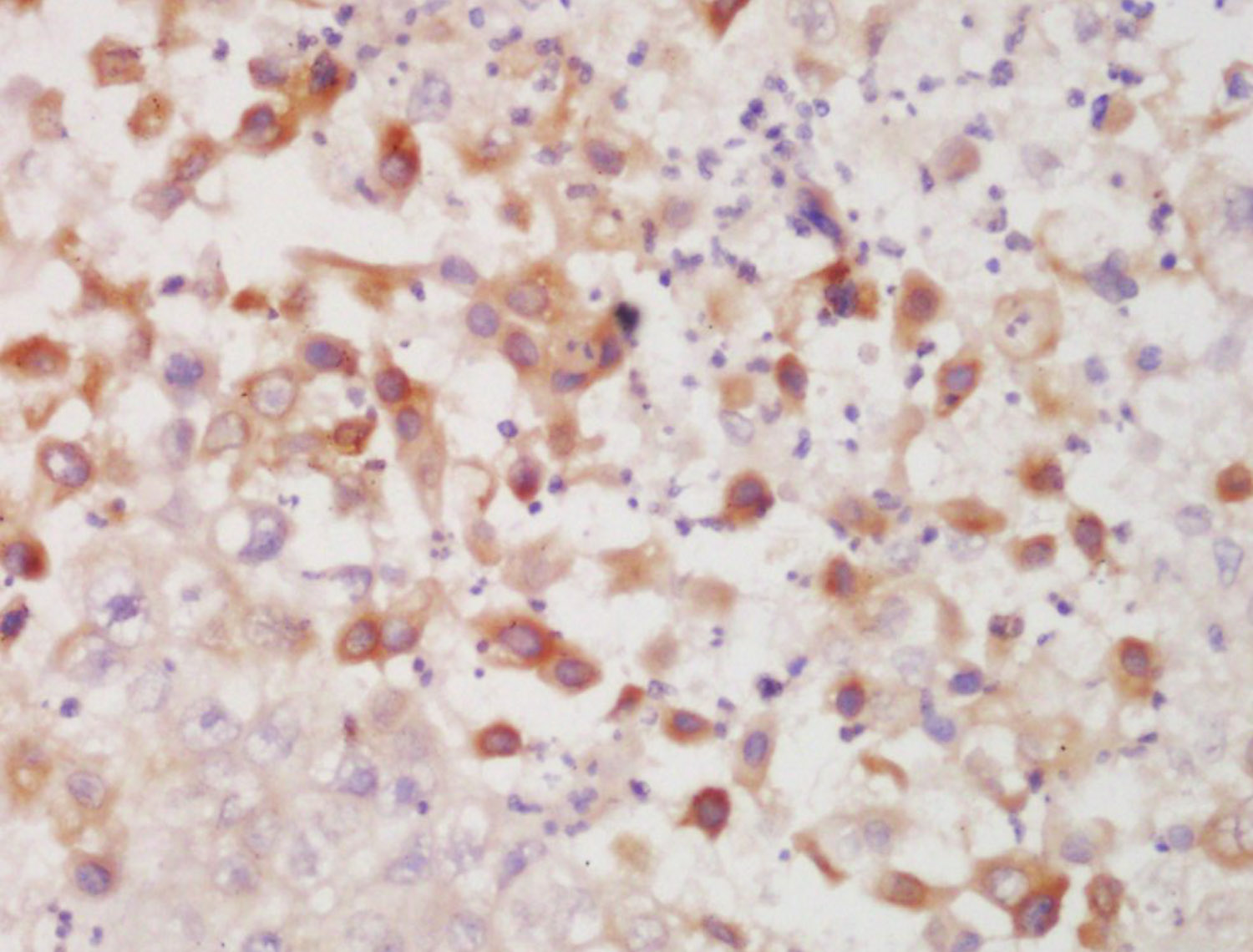
Tissue/cell: human lung carcinoma; 4% Paraformaldehyde-fixed and paraffin-embedded;
Antigen retrieval: citrate buffer ( 0.01M, pH 6.0 ), Boiling bathing for 15min; Block endogenous peroxidase by 3% Hydrogen peroxide for 30min; Blocking buffer (normal goat serum,C-0005) at 37℃ for 20 min;
Incubation: Anti-Cytokeratin 7 Polyclonal Antibody, Unconjugated(bs-1744R) 1:200, overnight at 4°C, followed by conjugation to the secondary antibody(SP-0023) and DAB(C-0010) staining
-
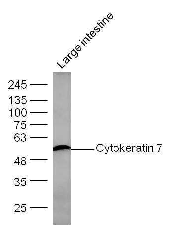
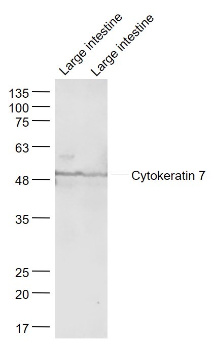
Sample: Large intestine (Mouse) Lysate at 30 ug
Primary: Anti- Cytokeratin 7 (bs-1744R) at 1/300 dilution
Secondary: IRDye800CW Goat Anti-Mouse IgG at 1/10000 dilution
Predicted band size: 54 kD
Observed band size: 54 kD
-
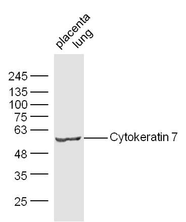
Sample:
Placenta (Mouse) Lysate at 30 ug
Lung (Mouse) Lysate at 30 ug
Primary: Anti-Cytokeratin 7 (bs-1744R) at 1/300 dilution
Secondary: IRDye800CW Goat Anti-Mouse IgG at 1/10000 dilution
Predicted band size: 54 kD
Observed band size: 54 kD
-
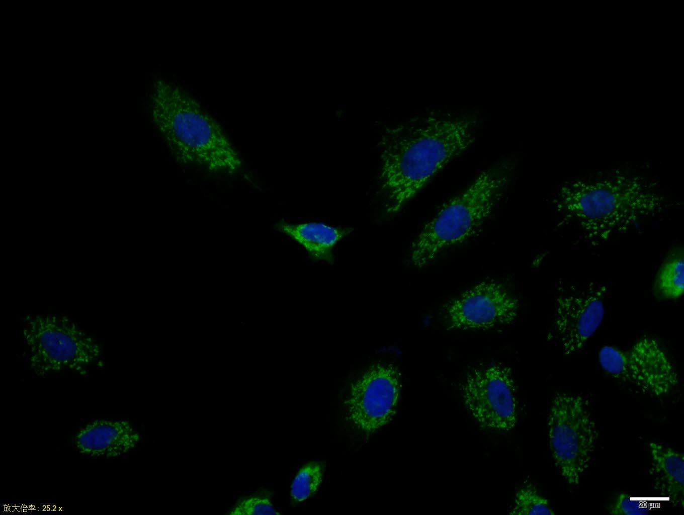
A549 cell; 4% Paraformaldehyde-fixed; Triton X-100 at room temperature for 20 min; Blocking buffer (normal goat serum, C-0005) at 37°C for 20 min; Antibody incubation with (Cytokeratin 7) polyclonal Antibody, Unconjugated (bs-1744R) 1:100, 90 minutes at 37°C; followed by a conjugated Goat Anti-Rabbit IgG antibody at 37°C for 90 minutes, DAPI (blue, C02-04002) was used to stain the cell nuclei.
-
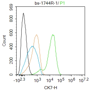
Blank control:Hela.
Primary Antibody (green line): Rabbit Anti-Cytokeratin 7 antibody (bs-1744R)
Dilution: 1ug/Test;
Secondary Antibody : Goat anti-rabbit IgG-FITC
Dilution: 0.5ug/Test.
Protocol
The cells were fixed with 4% PFA (10min at room temperature)and then permeabilized with 90% ice-cold methanol for 20 min at -20℃.The cells were then incubated in 5%BSA to block non-specific protein-protein interactions for 30 min at room temperature .Cells stained with Primary Antibody for 30 min at room temperature. The secondary antibody used for 40 min at room temperature. Acquisition of 20,000 events was performed.
-
Sample:
Large intestine (Mouse) Lysate at 40 ug
Large intestine(Rat) Lysate at 40 ug
Primary: Anti- Cytokeratin 7 (bs-1744R) at 1/1000 dilution
Secondary: IRDye800CW Goat Anti-Rabbit IgG at 1/20000 dilution
Predicted band size: 54 kD
Observed band size: 54 kD
RRID:AB_10856673
产品名称:Rabbit Anti-CK7 antibody
别名: CK 7; CK-7; Cytokeratin 7; Cytokeratin-7; Cytokeratin7; D15Wsu77e; K2C7; K2C7_HUMAN; K7; Keratin 55k type ii cytoskeletal; Keratin 7; Keratin simple epithelial type 1 k7; Keratin type II cytoskeletal 7; Keratin type ii cytoskletal 7; Keratin, 55K type II
中文名称:细胞角蛋白7抗体
英文名称:Rabbit Anti-CK7 antibody
抗体来源: Rabbit
克隆类型:多克隆
细胞定位:细胞浆
性 状:Liquid
亚 型:IgG
纯化方法:affinity purified by Protein A
保存条件:Shipped at 4℃. Store at -20 °C for one year. Avoid repeated freeze/thaw cycles.
免 疫 原:KLH conjugated synthetic peptide derived from the middle of mouse CK7
抗原表位:251-350/469
SWISS:Q9DCV7
Gene ID :110310
Human Gene ID:3855
The protein encoded by this gene is a member of the keratin gene family. The type II cytokeratins consist of basic or neutral proteins which are arranged in pairs of heterotypic keratin chains coexpressed during differentiation of simple and stratified epithelial tissues. This type II cytokeratin is specifically expressed in the simple epithelia lining the cavities of the internal organs and in the gland ducts and blood vessels. The genes encoding the type II cytokeratins are clustered in a region of chromosome 12q12-q13. Alternative splicing may result in several transcript variants; however, not all variants have been fully described. [provided by RefSeq, Jul 2008]
Function:Blocks interferon-dependent interphase and stimulates DNA synthesis in cells. Involved in the translational regulation of the human papillomavirus type 16 E7 mRNA (HPV16 E7).
Subunit:Heterotetramer of two type I and two type II keratins. Interacts with eukaryotic translation initiator factor 3 (eIF3) subunit EIF3S10 and with HPV16 E7.
Subcellular Location:Cytoplasm.
Tissue Specificity:Expressed in cultured epidermal, bronchial and mesothelial cells but absent in colon, ectocervix and liver. Observed throughout the glandular cells in the junction between stomach and esophagus but is absent in the esophagus.
Post-translational modifications:Arg-20 is dimethylated, probably to asymmetric dimethylarginine.
Similarity:Belongs to the intermediate filament family.
Important Note:This product as supplied is intended for research use only, not for use in human, therapeutic or diagnostic applications.
 400-901-9800


 说明书
说明书 联系我们
联系我们 打印此页面
打印此页面 收藏
收藏