| Rabbit Anti-GFAP antibody |
| 反应物种(预测) |
Chicken,Pig,Cow,Rabbit,Sheep |
| 产品应用(已验证) |
WB,ICC,FCM |
| 产品应用(可尝试) |
IHC,ELISA,IP |
| 推荐稀释比例 |
WB=1:500-2000,Elisa=1:5000-10000,IP=1:20-100,IHC-P=1:100-500,Flow Cyt=3μg/Test,ICC=1:100, |
| 标签 |
Array |
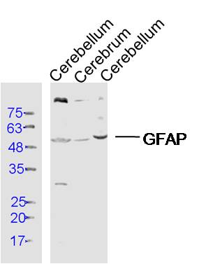
-
Sample:
Cerebellum (Rat) Lysate at 40 ug
Cerebrum (Mouse) Lysate at 40 ug
Cerebellum (Mouse) Lysate at 40 ug
Primary: Anti- GFAP (bs-10950R) at 1/300 dilution
Secondary: IRDye800CW Goat Anti-Rabbit IgG at 1/20000 dilution
Predicted band size: 48 kD
Observed band size: 50 kD
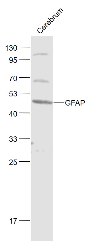
-
Sample:
Cerebrum (Mouse) Lysate at 40 ug
Primary: Anti-GFAP (bs-10950R) at 1/1000 dilution
Secondary: IRDye800CW Goat Anti-Rabbit IgG at 1/20000 dilution
Predicted band size: 48 kD
Observed band size: 48 kD
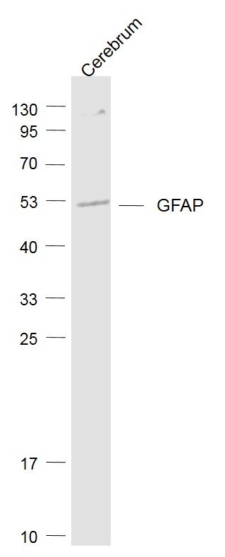
-
Sample:
Cerebrum (Rat) Lysate at 40 ug
Primary: Anti- GFAP (bs-10950R) at 1/1000 dilution
Secondary: IRDye800CW Goat Anti-Rabbit IgG at 1/20000 dilution
Predicted band size: 48 kD
Observed band size: 48 kD
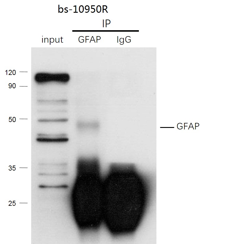
-
GFAP was immunoprecipitated from human hela cells lysate with bs-10950R at 1/150 dilution. Western blot was performed from the immunoprecipitate using protein A/G beads. HRP Conjugated Mouse anti-Rabbit IgG (Light Chain specific) was used as secondary antibody at 1:5000 dilution.
Lane 1: human hela cells lysate 10 µg (Input).
Lane 2: bs-10950R IP in human hela cells lysate.
Lane 3: native rabbit IgG IP in human hela cells lysate (negative control).
Secondary
All lanes : Mouse anti-Rabbit IgG (Light Chain specific), HRP Conjugated, 1:5000
-
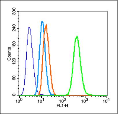
Blank control (blue line): Hela (fixed with 80% methanol (5 min at -20℃) and then permeabilized with 0.1% PBS-Tween for 20 min at room temperature ).
Primary Antibody (green line): Rabbit Anti-GFAP antibody (bs-10950R),dilution: 3μg /10^6 cells;
Isotype Control Antibody (orange line): Rabbit IgG .
Secondary Antibody (white blue line): Goat anti-rabbit IgG-PE,Dilution: 1μg /test.
-
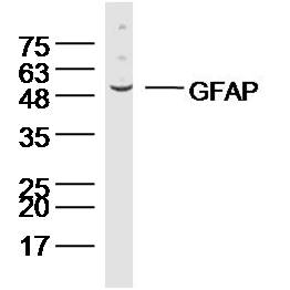
Sample:
U251(human) Cell Lysate at 40 ug
U87MG(human) Cell Lysate at 40 ug
BV2(mouse) Cell Lysate at 40 ug
Primary: Anti-GFAP (bs-10950R) at 1/300 dilution
Secondary: IRDye800CW Goat Anti-Rabbit IgG at 1/20000 dilution
Predicted band size: 48 kD
Observed band size: 48 kD
-
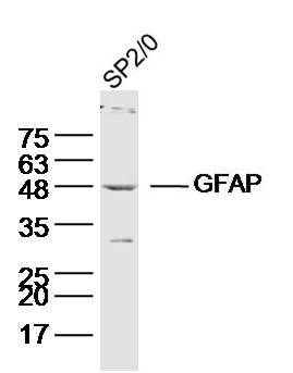
Sample:SP2/0(human) Cell Lysate at 40 ug
Primary: Anti-GFAP (bs-10950R) at 1/300 dilution
Secondary: IRDye800CW Goat Anti-Rabbit IgG at 1/20000 dilution
Predicted band size: 48 kD
Observed band size: 48 kD
-
Sample:Cerebellum (Mouse) Lysate at 40 ug
Primary: Anti-GFAP (bs-10950R) at 1/300 dilution
Secondary: IRDye800CW Goat Anti-Rabbit IgG at 1/20000 dilution
Predicted band size: 48 kD
Observed band size: 48 kD
-
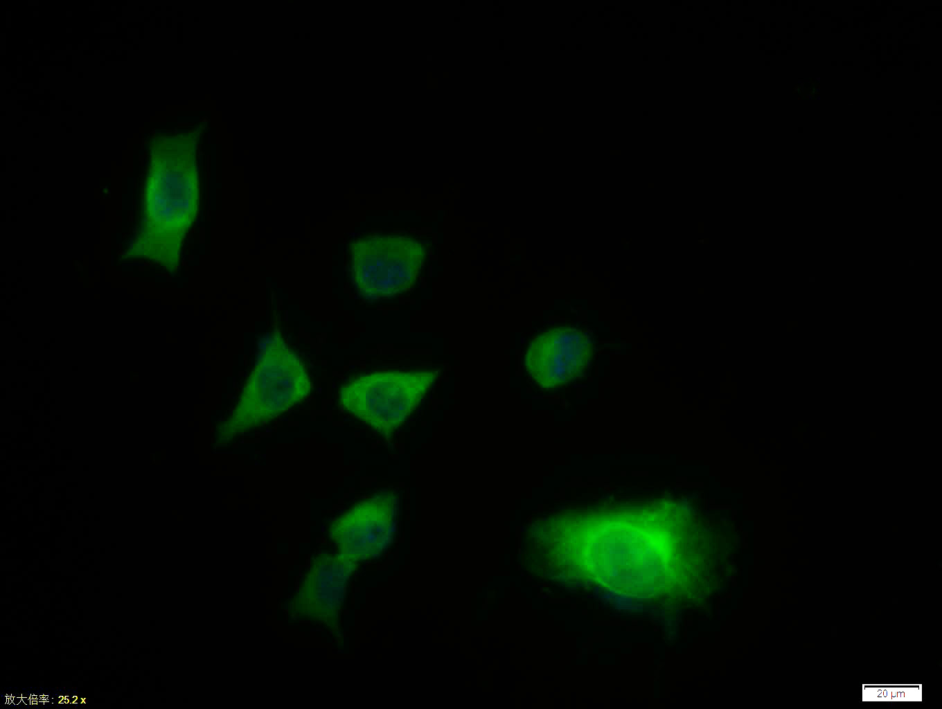
Tissue/cell:SH-SY5Y cell; 4% Paraformaldehyde-fixed; Triton X-100 at room temperature for 20 min; Blocking buffer (normal goat serum, C-0005) at 37°C for 20 min; Antibody incubation with (GFAP) polyclonal Antibody, Unconjugated (bs-10950R) 1:100, 90 minutes at 37°C; followed by a FITC conjugated Goat Anti-Rabbit IgG antibody at 37°C for 90 minutes, DAPI (blue, C02-04002) was used to stain the cell nuclei.
RRID:RRID
产品名称:Rabbit Anti-GFAP antibody
别名: Astrocyte; FLJ45472; GFAP; Glial Fibrillary Acidic Protein; Intermediate filament protein; GFAP_HUMAN.
中文名称:胶质纤维酸性蛋白抗体
英文名称:Rabbit Anti-GFAP antibody
抗体来源: Rabbit
克隆类型:多克隆
细胞定位:细胞浆
性 状:Liquid
亚 型:IgG
纯化方法:affinity purified by Protein A
保存条件:Shipped at 4℃. Store at -20 °C for one year. Avoid repeated freeze/thaw cycles.
免 疫 原:KLH conjugated synthetic peptide derived from human GFAP
抗原表位:341-432/432
SWISS:P14136
Gene ID :2670
Human Gene ID:2670
This gene encodes one of the major intermediate filament proteins of mature astrocytes. It is used as a marker to distinguish astrocytes from other glial cells during development. Mutations in this gene cause Alexander disease, a rare disorder of astrocytes in the central nervous system. Alternative splicing results in multiple transcript variants encoding distinct isoforms. [provided by RefSeq, Oct 2008]
Function:GFAP, a class-III intermediate filament, is a cell-specific marker that, during the development of the central nervous system, distinguishes astrocytes from other glial cells.
Subunit:Interacts with SYNM. Isoform 3 interacts with PSEN1 (via N-terminus).
Subcellular Location:Cytoplasm. Note=Associated with intermediate filaments.
Tissue Specificity:Expressed in cells lacking fibronectin.
Post-translational modifications:Phosphorylated by PKN1.
DISEASE:Defects in GFAP are a cause of Alexander disease (ALEXD) [MIM:203450]. Alexander disease is a rare disorder of the central nervous system. It is a progressive leukoencephalopathy whose hallmark is the widespread accumulation of Rosenthal fibers which are
Similarity:Belongs to the intermediate filament family.
Important Note:This product as supplied is intended for research use only, not for use in human, therapeutic or diagnostic applications.
 400-901-9800


 说明书
说明书 联系我们
联系我们 打印此页面
打印此页面 收藏
收藏