| Rabbit Anti-CIDEC antibody |
| 反应物种(预测) |
Pig |
| 产品应用(已验证) |
WB,IHC,FCM |
| 产品应用(可尝试) |
IF,ELISA |
| 推荐稀释比例 |
WB=1:500-2000,Elisa=1:5000-10000,IHC-P=1:100-500,IHC-F=1:100-500,Flow Cyt=1ug/Test,IF=1:100-500, |
| 研究领域 |
肿瘤,心血管,细胞生物,免疫学,信号转导,细胞凋亡,表观遗传学, |
| 标签 |
Array |
-
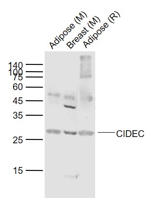
Sample:
Lane 1: Adipose (Mouse) Lysate at 40 ug
Lane 2: Breast (Mouse) Lysate at 40 ug
Lane 3: Adipose (Rat) Lysate at 40 ug
Primary:
Anti-CIDEC (bs-6796R) at 1/1000 dilution
Secondary: IRDye800CW Goat Anti-Rabbit IgG at 1/20000 dilution
Predicted band size: 27-30 kD
Observed band size: 27 kD
-
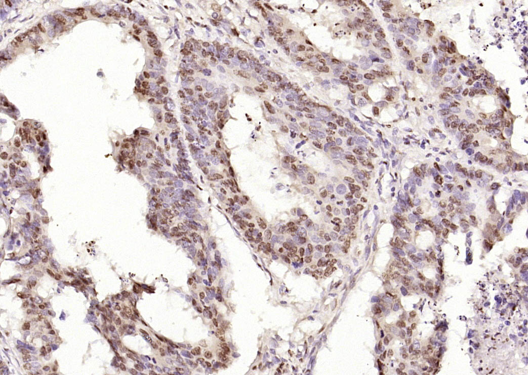
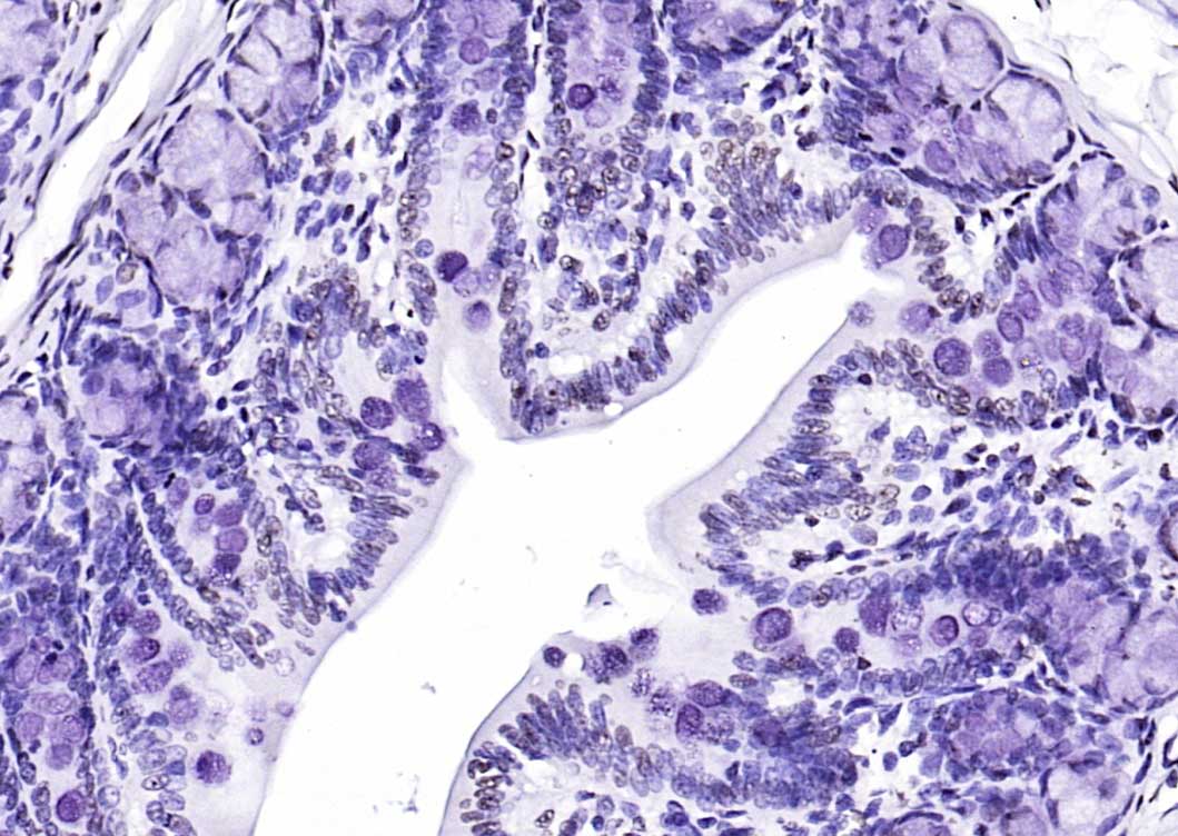
Paraformaldehyde-fixed, paraffin embedded (Human colon carcinoma); Antigen retrieval by boiling in sodium citrate buffer (pH6.0) for 15min; Block endogenous peroxidase by 3% hydrogen peroxide for 20 minutes; Blocking buffer (normal goat serum) at 37°C for 30min; Antibody incubation with (CIDEC) Polyclonal Antibody, Unconjugated (bs-6796R) at 1:400 overnight at 4°C, followed by operating according to SP Kit(Rabbit) (sp-0023) instructionsand DAB staining.
-
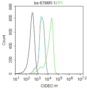
Blank control: A431.
Primary Antibody (green line): Rabbit Anti-CIDEC antibody (bs-6796R)
Dilution: 1ug/Test;
Secondary Antibody : Goat anti-rabbit IgG-FITC
Dilution: 0.5ug/Test.
Protocol
The cells were fixed with 4% PFA (10min at room temperature)and then permeabilized with 0.1% PBST for 20 min at room temperature.The cells were then incubated in 5%BSA to block non-specific protein-protein interactions for 30 min at room temperature .Cells stained with Primary Antibody for 30 min at room temperature. The secondary antibody used for 40 min at room temperature. Acquisition of 20,000 events was performed.
-
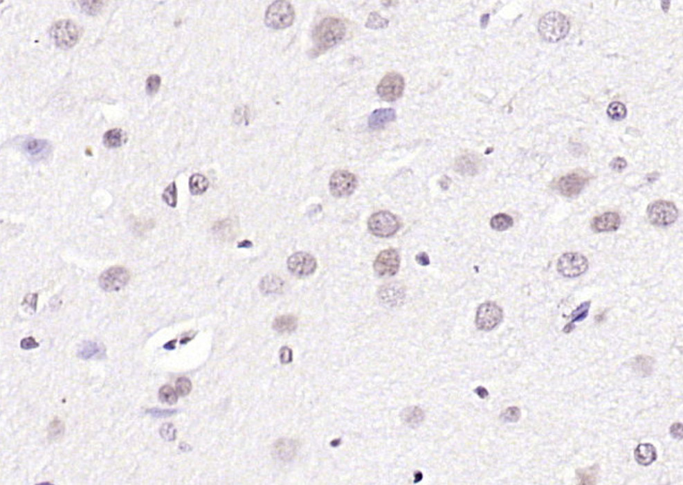
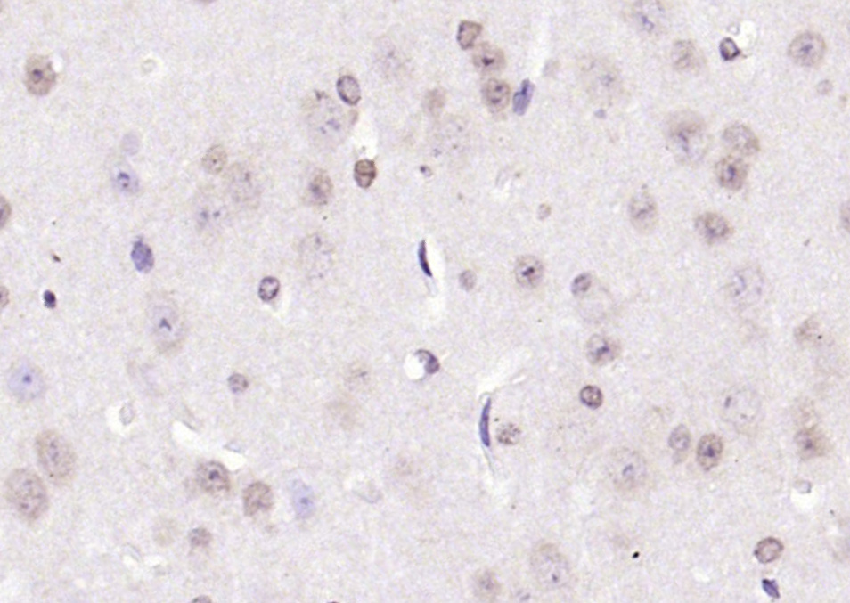
Paraformaldehyde-fixed, paraffin embedded (rat brain); Antigen retrieval by boiling in sodium citrate buffer (pH6.0) for 15min; Block endogenous peroxidase by 3% hydrogen peroxide for 20 minutes; Blocking buffer (normal goat serum) at 37°C for 30min; Antibody incubation with (CIDEC) Polyclonal Antibody, Unconjugated (bs-6796R) at 1:200 overnight at 4°C, followed by operating according to SP Kit(Rabbit) (sp-0023) instructionsand DAB staining.
-
Paraformaldehyde-fixed, paraffin embedded (mouse brain); Antigen retrieval by boiling in sodium citrate buffer (pH6.0) for 15min; Block endogenous peroxidase by 3% hydrogen peroxide for 20 minutes; Blocking buffer (normal goat serum) at 37°C for 30min; Antibody incubation with (CIDEC) Polyclonal Antibody, Unconjugated (bs-6796R) at 1:200 overnight at 4°C, followed by operating according to SP Kit(Rabbit) (sp-0023) instructionsand DAB staining.
-
Paraformaldehyde-fixed, paraffin embedded (rat breast); Antigen retrieval by boiling in sodium citrate buffer (pH6.0) for 15min; Block endogenous peroxidase by 3% hydrogen peroxide for 20 minutes; Blocking buffer (normal goat serum) at 37°C for 30min; Antibody incubation with (CIDEC) Polyclonal Antibody, Unconjugated (bs-6796R) at 1:200 overnight at 4°C, followed by operating according to SP Kit(Rabbit) (sp-0023) instructionsand DAB staining.
-
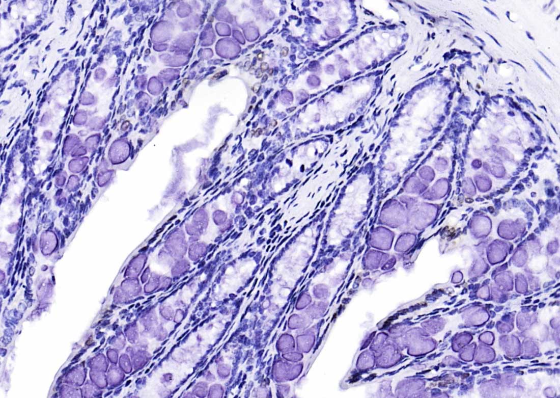
Paraformaldehyde-fixed, paraffin embedded (rat colon); Antigen retrieval by boiling in sodium citrate buffer (pH6.0) for 15min; Block endogenous peroxidase by 3% hydrogen peroxide for 20 minutes; Blocking buffer (normal goat serum) at 37°C for 30min; Antibody incubation with (CIDEC) Polyclonal Antibody, Unconjugated (bs-6796R) at 1:200 overnight at 4°C, followed by operating according to SP Kit(Rabbit) (sp-0023) instructionsand DAB staining.
-
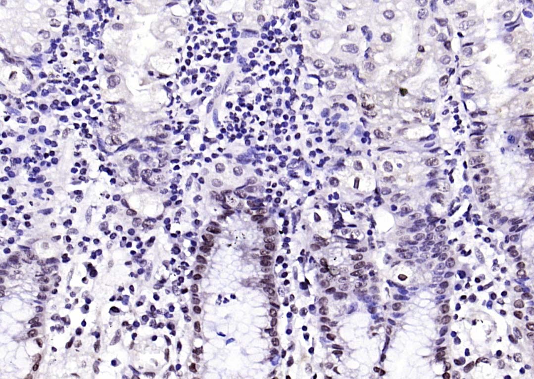
Paraformaldehyde-fixed, paraffin embedded (human gastric); Antigen retrieval by boiling in sodium citrate buffer (pH6.0) for 15min; Block endogenous peroxidase by 3% hydrogen peroxide for 20 minutes; Blocking buffer (normal goat serum) at 37°C for 30min; Antibody incubation with (CIDEC) Polyclonal Antibody, Unconjugated (bs-6796R) at 1:200 overnight at 4°C, followed by operating according to SP Kit(Rabbit) (sp-0023) instructionsand DAB staining.
-
Paraformaldehyde-fixed, paraffin embedded (MOUSE colon); Antigen retrieval by boiling in sodium citrate buffer (pH6.0) for 15min; Block endogenous peroxidase by 3% hydrogen peroxide for 20 minutes; Blocking buffer (normal goat serum) at 37°C for 30min; Antibody incubation with (CIDEC) Polyclonal Antibody, Unconjugated (bs-6796R) at 1:200 overnight at 4°C, followed by operating according to SP Kit(Rabbit) (sp-0023) instructionsand DAB staining.
-
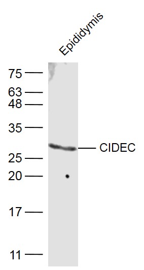
Sample:
Epididymis (Mouse) Lysate at 40 ug
Primary: Anti-CIDEC (bs-6796R) at 1/500 dilution
Secondary: IRDye800CW Goat Anti-Rabbit IgG at 1/20000 dilution
Predicted band size: 27 kD
Observed band size: 27 kD
-
Tissue/cell: mouse stomach wall; 4% Paraformaldehyde-fixed and paraffin-embedded;
Antigen retrieval: citrate buffer ( 0.01M, pH 6.0 ), Boiling bathing for 15min; Block endogenous peroxidase by 3% Hydrogen peroxide for 30min; Blocking buffer (normal goat serum,C-0005) at 37℃ for 20 min;
Incubation: Anti-CIDEC Polyclonal Antibody, Unconjugated(bs-6796R) 1:200, overnight at 4°C, followed by conjugation to the secondary antibody(SP-0023) and DAB(C-0010) staining
-
Tissue/cell: rat heart tissue; 4% Paraformaldehyde-fixed and paraffin-embedded;
Antigen retrieval: citrate buffer ( 0.01M, pH 6.0 ), Boiling bathing for 15min; Block endogenous peroxidase by 3% Hydrogen peroxide for 30min; Blocking buffer (normal goat serum,C-0005) at 37℃ for 20 min;
Incubation: Anti-CIDEC Polyclonal Antibody, Unconjugated(bs-6796R) 1:200, overnight at 4°C, followed by conjugation to the secondary antibody(SP-0023) and DAB(C-0010) staining
-
Paraformaldehyde-fixed, paraffin embedded (Rat brain); Antigen retrieval by boiling in sodium citrate buffer (pH6.0) for 15min; Block endogenous peroxidase by 3% hydrogen peroxide for 20 minutes; Blocking buffer (normal goat serum) at 37°C for 30min; Antibody incubation with (CIDEC) Polyclonal Antibody, Unconjugated (bs-6796R) at 1:400 overnight at 4°C, followed by operating according to SP Kit(Rabbit) (sp-0023) instructionsand DAB staining.
-
Blank control:Mouse spleen.
Primary Antibody (green line): Rabbit Anti-CIDEC antibody (bs-6796R)
Dilution: 2μg /10^6 cells;
Isotype Control Antibody (orange line): Rabbit IgG .
Secondary Antibody : Goat anti-rabbit IgG-AF647
Dilution: 1μg /test.
Protocol
The cells were fixed with 4% PFA (10min at room temperature)and then permeabilized with 90% ice-cold methanol for 20 min at-20℃. The cells were then incubated in 5%BSA to block non-specific protein-protein interactions for 30 min at room temperature .Cells stained with Primary Antibody for 30 min at room temperature. The secondary antibody used for 40 min at room temperature. Acquisition of 20,000 events was performed.
RRID:RRID
产品名称:Rabbit Anti-CIDEC antibody
别名: Cell Death Activator; Cell death activator CIDE-3; Cell Death Inducing DFFA Like Effector C; Cell death inducing DFFA like effector protein C; Cell death-inducing DFFA-like effector protein C; CIDE 3; CIDE3; CIDE C; CIDEC_HUMAN; Fat specific protein 27; F
中文名称:细胞死亡活化蛋白抗体
英文名称:Rabbit Anti-CIDEC antibody
抗体来源: Rabbit
克隆类型:多克隆
细胞定位:细胞核,细胞浆
性 状:Liquid
亚 型:IgG
纯化方法:affinity purified by Protein A
保存条件:Shipped at 4℃. Store at -20 °C for one year. Avoid repeated freeze/thaw cycles.
免 疫 原:KLH conjugated synthetic peptide derived from human CIDEC
抗原表位:101-200/238
SWISS:Q96AQ7
Gene ID :63924
Human Gene ID:63924
This gene encodes a member of the cell death-inducing DNA fragmentation factor-like effector family. Members of this family play important roles in apoptosis. The encoded protein promotes lipid droplet formation in adipocytes and may mediate adipocyte apoptosis. This gene is regulated by insulin and its expression is positively correlated with insulin sensitivity. Mutations in this gene may contribute to insulin resistant diabetes. A pseudogene of this gene is located on the short arm of chromosome 3. Alternatively spliced transcript variants that encode different isoforms have been observed for this gene. [provided by RefSeq, Dec 2010].
Tissue specificity: Expressed mainly in small intestine, heart, colon and stomach and, at lower levels, in brain, kidney and liver.
Function:May act as a CEBPB coactivator in white adipose tissueto control the expression of a subset of CEBPB downstream targetgenes, including SOCS1, SOCS3, TGFB1, TGFBR1, ID2 and XDH (Bysimilarity). Binds to lipid droplets and regulates theirenlargement, thereby
Subunit:Interacts with CEBPB (By similarity). Interacts withCIDEA.
Subcellular Location:Nucleus (By similarity). Endoplasmicreticulum (By similarity). Lipid droplet. Note=Diffuses quickly onlipid droplet surface, but becomes trapped and clustered at lipiddroplet contact sites, thereby enabling its rapid enrichment atlipid droplet contact sit
Tissue Specificity:Expressed mainly in adipose tissue, smallintestine, heart, colon and stomach and, at lower levels, in brain,kidney and liver.
Post-translational modifications:Ubiquitinated and targeted to proteasomal degradation,resulting in a short half-life. Protein stability depends ontriaclyglycerol synthesis, fatty acid availability and lipiddroplet formation (By similarity).
DISEASE:Note=In omental adipose tissue of obese patients matchedfor BMI, expression levels tend to correlate with insulinsensitivity. Expression is increased 2-3 fold in the group ofpatients with high insulin sensitivity, compared to theinsulin-resistant group. T
Similarity:Contains 1 CIDE-N domain.
Important Note:This product as supplied is intended for research use only, not for use in human, therapeutic or diagnostic applications.
 400-901-9800


 说明书
说明书 联系我们
联系我们 打印此页面
打印此页面 收藏
收藏