| Rabbit Anti-phospho-E2F1 (Ser364) antibody |
| 反应物种(预测) |
Mouse,Rat |
| 产品应用(已验证) |
WB,IHC,FCM |
| 产品应用(可尝试) |
ICC,IF,ELISA |
| 推荐稀释比例 |
WB=1:500-2000,Elisa=1:5000-10000,IHC-P=1:100-500,IHC-F=1:100-500,Flow Cyt=1ug/Test,IF=1:100-500,ICC=1:100, |
| 研究领域 |
肿瘤,细胞生物,免疫学,转录调节因子 |
| 标签 |
Array |
-
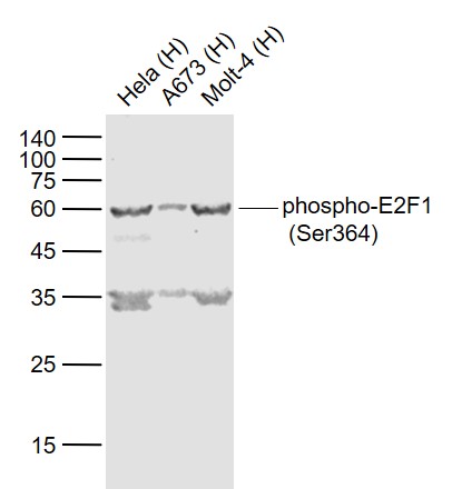
Sample:
Lane 1: Hela (Human) Cell Lysate at 30 ug
Lane 2: A673 (Human) Cell Lysate at 30 ug
Lane 3: Molt-4 (Human) Cell Lysate at 30 ug
Primary: Anti-phospho-E2F1 (Ser364) (bs-4076R) at 1/1000 dilution
Secondary: IRDye800CW Goat Anti-Rabbit IgG at 1/20000 dilution
Predicted band size: 55-60 kD
Observed band size: 60 kD
-
Blank control:HepG2.
Primary Antibody (green line): Rabbit Anti-phospho-E2F1 (Ser364) antibody (bs-4076R)
Dilution: 1ug/Test;
Secondary Antibody : Goat anti-rabbit IgG-FITC
Dilution: 0.5ug/Test.
Protocol
The cells were fixed with 4% PFA (10min at room temperature)and then permeabilized with 0.1% PBST for 20 min at room temperature.The cells were then incubated in 5%BSA to block non-specific protein-protein interactions for 30 min at room temperature .Cells stained with Primary Antibody for 30 min at room temperature. The secondary antibody used for 40 min at room temperature. Acquisition of 20,000 events was performed.
-
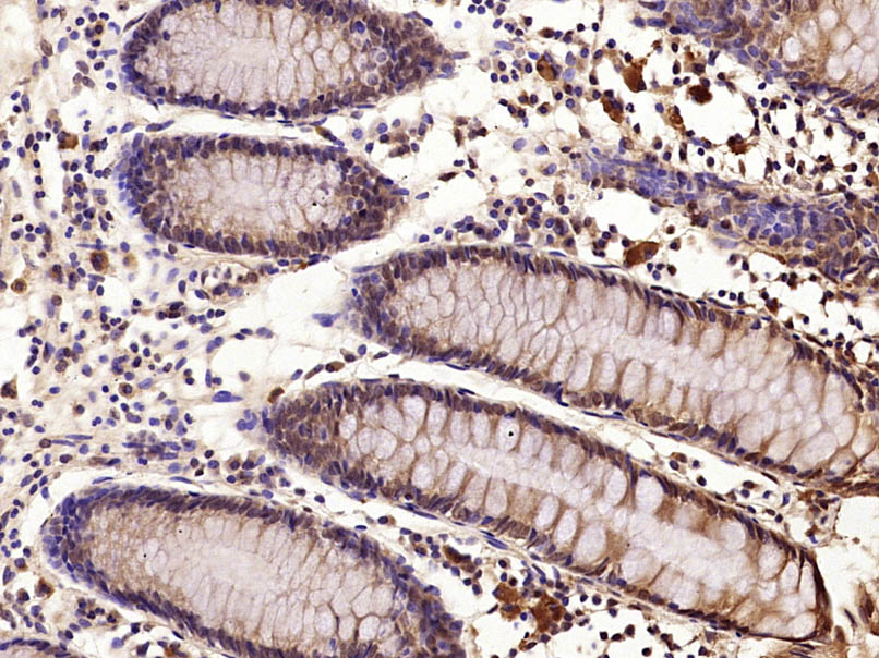
Paraformaldehyde-fixed, paraffin embedded (Human colon carcinoma); Antigen retrieval by microwave in sodium citrate buffer (pH6.0) ; Block endogenous peroxidase by 3% hydrogen peroxide for 30 minutes; Blocking buffer (3% BSA) at RT for 30min; Antibody incubation with (phospho-E2F1 (Ser364)) Polyclonal Antibody, Unconjugated (bs-4076R) at 1:400 overnight at 4°C, followed by conjugation to the secondary antibody (labeled with HRP)and DAB staining.
-
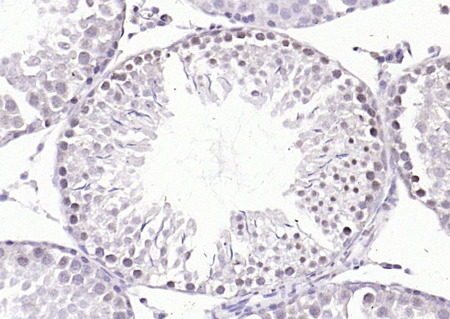
Paraformaldehyde-fixed, paraffin embedded (rat testis); Antigen retrieval by boiling in sodium citrate buffer (pH6.0) for 15min; Block endogenous peroxidase by 3% hydrogen peroxide for 20 minutes; Blocking buffer (normal goat serum) at 37°C for 30min; Antibody incubation with (phospho-E2F1 (Ser364)) Polyclonal Antibody, Unconjugated (bs-4076R) at 1:200 overnight at 4°C, followed by operating according to SP Kit(Rabbit) (sp-0023) instructionsand DAB staining.
-
Paraformaldehyde-fixed, paraffin embedded (rat colon); Antigen retrieval by boiling in sodium citrate buffer (pH6.0) for 15min; Block endogenous peroxidase by 3% hydrogen peroxide for 20 minutes; Blocking buffer (normal goat serum) at 37°C for 30min; Antibody incubation with (phospho-E2F1 (Ser364)) Polyclonal Antibody, Unconjugated (bs-4076R) at 1:200 overnight at 4°C, followed by operating according to SP Kit(Rabbit) (sp-0023) instructionsand DAB staining.
-
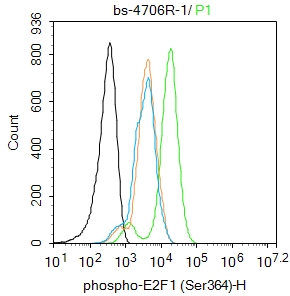
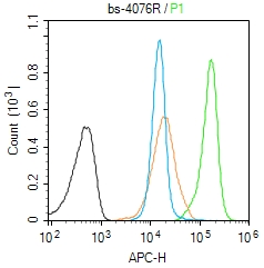
Blank control (Black line):Molt4 (Black).
Primary Antibody (green line): Rabbit Anti-phospho-E2F1 (Ser364) antibody (bs-4076R)
Dilution: 1μg /10^6 cells;
Isotype Control Antibody (orange line): Rabbit IgG .
Secondary Antibody (white blue line): Goat anti-rabbit IgG-AF647
Dilution: 1μg /test.
Protocol
The cells were fixed with 4% PFA (10min at room temperature)and then permeabilized with 90% ice-cold methanol for 20 min at room temperature. The cells were then incubated in 5%BSA to block non-specific protein-protein interactions for 30 min at room temperature .Cells stained with Primary Antibody for 30 min at room temperature. The secondary antibody used for 40 min at room temperature. Acquisition of 20,000 events was performed.
RRID:AB_11053575
产品名称:Rabbit Anti-phospho-E2F1 (Ser364) antibody
别名: E2F1 (phospho S364); E2F1 (phospho Ser364); p-E2F1 (S364); p-E2F1 (Ser364); E2F 1; E2F transcription factor 1; E2F-1; E2f1 E2F transcription factor 1; KIAA4009; mKIAA4009; OTTHUMP00000030661; PBR 3; PBR3; PRB binding protein E2F 1; PRB-binding protein E2F
中文名称:磷酸化转录因子E2F-1抗体
英文名称:Rabbit Anti-phospho-E2F1 (Ser364) antibody
抗体来源: Rabbit
克隆类型:多克隆
细胞定位:细胞核
性 状:Liquid
亚 型:IgG
纯化方法:affinity purified by Protein A
保存条件:Shipped at 4℃. Store at -20 °C for one year. Avoid repeated freeze/thaw cycles.
免 疫 原:KLH conjugated Synthesised phosphopeptide derived from human E2F1 around the phosphorylation site of Ser364
抗原表位:MG(p-S)LR
SWISS:Q01094
Gene ID :1869
Human Gene ID:1869
E2F's are DNA binding proteins, which associate with negative regulators, such as the retinoblastoma p107 protein, resulting in an altered rate of gene transcription. The E2F proteins contain several evolutionally conserved domains found in most members of the family. These domains include a DNA binding domain, a dimerization domain which determines interaction with the differentiation regulated transcription factor proteins (DP), a transactivation domain enriched in acidic amino acids, and a tumor suppressor protein association domain which is embedded within the transactivation domain. This protein and another 2 members, E2F2 and E2F3, have an additional cyclin binding domain. E2F1 is proposed to be involved in several cellular processes that range from tumor suppressor, cell progression and oncogenesis. E2F1 overexpression can also drive cells into apoptosis.
Subunit:Component of the DRTF1/E2F transcription factor complex. Forms heterodimers with DP family members. The E2F-1 complex binds specifically hypophosphorylated retinoblastoma protein RB1. During the cell cycle, RB1 becomes phosphorylated in mid-to-late G1 pha
Subcellular Location:Nucleus.
Post-translational modifications:Phosphorylated by CDK2 and cyclin A-CDK2 in the S-phase.
Similarity:Belongs to the E2F/DP family.
Important Note:This product as supplied is intended for research use only, not for use in human, therapeutic or diagnostic applications.
 400-901-9800


 说明书
说明书 联系我们
联系我们 打印此页面
打印此页面 收藏
收藏