| Rabbit Anti-phospho-FSCN1 (Ser39) antibody |
| 反应物种(预测) |
Dog,Pig |
| 产品应用(已验证) |
WB,IHC,ICC,FCM |
| 产品应用(可尝试) |
IF,ELISA |
| 推荐稀释比例 |
WB=1:500-2000,Elisa=1:5000-10000,IHC-P=1:100-500,IHC-F=1:100-500,Flow Cyt=1μg/Test,IF=1:100-500,ICC=1:100, |
| 研究领域 |
细胞生物,信号转导,结合蛋白, |
| 标签 |
Array |
-
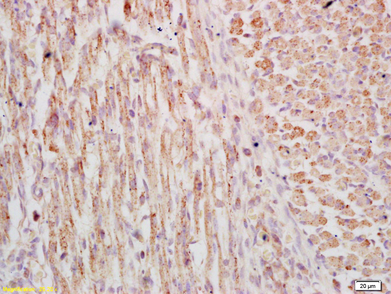
Tissue/cell: musle of mouse embryo; 4% Paraformaldehyde-fixed and paraffin-embedded;
Antigen retrieval: citrate buffer ( 0.01M, pH 6.0 ), Boiling bathing for 15min; Block endogenous peroxidase by 3% Hydrogen peroxide for 30min; Blocking buffer (normal goat serum,C-0005) at 37℃ for 20 min;
Incubation: Anti-phospho-FSCN1(Ser39) Polyclonal Antibody, Unconjugated(bs-0772R) 1:200, overnight at 4°C, followed by conjugation to the secondary antibody(SP-0023) and DAB(C-0010) staining
-
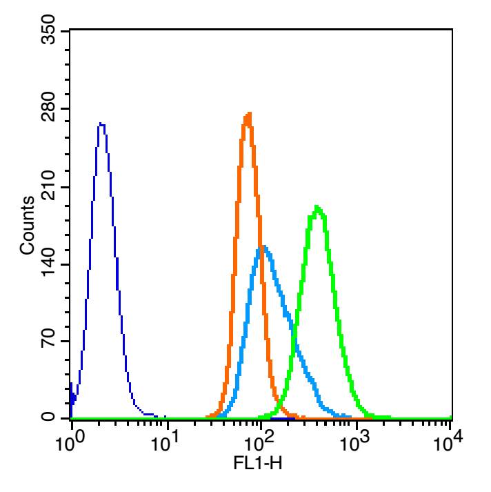
Blank control: U937(blue)
Isotype Control Antibody: Rabbit IgG(orange) ;
Secondary Antibody: Goat anti-rabbit IgG-FITC(white blue),
Dilution: 1:100 in 1 X PBS containing 0.5% BSA ;
Primary Antibody Dilution: 3μl in 100 μL1X PBS containing 0.5% BSA(green).
-
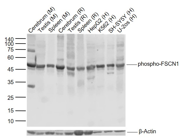
Sample:
Lane 1: Mouse Cerebrum tissue lysates
Lane 2: Mouse Testis tissue lysates
Lane 3: Mouse Spleen tissue lysates
Lane 4: Rat Cerebrum tissue lysates
Lane 5: Rat Testis tissue lysates
Lane 6: Rat Spleen tissue lysates
Lane 7: Human HepG2 cell lysates
Lane 8: Human K562 cell lysates
Lane 9: Human SH-SY5Y cell lysates
Lane 10: Human U-2os cell lysates
Primary: Anti- phospho-FSCN1 (Ser39) (bs-0772R) at 1/1000 dilution
Secondary: IRDye800CW Goat Anti-Rabbit IgG at 1/20000 dilution
Predicted band size: 55 kDa
Observed band size: 51 kDa
-
A549 cell; 4% Paraformaldehyde-fixed; Triton X-100 at room temperature for 20 min; Blocking buffer (normal goat serum, C-0005) at 37°C for 20 min; Antibody incubation with (phospho-FSCN1 (Ser39)) polyclonal Antibody, Unconjugated (bs-0772R) 1:100, 90 minutes at 37°C; followed by a conjugated Goat Anti-Rabbit IgG antibody at 37°C for 90 minutes, DAPI (blue, C02-04002) was used to stain the cell nuclei.
-
Sample:
U87MG(Human) Cell Lysate at 30 ug
Primary: Anti-phospho-FSCN1(Ser39) (bs-0772R) at 1/1000 dilution
Secondary: IRDye800CW Goat Anti-Rabbit IgG at 1/20000 dilution
Predicted band size: 55 kD
Observed band size: 55 kD
-
Paraformaldehyde-fixed, paraffin embedded (human brain glioma); Antigen retrieval by boiling in sodium citrate buffer (pH6.0) for 15min; Block endogenous peroxidase by 3% hydrogen peroxide for 20 minutes; Blocking buffer (normal goat serum) at 37°C for 30min; Antibody incubation with (FSCN1(Ser39)) Polyclonal Antibody, Unconjugated (bs-0772R) at 1:400 overnight at 4°C, followed by operating according to SP Kit(Rabbit) (sp-0023) instructionsand DAB staining.
RRID:AB_10855789
产品名称:Rabbit Anti-phospho-FSCN1 (Ser39) antibody
别名: Fascin (phospho Ser39); Fascin (phospho S39); 55 kDa actin bundling protein; Actin bundling protein; FAN 1; FAN1; Fascin 1; Fascin homolog 1 actin bundling protein (Strongylocentrotus purpuratus); Fascin homolog 1; Fascin1; FLJ38511; FSCN 1; FSCN1; HSN; p
中文名称:磷酸化纤维束蛋白同源物1抗体
英文名称:Rabbit Anti-phospho-FSCN1 (Ser39) antibody
抗体来源: Rabbit
克隆类型:多克隆
细胞定位:细胞浆
性 状:Liquid
亚 型:IgG
纯化方法:affinity purified by Protein A
保存条件:Shipped at 4℃. Store at -20 °C for one year. Avoid repeated freeze/thaw cycles.
免 疫 原:KLH conjugated Synthesised phosphopeptide derived from human FSCN1 around the phosphorylation site of Ser39
抗原表位:AS(p-S)LK
SWISS:Q16658
Gene ID :6624
Human Gene ID:6624
Human fascin is a highly conserved actin-bundling protein. Fascin, encoded by the human homolog for sn (hsn) gene, has been localized to microspikes and stress fibers of cultured cells where it is thought to be involved in the formation of microfilament bundles. It is expressed predominantly in dendritic cells. Lymphoid cells, myeloid cells and plasma cells are negative. However, Reed Sternberg cells in Hodgkin’s lymphoma are positive for fascin staining. Epstein-Barr virus may induce expression of fascin in B cells.
Function:Organizes filamentous actin into bundles with a minimum of 4.1:1 actin/fascin ratio. Plays a role in the organization of actin filament bundles and the formation of microspikes, membrane ruffles, and stress fibers. Important for the formation of a diverse
Subcellular Location:Cytoplasm > cytoskeleton. Cell projection > filopodium. Cell projection > invadopodium.
Tissue Specificity:Ubiquitous.
Post-translational modifications:Phosphorylation on Ser-39 inhibits the actin-binding ability of fascin.
Similarity:Belongs to the fascin family.
Important Note:This product as supplied is intended for research use only, not for use in human, therapeutic or diagnostic applications.
 400-901-9800


 说明书
说明书 联系我们
联系我们 打印此页面
打印此页面 收藏
收藏