| Rabbit Anti-Phospho-MAP2 (Ser136) antibody |
| 反应物种(预测) |
Mouse,Dog,Pig,Cow,Rabbit |
| 产品应用(已验证) |
IHC,IF,FCM |
| 产品应用(可尝试) |
ELISA |
| 推荐稀释比例 |
Elisa=1:5000-10000,IHC-P=1:100-500,IHC-F=1:100-500,Flow Cyt=0.2μg /Test,IF=1:100-500, |
| 研究领域 |
免疫学,神经生物学,信号转导,干细胞,细胞凋亡,细胞类型标志物,细胞骨架, |
| 标签 |
Array |
-
Blank control: SH-SY5Y.
Primary Antibody (green line): Rabbit Anti-Phospho-MAP2 (Ser136) antibody (bs-3259R)
Dilution: 1μg /10^6 cells;
Isotype Control Antibody (orange line): Rabbit IgG .
Secondary Antibody : Goat anti-rabbit IgG-AF647
Dilution: 1μg /test.
Protocol
The cells were fixed with 4% PFA (10min at room temperature)and then permeabilized with 90% ice-cold methanol for 20 min at-20℃.The cells were then incubated in 5%BSA to block non-specific protein-protein interactions for 30 min at room temperature .Cells stained with Primary Antibody for 30 min at room temperature. The secondary antibody used for 40 min at room temperature. Acquisition of 20,000 events was performed.
-
Tissue/cell: rat testis tissue; 4% Paraformaldehyde-fixed and paraffin-embedded;
Antigen retrieval: citrate buffer ( 0.01M, pH 6.0 ), Boiling bathing for 15min; Block endogenous peroxidase by 3% Hydrogen peroxide for 30min; Blocking buffer (normal goat serum,C-0005) at 37℃ for 20 min;
Incubation: Anti-Phospho-MAP2 (Ser136) Polyclonal Antibody, Unconjugated(bs-3259R) 1:200, overnight at 4°C, followed by conjugation to the secondary antibody(SP-0023) and DAB(C-0010) staining
-
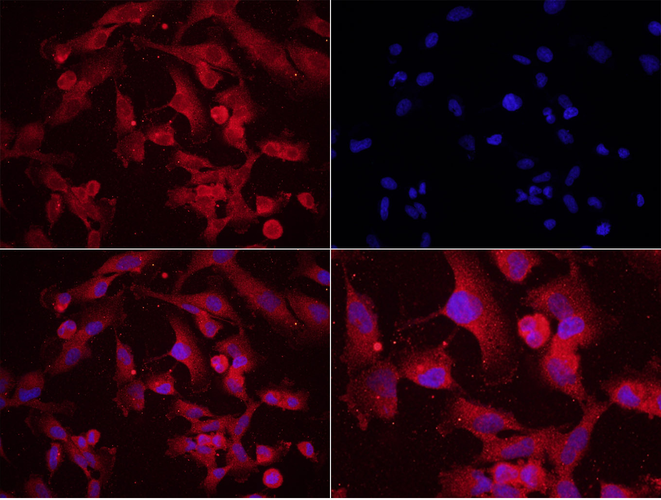
Tissue/cell: human glioma cells, U251;4% Paraformaldehyde-fixed;
Blocking buffer (normal goat serum,C-0005) at 37℃ for 20 min;
Incubation: Anti-Phospho-MAP2(Ser136) Polyclonal Antibody, Unconjugated(bs-3259R) 1:200, overnight at 4°C; The secondary antibody was Goat Anti-Rabbit IgG, Cy3 conjugated (bs-0295G-Cy3)used at 1:200 dilution for 40 minutes at 37°C. DAPI(5ug/ml,blue,C-0033) was used to stain the cell nuclei
-
Blank control: RSC96 Cells(blue).
Primary Antibody: Rabbit Anti-hospho-MAP2(Ser136)/FITC Conjugated antibody (bs-3259R-FITC), Dilution: 0.2μg in 100 μL 1X PBS containing 0.5% BSA;
Isotype Control Antibody: Rabbit IgG/FITC(orange) ,used under the same conditions.
RRID:AB_10882043
产品名称:Rabbit Anti-Phospho-MAP2 (Ser136) antibody
别名: MAP2(Phospho Ser136); MAP2 (phospho S136); p-MAP2 (phospho S136); MAP2(Phospho S136); MAP2(Phospho-Ser136); p-MAP2(Phospho-Ser136); DKFZp686I2148; Dendrite specific MAP; DKFZp686I2148; MAP 2; MAP-2; MAP2; MAP2_HUMAN; MAP2A; MAP2B; MAP2C; Microtubule assoc
中文名称:磷酸化微管相关蛋白2抗体
英文名称:Rabbit Anti-Phospho-MAP2 (Ser136) antibody
抗体来源: Rabbit
克隆类型:多克隆
细胞定位:细胞核,细胞浆
性 状:Liquid
亚 型:IgG
纯化方法:affinity purified by Protein A
保存条件:Shipped at 4℃. Store at -20 °C for one year. Avoid repeated freeze/thaw cycles.
免 疫 原:KLH conjugated synthesised phosphopeptide derived from human MAP2 around the phosphorylation site of Ser136
抗原表位:PP(p-S)P
SWISS:P11137
Gene ID :4133
Human Gene ID:4133
MAP2 is the major microtubule associated protein of brain tissue. There are three forms of MAP2; two are similarily sized with apparent molecular weights of 280 kDa (MAP2a and MAP2b) and the third with a lower molecular weight of 70 kDa (MAP2c). In the newborn rat brain, MAP2b and MAP2c are present, while MAP2a is absent. Between postnatal days 10 and 20, MAP2a appears. At the same time, the level of MAP2c drops by 10-fold. This change happens during the period when dendrite growth is completed and when neurons have reached their mature morphology. MAP2 is degraded by a Cathepsin D-like protease in the brain of aged rats. There is some indication that MAP2 is expressed at higher levels in some types of neurons than in other types. MAP2 is known to promote microtubule assembly and to form side-arms on microtubules. It also interacts with neurofilaments, actin, and other elements of the cytoskeleton.
Function:The exact function of MAP2 is unknown but MAPs may stabilize the microtubules against depolymerization. They also seem to have a stiffening effect on microtubules.
Subcellular Location:Cytoplasm, cytoskeleton (Probable).
Post-translational modifications:Phosphorylated at serine residues in K-X-G-S motifs by MAP/microtubule affinity-regulating kinase (MARK1 or MARK2), causing detachment from microtubules, and their disassembly (By similarity). MAP2A/c is phosphorylated. Isoform MAP2c is phosphorylated by
Similarity:Contains 3 Tau/MAP repeats.
Important Note:This product as supplied is intended for research use only, not for use in human, therapeutic or diagnostic applications.
 400-901-9800


 说明书
说明书 联系我们
联系我们 打印此页面
打印此页面 收藏
收藏