| Rabbit Anti-Phospho-TAK1 (Ser192) antibody |
| 反应物种(预测) |
Mouse,Chicken,Pig,Cow,Horse,Rabbit |
| 产品应用(已验证) |
ICC |
| 产品应用(可尝试) |
IHC,IF,ELISA |
| 推荐稀释比例 |
Elisa=1:5000-10000,IHC-P=1:100-500,IHC-F=1:100-500,IF=1:100-500,ICC=1:100, |
| 研究领域 |
肿瘤,心血管,免疫学,信号转导,激酶和磷酸酶 |
| 标签 |
Array |
-
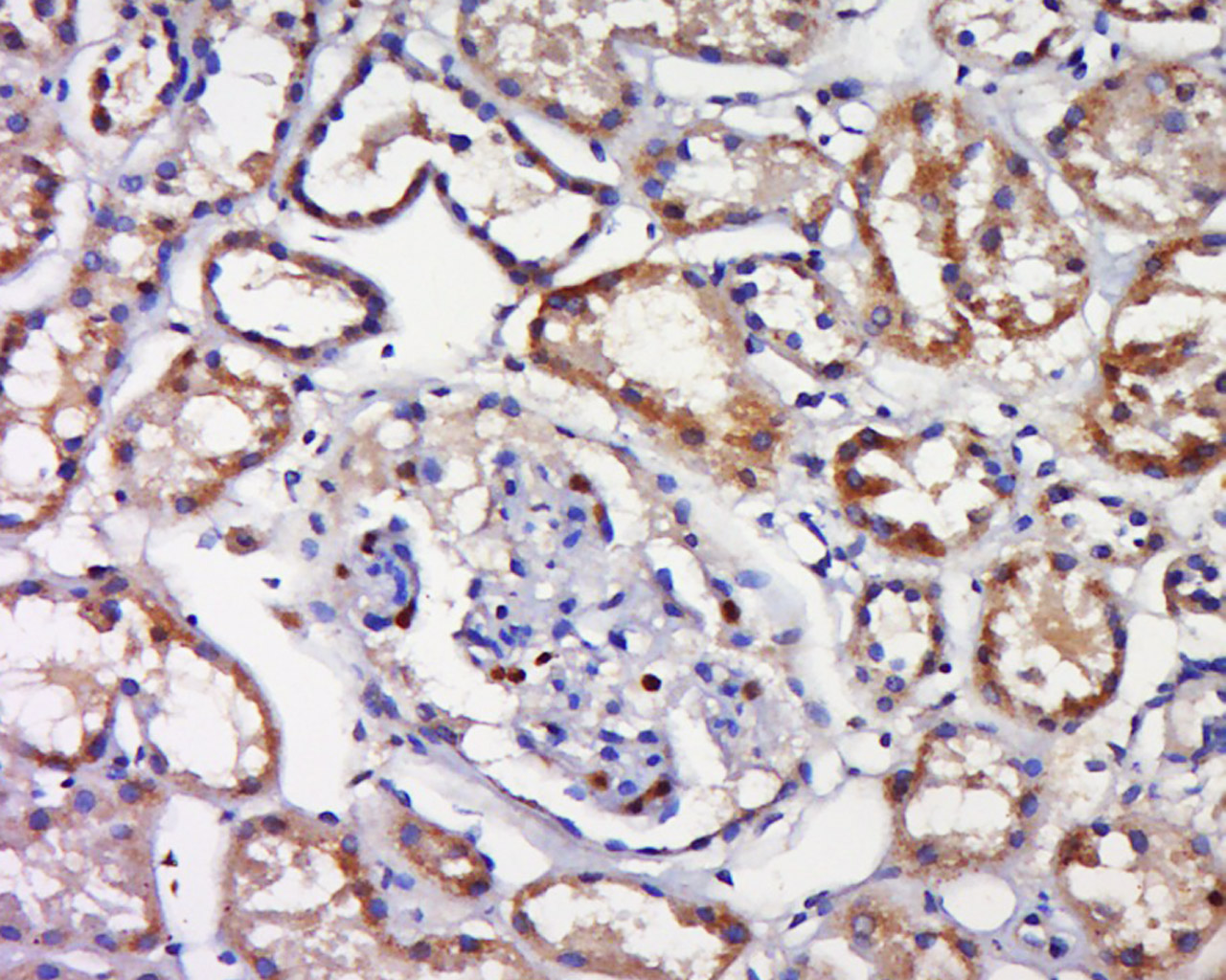
Tissue/cell: human kidney tissue; 4% Paraformaldehyde-fixed and paraffin-embedded;
Antigen retrieval: citrate buffer ( 0.01M, pH 6.0 ), Boiling bathing for 15min; Block endogenous peroxidase by 3% Hydrogen peroxide for 30min; Blocking buffer (normal goat serum,C-0005) at 37℃ for 20 min;
Incubation: Anti-Phospho-TAK1(Ser192)Polyclonal Antibody, Unconjugated(bs-5435R) 1:500, overnight at 4°C, followed by conjugation to the secondary antibody(SP-0023) and DAB(C-0010) staining
-
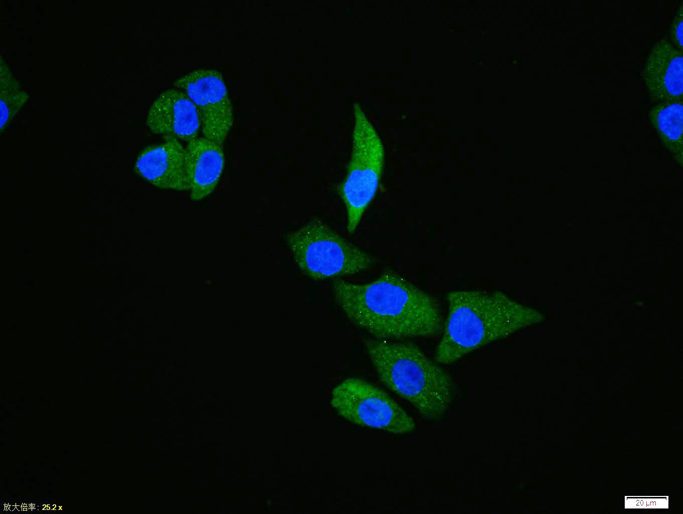
Hela cell; 4% Paraformaldehyde-fixed; Triton X-100 at room temperature for 20 min; Blocking buffer (normal goat serum, C-0005) at 37°C for 20 min; Antibody incubation with (Phospho-TAK1 (Ser192)) polyclonal Antibody, Unconjugated (bs-5435R) 1:100, 90 minutes at 37°C; followed by a conjugated Goat Anti-Rabbit IgG antibody at 37°C for 90 minutes, DAPI (blue, C02-04002) was used to stain the cell nuclei.
-
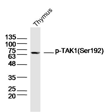
Sample: Thymus (Rat) Lysate at 40 ug
Primary: Anti-Phospho-TAK1(Ser192) (bs-5435R) at 1/300 dilution
Secondary: IRDye800CW Goat Anti-Rabbit IgG at 1/20000 dilution
Predicted band size: 67 kD
Observed band size: 67 kD
-
Blank control (Black line): Raji (Black).
Primary Antibody (green line): Rabbit Anti- Phospho-c-Fos (Ser32) antibody (bs-3152R)
Dilution: 1μg /10^6 cells;
Isotype Control Antibody (orange line): Rabbit IgG .
Secondary Antibody (white blue line): Goat anti-rabbit IgG-PE
Dilution: 1μg /test.
Protocol
The cells were fixed with 70% ice-cold methanol overnight at 4℃ and then permeabilized with 0.1% PBS-Tween for 20 min at room temperature. Cells stained with Primary Antibody for 30 min at room temperature. The cells were then incubated in 1 X PBS/2%BSA/10% goat serum to block non-specific protein-protein interactions followed by the antibody for 15 min at room temperature. The secondary antibody used for 40 min at room temperature. Acquisition of 20,000 events was performed.
RRID:AB_11085100
产品名称:Rabbit Anti-Phospho-TAK1 (Ser192) antibody
别名: MAP3K7(phospho S192); TAK1(Phospho Ser192); MAP3K7; Mitogen-activated protein kinase kinase kinase 7; Transforming growth factor-beta-activated kinase 1; TGF-beta-activated kinase 1; MAP3K 7; MAPKKK7; Mitogen activated protein kinase kinase kinase 7; TAK1
中文名称:磷酸化转化生长因子β活化激酶1
英文名称:Rabbit Anti-Phospho-TAK1 (Ser192) antibody
抗体来源: Rabbit
克隆类型:多克隆
细胞定位:细胞浆,细胞膜
性 状:Liquid
亚 型:IgG
纯化方法:affinity purified by Protein A
保存条件:Shipped at 4℃. Store at -20 °C for one year. Avoid repeated freeze/thaw cycles.
免 疫 原:KLH conjugated Synthesised phosphopeptide derived from human TAK1 around the phosphorylation site of Ser192
抗原表位:KG(p-S)AA
SWISS:O43318
Gene ID :6885
Human Gene ID:6885
TAK1 (or MAP3K7) was shown to participate in regulation of transcription by transforming growth factor beta (TGF beta). TAK1 is stimulated in response to TGF beta and bone morphogenetic protein. These results suggest that TAK1 functions as a mediator in the signaling pathway of TGF beta superfamily members. TAB1 and TAB2 are TAK1 binding proteins that may function as activators of the TAK1 (TGF b activated kinase 1) MAPKKK in TGF b signal transduction. TAB1 induced TAK1 activation promoted the dissociation of active forms of IKKa and IKK b from active TAK1, whereas the IKK mutants remained to interact with active TAK1. TNF a activated endogenous TAK1, and the kinase negative TAK1 acted as a dominant negative inhibitor against TNF a induced NFkB activation. TAK1 was suggested to act as a regulatory kinase of IKKs.
Function:Serine/threonine kinase which acts as an essential component of the MAP kinase signal transduction pathway. Plays an important role in the cascades of cellular responses evoked by changes in the environment. Mediates signal transduction of TRAF6, various
Subunit:Binds both upstream activators and downstream substrates in multimolecular complexes. Interacts with TAB1/MAP3K7IP1, TAB2/MAP3K7IP2 and TAB3/MAP3K7IP3. Identified in the TRIKA2 complex composed of MAP3K7/TAK1, TAB1/MAP3K7IP1 and TAB2/MAP3K7IP2. Interacts
Subcellular Location:Cytoplasm. Cell membrane; Peripheral membrane protein; Cytoplasmic side. Note=Although the majority of MAP3K7/TAK1 is found in the cytosol, when complexed with TAB1/MAP3K7IP1 and TAB2/MAP3K7IP2, it is also localized at the cell membrane.
Tissue Specificity:Isoform 1A is the most abundant in ovary, skeletal muscle, spleen and blood mononuclear cells. Isoform 1B is highly expressed in brain, kidney and small intestine. Isoform 1C is the major form in prostate. Isoform 1D is the less abundant form.
Post-translational modifications:Association with TAB1/MAP3K7IP1 promotes autophosphorylation at Ser-192 and subsequent activation. Association with TAB2/MAP3K7IP2, itself associated with free unanchored Lys-63 polyubiquitin chain, promotes autophosphorylation and subsequent activation o
Similarity:Belongs to the protein kinase superfamily. STE Ser/Thr protein kinase family. MAP kinase kinase kinase subfamily.
Contains 1 protein kinase domain.
Important Note:This product as supplied is intended for research use only, not for use in human, therapeutic or diagnostic applications.
 400-901-9800


 说明书
说明书 联系我们
联系我们 打印此页面
打印此页面 收藏
收藏