| Rabbit Anti-Phospho-c-Fos (Ser362) antibody |
| 反应物种(预测) |
Chicken,Dog,Pig,Cow,Horse,Rabbit,Sheep |
| 产品应用(已验证) |
WB,IHC |
| 产品应用(可尝试) |
ICC,IF |
| 推荐稀释比例 |
WB=1:500-2000,IHC-P=1:100-500,IHC-F=1:100-500,IF=1:100-500,ICC=1:100-500, |
| 研究领域 |
肿瘤,细胞生物,免疫学,神经生物学,信号转导,转录调节因子,肿瘤细胞生物标志物,表观遗传学, |
| 标签 |
Array |
-
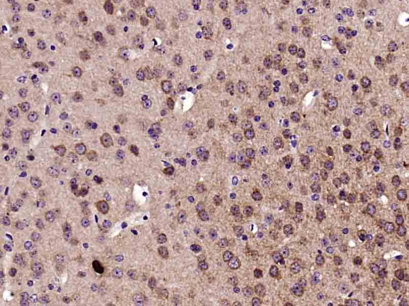
Paraformaldehyde-fixed, paraffin embedded (Mouse brain); Antigen retrieval by boiling in sodium citrate buffer (pH6.0) for 15min; Block endogenous peroxidase by 3% hydrogen peroxide for 20 minutes; Blocking buffer (normal goat serum) at 37°C for 30min; Antibody incubation with (Phospho-c-Fos (Ser362)) Polyclonal Antibody, Unconjugated (bs-12910R) at 1:400 overnight at 4°C, followed by operating according to SP Kit(Rabbit) (sp-0023) instructionsand DAB staining.
-
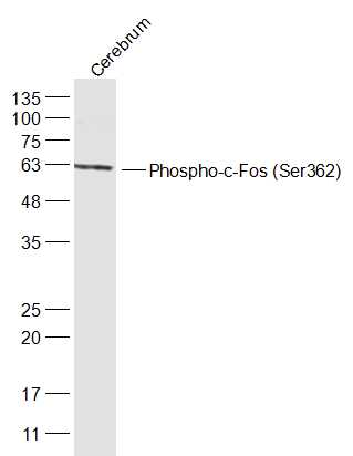
Sample:
Cerebrum (Mouse) Lysate at 40 ug
Primary: Anti-Phospho-c-Fos (Ser362) (bs-12910R) at 1/300 dilution
Secondary: IRDye800CW Goat Anti-Rabbit IgG at 1/20000 dilution
Predicted band size: 41 kD
Observed band size: 41 kD
-
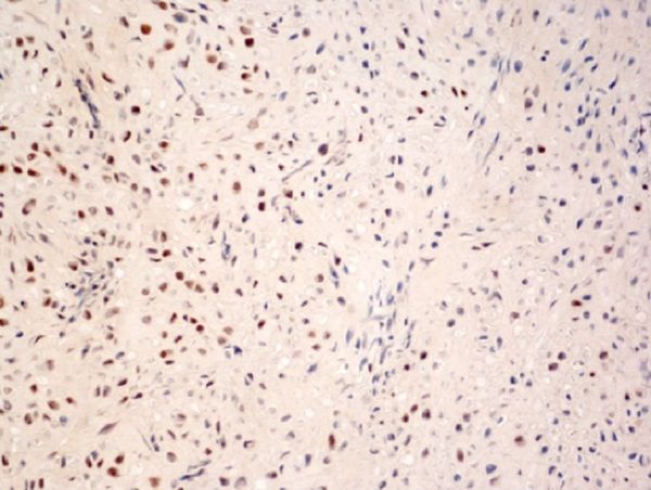
Generously provided by Markus Linder from Medical University Vienna as part of the Bioss Discovery Program. Formalin-fixed, paraffin embedded, and decalcified in EDTA mouse osteosarcoma labeled with Anti-Phospho-c-Fos (Ser362) Polyclonal Antibody, Unconjugated (bs-12910R) at 1:100 followed by conjugation to the secondary antibody and DAB staining
-
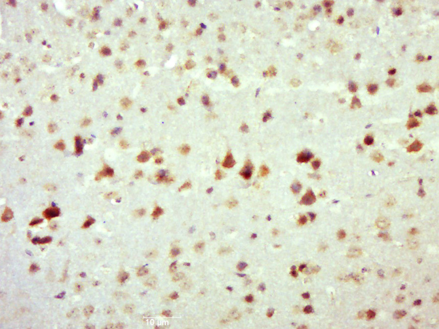
Paraformaldehyde-fixed, paraffin embedded (Mouse brain); Antigen retrieval by boiling in sodium citrate buffer (pH6.0) for 15min; Block endogenous peroxidase by 3% hydrogen peroxide for 20 minutes; Blocking buffer (normal goat serum) at 37°C for 30min; Antibody incubation with (Phospho-c-Fos (Ser362)) Polyclonal Antibody, Unconjugated (bs-12910R) at 1:500 overnight at 4°C, followed by a conjugated secondary (sp-0023) for 20 minutes and DAB staining.
-
Sample:
Cerebrum (Mouse) Lysate at 40 ug
Cerebrum (Rat) Lysate at 40 ug
A431 (Human) Cell Lysate at 30 ug
Adrenal glands (Mouse) Lysate at 40 ug
Adrenal glands (Rat) Lysate at 40 ug
Uterus (Mouse) Lysate at 40 ug
Primary: Anti-Phospho-c-Fos (Ser362) (bs-12910R) at 1/1000 dilution
Secondary: IRDye800CW Goat Anti-Rabbit IgG at 1/20000 dilution
Predicted band size: 62/46 kD
Observed band size: 46 kD
RRID:RRID
产品名称:Rabbit Anti-Phospho-c-Fos (Ser362) antibody
别名: c-Fos (phospho S362); c-Fos (phospho-Ser362); c-Fos (phospho Ser362); p-c-Fos (Ser362); Cellular oncogene fos; FBJ murine osteosarcoma viral v fos oncogene homolog antibody FBJ Osteosarcoma Virus; FOS; FOS protein; G0 G1 switch regulatory protein 7; G0S7
中文名称:磷酸化c-fos抗体
英文名称:Rabbit Anti-Phospho-c-Fos (Ser362) antibody
抗体来源: Rabbit
克隆类型:多克隆
细胞定位:细胞核
性 状:Liquid
亚 型:IgG
纯化方法:affinity purified by Protein A
保存条件:Shipped at 4℃. Store at -20 °C for one year. Avoid repeated freeze/thaw cycles.
免 疫 原:KLH conjugated Synthesised phosphopeptide derived from human c-Fos around the phosphorylation site of Ser362
抗原表位:KG(p-S)SS
SWISS:P01100
Gene ID :2353
Human Gene ID:2353
The Fos gene family consists of 4 members: FOS, FOSB, FOSL1, and FOSL2. These genes encode leucine zipper proteins that can dimerize with proteins of the JUN family, thereby forming the transcription factor complex AP-1. As such, the FOS proteins have been implicated as regulators of cell proliferation, differentiation, and transformation. In some cases, expression of the FOS gene has also been associated with apoptotic cell death. [provided by RefSeq, Jul 2008].
Function:Nuclear phosphoprotein which forms a tight butnon-covalently linked complex with the JUN/AP-1 transcriptionfactor. In the heterodimer, FOS and JUN/AP-1 basic regions eachseems to interact with symmetrical DNA half sites. On TGF-betaactivation, forms a mul
Subunit:Heterodimer; with JUN (By similarity). Interacts withMAFB. Component of the SMAD3/SMAD4/JUN/FOS complexrequired for syngernistic TGF-beta-mediated transcription at theAP1 promoter site. Interacts with SMAD3; the interaction is weakeven on TGF-beta activat
Subcellular Location:Nucleus.
Post-translational modifications:Phosphorylated in the C-terminal upon stimulation by nerve growth factor (NGF) and epidermal growth factor (EGF). Phosphorylated, in vitro, by MAPK and RSK1. Phosphorylation on both Ser-362 and Ser-374 by MAPK1/2 and RSK1/2 leads to protein stabilization
Similarity:Belongs to the bZIP family. Fos subfamily.
Contains 1 bZIP domain
Important Note:This product as supplied is intended for research use only, not for use in human, therapeutic or diagnostic applications.
 400-901-9800


 说明书
说明书 联系我们
联系我们 打印此页面
打印此页面 收藏
收藏