| Rabbit Anti-phospho-AKT1 (Ser129) antibody |
| 反应物种(预测) |
Rat,Chicken,Dog,Horse |
| 产品应用(已验证) |
WB,IHC,ICC,FCM |
| 产品应用(可尝试) |
IF,ELISA |
| 推荐稀释比例 |
WB=1:500-2000,Elisa=1:5000-10000,IHC-P=1:100-500,IHC-F=1:100-500,Flow Cyt=2ug/Test,IF=1:100-500,ICC=1:100, |
| 标签 |
Array |
-
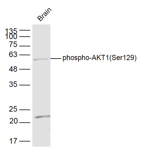
Sample:
Brain (Mouse) Lysate at 40 ug
Primary: Anti- phospho-AKT1(Ser129) (bs-10996R) at 1/300 dilution
Secondary: IRDye800CW Goat Anti-Rabbit IgG at 1/20000 dilution
Predicted band size: 56 kD
Observed band size: 56 kD
-
Sample:
Heart (Mouse) Lysate at 40 ug
Primary: Anti- phospho-AKT1(Ser129) (bs-10996R) at 1/300 dilution
Secondary: IRDye800CW Goat Anti-Rabbit IgG at 1/20000 dilution
Predicted band size: 56 kD
Observed band size: 56 kD
-
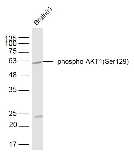
Sample:
Brain (Rat) Lysate at 40 ug
Primary: Anti- phospho-AKT1(Ser129) (bs-10996R) at 1/300 dilution
Secondary: IRDye800CW Goat Anti-Rabbit IgG at 1/20000 dilution
Predicted band size: 56 kD
Observed band size: 56 kD
-
Sample:
Heart (Rat) Lysate at 40 ug
Primary: Anti- phospho-AKT1(Ser129) (bs-10996R) at 1/300 dilution
Secondary: IRDye800CW Goat Anti-Rabbit IgG at 1/20000 dilution
Predicted band size: 56 kD
Observed band size: 56 kD
-
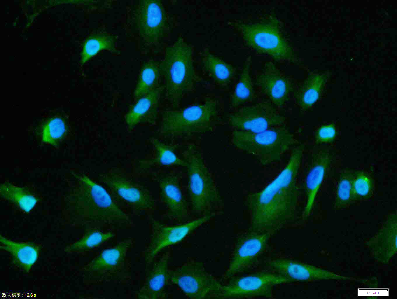
Tissue/cell: A549 cell; 4% Paraformaldehyde-fixed; Triton X-100 at room temperature for 20 min; Blocking buffer (normal goat serum, C-0005) at 37°C for 20 min; Antibody incubation with (phospho-AKT1(Ser129)) polyclonal Antibody, Unconjugated (bs-10996R) 1:100, 90 minutes at 37°C; followed by a FITC conjugated Goat Anti-Rabbit IgG antibody at 37°C for 90 minutes, DAPI (blue, C02-04002) was used to stain the cell nuclei.
-
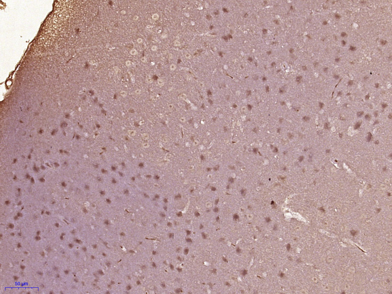
Paraformaldehyde-fixed, paraffin embedded (Mouse brain); Antigen retrieval by boiling in sodium citrate buffer (pH6.0) for 15min; Block endogenous peroxidase by 3% hydrogen peroxide for 20 minutes; Blocking buffer (normal goat serum) at 37°C for 30min; Antibody incubation with (phospho-AKT1(Ser129)) Polyclonal Antibody, Unconjugated (bs-10996R) at 1:400 overnight at 4°C, followed by operating according to SP Kit(Rabbit) (sp-0023) instructionsand DAB staining.
-
Blank control:A549.
Primary Antibody (green line): Rabbit Anti-phospho-AKT1(Ser129) antibody (bs-10996R)
Dilution: 2μg /10^6 cells;
Isotype Control Antibody (orange line): Rabbit IgG .
Secondary Antibody : Goat anti-rabbit IgG-FITC
Dilution: 1μg /test.
Protocol
The cells were incubated in 5%BSA to block non-specific protein-protein interactions for 30 min at room temperature .Cells stained with Primary Antibody for 30 min at room temperature. The secondary antibody used for 40 min at room temperature. Acquisition of 20,000 events was performed.
RRID:RRID
产品名称:Rabbit Anti-phospho-AKT1 (Ser129) antibody
别名: AKT1 (phospho S129); p-AKT1 (phospho S129); AKT 1; AKT; AKT1; AKT-1; AKT1_HUMAN; C AKT; cAKT; MGC9965; MGC99656; Oncogene AKT1; PKB; PKB alpha; PKB-ALPHA; PRKBA; Protein Kinase B Alpha; Protein kinase B; Proto-oncogene c-Akt; RAC Alpha; RAC alpha serine/t
中文名称:磷酸化蛋白激酶B抗体
英文名称:Rabbit Anti-phospho-AKT1 (Ser129) antibody
抗体来源: Rabbit
克隆类型:多克隆
细胞定位:细胞核,细胞浆,细胞膜
性 状:Liquid
亚 型:IgG
纯化方法:affinity purified by Protein A
保存条件:Shipped at 4℃. Store at -20 °C for one year. Avoid repeated freeze/thaw cycles.
免 疫 原:KLH conjugated Synthesised phosphopeptide derived from human Akt1 around the phosphorylation site of Ser129
抗原表位:DN(p-S)GA
SWISS:P31749
Gene ID :207
Human Gene ID:207
The serine-threonine protein kinase encoded by the AKT1 gene is catalytically inactive in serum-starved primary and immortalized fibroblasts. AKT1 and the related AKT2 are activated by platelet-derived growth factor. The activation is rapid and specific, and it is abrogated by mutations in the pleckstrin homology domain of AKT1. It was shown that the activation occurs through phosphatidylinositol 3-kinase. In the developing nervous system AKT is a critical mediator of growth factor-induced neuronal survival. Survival factors can suppress apoptosis in a transcription-independent manner by activating the serine/threonine kinase AKT1, which then phosphorylates and inactivates components of the apoptotic machinery. Mutations in this gene have been associated with the Proteus syndrome. Multiple alternatively spliced transcript variants have been found for this gene. [provided by RefSeq, Jul 2011]
Function:AKT1 is one of 3 closely related serine/threonine-protein kinases (AKT1, AKT2 and AKT3) called the AKT kinase, and which regulate many processes including metabolism, proliferation, cell survival, growth and angiogenesis. This is mediated through serine a
Subunit:Interacts (via the C-terminus) with CCDC88A (via its C-terminus). Interacts with GRB10; the interaction leads to GRB10 phosphorylation thus promoting YWHAE-binding. Interacts with AGAP2 (isoform 2/PIKE-A); the interaction occurs in the presence of guanine
Subcellular Location:Cytoplasm. Nucleus. Cell membrane. Note=Nucleus after activation by integrin-linked protein kinase 1 (ILK1). Nuclear translocation is enhanced by interaction with TCL1A. Phosphorylation on Tyr-176 by TNK2 results in its localization to the cell membrane w
Tissue Specificity:Expressed in prostate cancer and levels increase from the normal to the malignant state (at protein level). Expressed in all human cell types so far analyzed. The Tyr-176 phosphorylated form shows a significant increase in expression in breast cancers dur
Post-translational modifications:O-GlcNAcylation at Thr-305 and Thr-312 inhibits activating phosphorylation at Thr-308 via disrupting the interaction between AKT1 and PDPK1. O-GlcNAcylation at Ser-473 also probably interferes with phosphorylation at this site.
Phosphorylation on Thr-3
DISEASE:Defects in AKT1 are a cause of susceptibility to breast cancer (BC) [MIM:114480]. A common malignancy originating from breast epithelial tissue. Breast neoplasms can be distinguished by their histologic pattern. Invasive ductal carcinoma is by far the mos
Similarity:Belongs to the protein kinase superfamily. AGC Ser/Thr protein kinase family. RAC subfamily.
Contains 1 AGC-kinase C-terminal domain.
Contains 1 PH domain.
Contains 1 protein kinase domain.
Important Note:This product as supplied is intended for research use only, not for use in human, therapeutic or diagnostic applications.
 400-901-9800


 说明书
说明书 联系我们
联系我们 打印此页面
打印此页面 收藏
收藏