| Rabbit Anti-CD43 antibody |
| 产品应用(已验证) |
WB |
| 推荐稀释比例 |
WB=1:500-2000, |
| 研究领域 |
免疫学,干细胞,淋巴细胞,T-淋巴细胞,B-淋巴细胞, |
| 标签 |
Array |
-
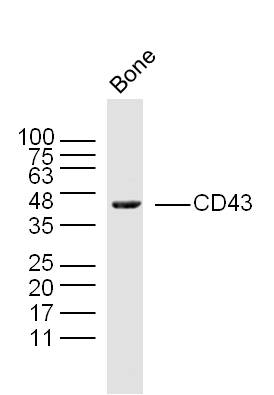
Sample:
Bone (Mouse) Lysate at 40 ug
Primary: Anti- CD43 (bs-20324R) at 1/300 dilution
Secondary: IRDye800CW Goat Anti-Rabbit IgG at 1/20000 dilution
Predicted band size: 38 kD
Observed band size: 38 kD
RRID:RRID
产品名称:Rabbit Anti-CD43 antibody
别名: LEUK_HUMAN; Leukosialin; SPN; GPL115; GALGP; GPL 115; Galactoglycoprotein (GALGP); Leukocyte sialoglycoprotein; Sialophorin; CD43 cytoplasmic tail; CD 43; CD43 antigen;
中文名称:CD43抗体
英文名称:Rabbit Anti-CD43 antibody
抗体来源: Rabbit
克隆类型:多克隆
细胞定位:细胞膜
性 状:Liquid
亚 型:IgG
纯化方法:affinity purified by Protein A
保存条件:Shipped at 4℃. Store at -20 °C for one year. Avoid repeated freeze/thaw cycles.
免 疫 原:KLH conjugated synthetic peptide derived from human CD43
抗原表位:101-200/400
抗原细胞定位:Extracellular
SWISS:P16150
Gene ID :6693
Human Gene ID:6693
This gene encodes a highly sialylated glycoprotein that functions in antigen-specific activation of T cells, and is found on the surface of thymocytes, T lymphocytes, monocytes, granulocytes, and some B lymphocytes. It contains a mucin-like extracellular domain, a transmembrane region and a carboxy-terminal intracellular region. The extracellular domain has a high proportion of serine and threonine residues, allowing extensive O-glycosylation, and has one potential N-glycosylation site, while the carboxy-terminal region has potential phosphorylation sites that may mediate transduction of activation signals. Different glycoforms of this protein have been described. In stimulated immune cells, proteolytic cleavage of the extracellular domain occurs in some cell types, releasing a soluble extracellular fragment. Defects in expression of this gene are associated with Wiskott-Aldrich syndrome. [provided by RefSeq, Sep 2017]
Function:One of the major glycoproteins of thymocytes and T lymphocytes. Plays a role in the physicochemical properties of the T-cell surface and in lectin binding. Presents carbohydrate ligands to selectins. Has an extended rodlike structure that could protrude a
Subunit:Interacts with HIPK2 via the cytoplasmic domain. Interacts with RDX.
Subcellular Location:Membrane; Single-pass type I membrane protein.
Tissue Specificity:Cell surface of thymocytes, T-lymphocytes, neutrophils, plasma cells and myelomas.
Post-translational modifications:Glycosylated; has a high content of sialic acid and O-linked carbohydrate structures.
Important Note:This product as supplied is intended for research use only, not for use in human, therapeutic or diagnostic applications.
 400-901-9800


 说明书
说明书 联系我们
联系我们 打印此页面
打印此页面 收藏
收藏