| Rabbit Anti-GAPDH (Loading Control) antibody |
| 产品应用(已验证) |
WB,IHC,ICC |
| 产品应用(可尝试) |
IF |
| 推荐稀释比例 |
WB=1:10000-200000,IHC-P=1:100-500,IHC-F=1:100-500,IF=1:100-500,ICC=1:100, |
| 研究领域 |
肿瘤,细胞生物,免疫学,信号转导,新陈代谢, |
| 标签 |
Array |
-
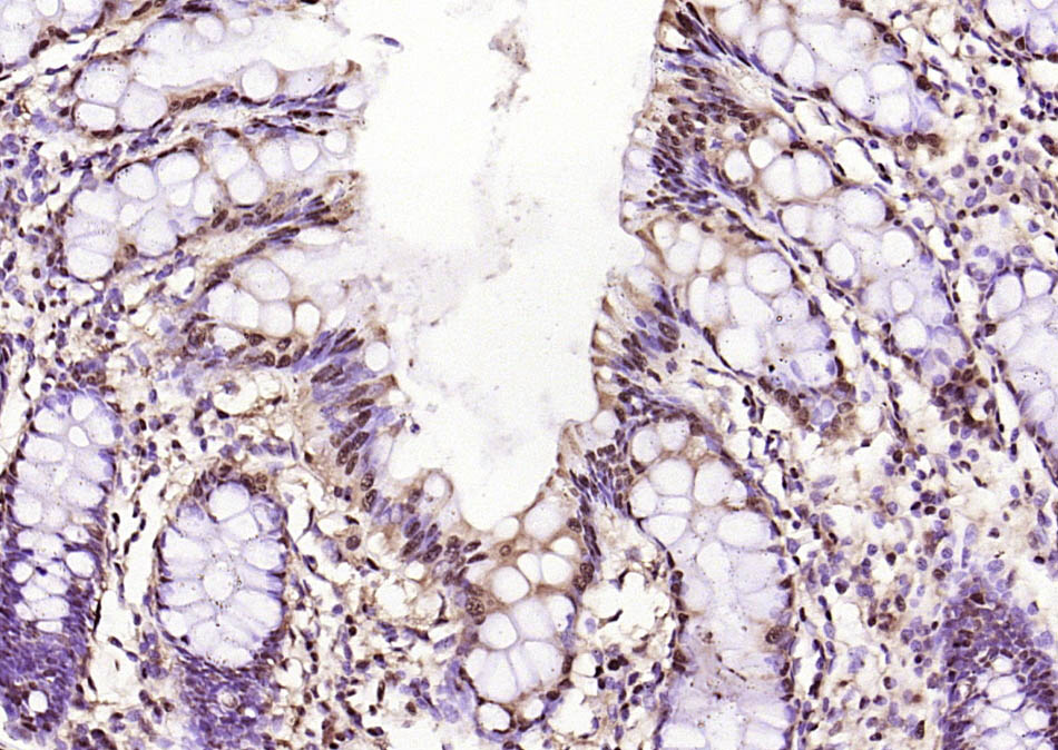
Paraformaldehyde-fixed, paraffin embedded (human colon); Antigen retrieval by boiling in sodium citrate buffer (pH6.0) for 15min; Block endogenous peroxidase by 3% hydrogen peroxide for 20 minutes; Blocking buffer (normal goat serum) at 37°C for 30min; Antibody incubation with (GAPDH (Loading Control)) Polyclonal Antibody, Unconjugated (bs-10900R) at 1:200 overnight at 4°C, followed by operating according to SP Kit(Rabbit) (sp-0023) instructionsand DAB staining.
-
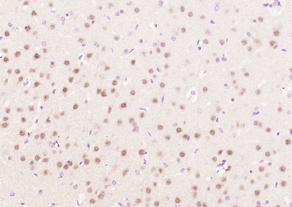
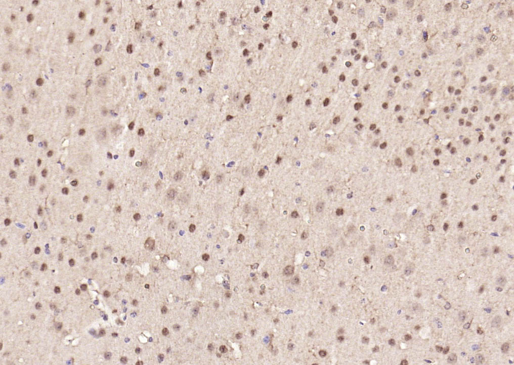
Paraformaldehyde-fixed, paraffin embedded (rat brain); Antigen retrieval by boiling in sodium citrate buffer (pH6.0) for 15min; Block endogenous peroxidase by 3% hydrogen peroxide for 20 minutes; Blocking buffer (normal goat serum) at 37°C for 30min; Antibody incubation with (GAPDH (Loading Control)) Polyclonal Antibody, Unconjugated (bs-10900R) at 1:200 overnight at 4°C, followed by operating according to SP Kit(Rabbit) (sp-0023) instructionsand DAB staining.
-
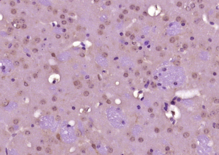
Paraformaldehyde-fixed, paraffin embedded (mouse brain); Antigen retrieval by boiling in sodium citrate buffer (pH6.0) for 15min; Block endogenous peroxidase by 3% hydrogen peroxide for 20 minutes; Blocking buffer (normal goat serum) at 37°C for 30min; Antibody incubation with (GAPDH (Loading Control)) Polyclonal Antibody, Unconjugated (bs-10900R) at 1:200 overnight at 4°C, followed by operating according to SP Kit(Rabbit) (sp-0023) instructionsand DAB staining.
-
Paraformaldehyde-fixed, paraffin embedded (human colon); Antigen retrieval by boiling in sodium citrate buffer (pH6.0) for 15min; Block endogenous peroxidase by 3% hydrogen peroxide for 20 minutes; Blocking buffer (normal goat serum) at 37°C for 30min; Antibody incubation with (GAPDH (Loading Control)) Polyclonal Antibody, Unconjugated (bs-10900R) at 1:200 overnight at 4°C, followed by operating according to SP Kit(Rabbit) (sp-0023) instructionsand DAB staining.
-
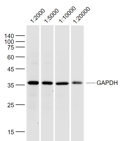
Sample:
293T (Human) Lysate at 40 ug
Primary:
Anti-GAPDH (bs-10900R) at 1/2000~1/20000 dilution
Secondary: IRDye800CW Goat Anti-Rabbit IgG at 1/20000 dilution
Predicted band size: 38 kD
Observed band size: 36 kD
-
Paraformaldehyde-fixed, paraffin embedded (mouse brain tissue); Antigen retrieval by boiling in sodium citrate buffer (pH6.0) for 15min; Block endogenous peroxidase by 3% hydrogen peroxide for 20 minutes; Blocking buffer (normal goat serum) at 37°C for 30min; Antibody incubation with (GAPDH-Loading Contro) Polyclonal Antibody, Unconjugated (bs-10900R) at 1:400 overnight at 4°C, followed by operating according to SP Kit(Rabbit) (sp-0023) instructionsand DAB staining.
-
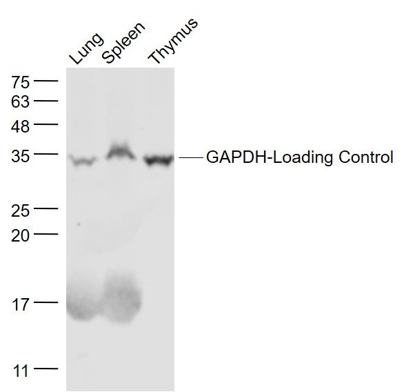
Sample:
Lung (Mouse) Lysate at 40 ug
Spleen (Mouse) Lysate at 40 ug
Thymus (Mouse) Lysate at 40 ug
Primary: Anti- GAPDH-Loading Control (bs-10900R) at 1/1000 dilution
Secondary: IRDye800CW Goat Anti-Rabbit IgG at 1/20000 dilution
Predicted band size: 38 kD
Observed band size: 35 kD
-
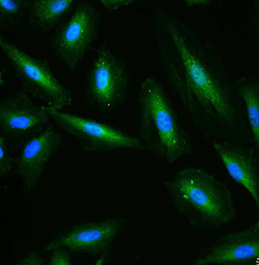
Tissue/cell: A549 cell; 4% Paraformaldehyde-fixed; Triton X-100 at room temperature for 20 min; Blocking buffer (normal goat serum, C-0005) at 37°C for 20 min; Antibody incubation with (GAPDH (Loading Control)) polyclonal Antibody, Unconjugated (bs-10900R) 1:100, 90 minutes at 37°C; followed by a FITC conjugated Goat Anti-Rabbit IgG antibody at 37°C for 90 minutes, DAPI (blue, C02-04002) was used to stain the cell nuclei.
-
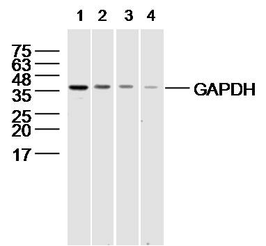
Sample:
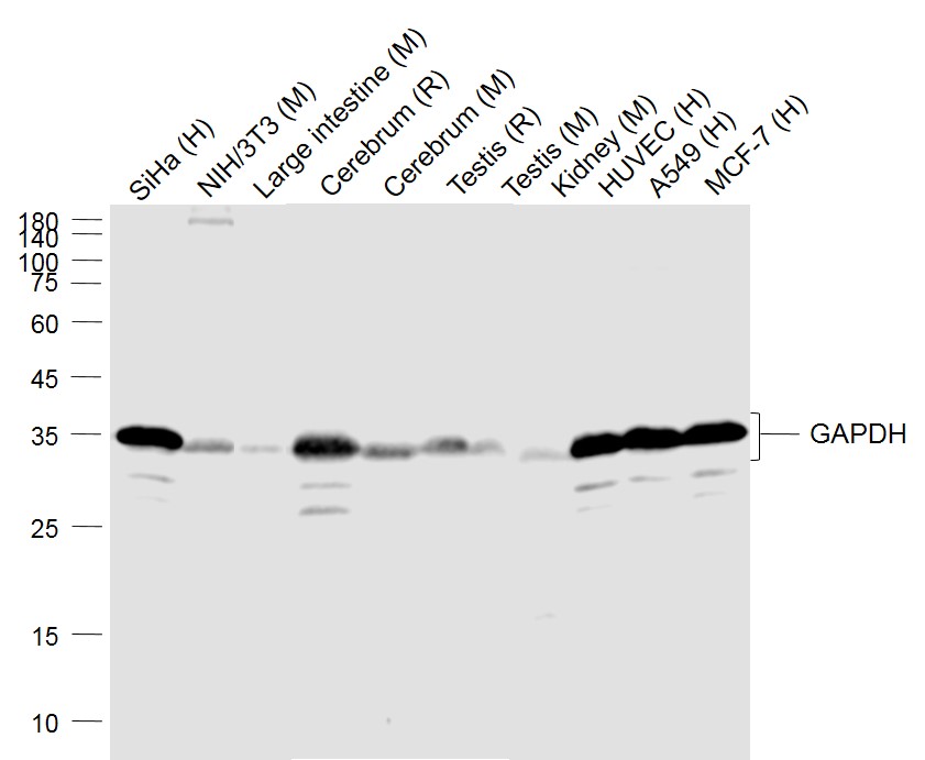
Lane 1: SiHa (Human) Cell Lysate at 30 ug
Lane 2: NIH/3T3(Mouse) Cell Lysate at 30 ug
Lane 3: Large intestine (Mouse) Lysate at 40 ug
Lane 4: Cerebrum (Rat) Lysate at 40 ug
Lane 5: Cerebrum (Mouse) Lysate at 40 ug
Lane 6: Testis (Rat) Lysate at 40 ug
Lane 7: Testis (Mouse) Lysate at 40 ug
Lane 8: Kidney (Mouse) Lysate at 40 ug
Lane 9: HUVEC (Human) Cell Lysate at 30 ug
Lane 10: A549 (Human) Cell Lysate at 30 ug
Lane 11: MCF-7 (Human) Cell Lysate at 30 ug
Primary: Anti-GAPDH (bs-10900R) at 1/1000 dilution
Secondary: IRDye800CW Goat Anti-Rabbit IgG at 1/20000 dilution
Predicted band size: 36 kD
Observed band size: 36 kD
-
Sample: 293T(human) cell lysate at 30ug;
Primary:
Lane1: Anti-GAPDH (bs-10900R) at 1/2000 dilution
Lane2: Anti-GAPDH (bs-10900R) at 1/10000 dilution
Lane3: Anti-GAPDH (bs-10900R) at 1/40000 dilution
Lane4: Anti-GAPDH (bs-10900R) at 1/80000 dilution
Secondary: IRDye800CW Goat Anti-Rabbit IgG at 1/20000 dilution
Predicted band size: 38 kD
Observed band size: 38kD
-
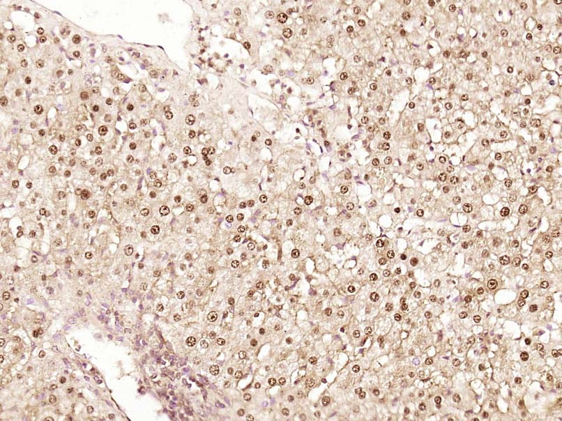
Paraformaldehyde-fixed, paraffin embedded (Human liver cancer); Antigen retrieval by boiling in sodium citrate buffer (pH6.0) for 15min; Block endogenous peroxidase by 3% hydrogen peroxide for 20 minutes; Blocking buffer (normal goat serum) at 37°C for 30min; Antibody incubation with (GAPDH) Polyclonal Antibody, Unconjugated (bs-10900R) at 1:500 overnight at 4°C, followed by a conjugated secondary (sp-0023) for 20 minutes and DAB staining.
-
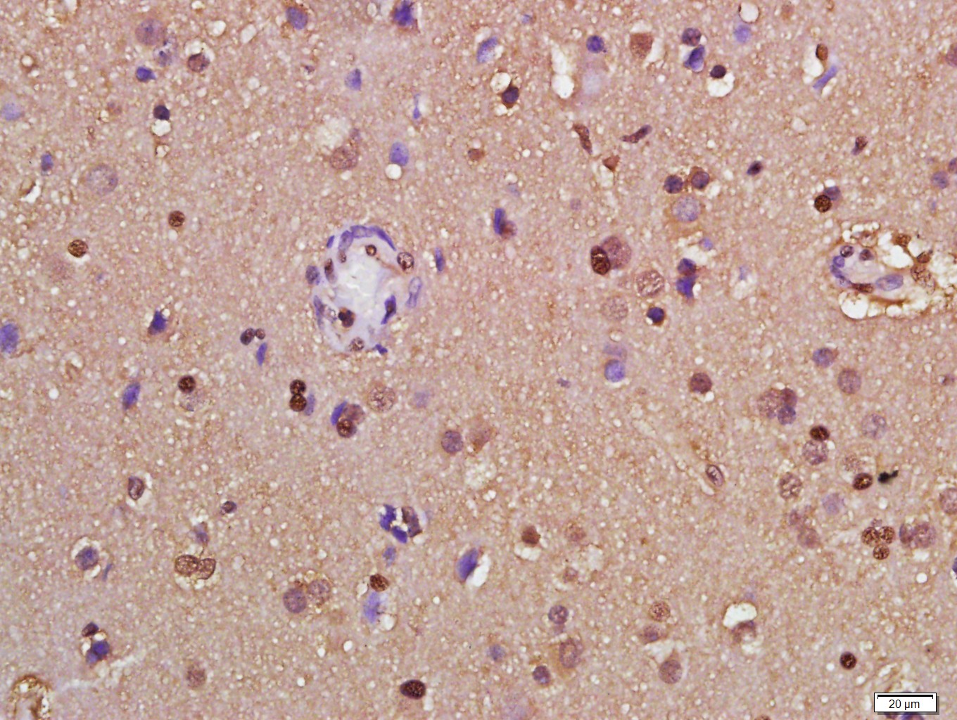
Paraformaldehyde-fixed, paraffin embedded (Human glioma); Antigen retrieval by boiling in sodium citrate buffer (pH6.0) for 15min; Block endogenous peroxidase by 3% hydrogen peroxide for 20 minutes; Blocking buffer (normal goat serum) at 37°C for 30min; Antibody incubation with (GAPDH) Polyclonal Antibody, Unconjugated (bs-10900R) at 1:500 overnight at 4°C, followed by a conjugated secondary (sp-0023) for 20 minutes and DAB staining.
-
Paraformaldehyde-fixed, paraffin embedded (Human kidney); Antigen retrieval by boiling in sodium citrate buffer (pH6.0) for 15min; Block endogenous peroxidase by 3% hydrogen peroxide for 20 minutes; Blocking buffer (normal goat serum) at 37°C for 30min; Antibody incubation with (GAPDH) Polyclonal Antibody, Unconjugated (bs-10900R) at 1:2000 overnight at 4°C, followed by a conjugated secondary (sp-0023) for 20 minutes and DAB staining.
-
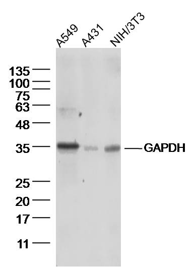
Sample:
A549 Cell (Human) Lysate at 40 ug
A431 Cell (Human) Lysate at 40 ug
NIH/3T3 Cell (Mouse) Lysate at 40 ug
Primary: Anti-GAPDH (bs-10900R) at 1/300 dilution
Secondary: IRDye800CW Goat Anti-Rabbit IgG at 1/20000 dilution
Predicted band size: 38 kD
Observed band size: 36 kD
-
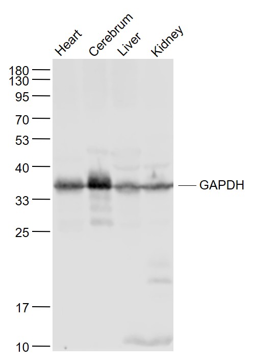
Sample:
Heart (Mouse) Lysate at 40 ug
Cerebrum (Mouse) Lysate at 40 ug
Liver (Mouse) Lysate at 40 ug
Kidney (Mouse) Lysate at 40 ug
Primary: Anti- GAPDH (bs-10900R) at 1/1000 dilution
Secondary: IRDye800CW Goat Anti-Rabbit IgG at 1/20000 dilution
Predicted band size: 38 kD
Observed band size: 38 kD
-
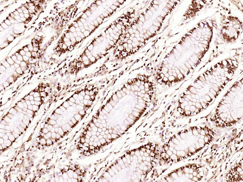
Paraformaldehyde-fixed, paraffin embedded (Human colon carcinoma); Antigen retrieval by boiling in sodium citrate buffer (pH6.0) for 15min; Block endogenous peroxidase by 3% hydrogen peroxide for 20 minutes; Blocking buffer (normal goat serum) at 37°C for 30min; Antibody incubation with (GAPDH) Polyclonal Antibody, Unconjugated (bs-10900R) at 1:500 overnight at 4°C, followed by a conjugated secondary (sp-0023) for 20 minutes and DAB staining.
-
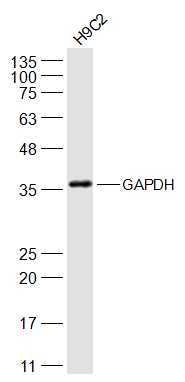
Sample:
H9C2(Rat) Cell Lysate at 30 ug
Primary: Anti-GAPDH (bs-10900R) at 1/2000 dilution
Secondary: IRDye800CW Goat Anti-Rabbit IgG at 1/20000 dilution
Predicted band size: 38 kD
Observed band size: 38 kD
-
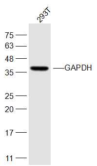
Sample:
293T(Human) Cell Lysate at 30 ug
Primary: Anti-GAPDH (bs-10900R) at 1/1000 dilution
Secondary: IRDye800CW Goat Anti-Rabbit IgG at 1/20000 dilution
Predicted band size: 38 kD
Observed band size: 38 kD
RRID:RRID
产品名称:Rabbit Anti-GAPDH (Loading Control) antibody
别名: 38 kDa BFA-dependent ADP-ribosylation substrate; Aging-associated gene 9 protein; BARS-38; cb609; EC 1.2.1.12; G3PD; G3PDH; GAPD; Glyceraldehyde 3 phosphate dehydrogenase;Glyceraldehyde 3 phosphate dehydrogenase liver;Glyceraldehyde 3 phosphate dehydrogen
中文名称:3-磷酸甘油醛脱氢酶(内参)抗体
英文名称:Rabbit Anti-GAPDH (Loading Control) antibody
抗体来源: Rabbit
克隆类型:多克隆
细胞定位:细胞核,细胞浆,细胞膜
性 状:Liquid
亚 型:IgG
纯化方法:affinity purified by Protein A
保存条件:Shipped at 4℃. Store at -20 °C for one year. Avoid repeated freeze/thaw cycles.
免 疫 原:Recombinant human GAPDH full length protein
SWISS:P04406
Gene ID :2597
Human Gene ID:2597
Glyceraldehyde 3 phosphate dehydrogenase (GAPDH) is well known as one of the key enzymes involved in glycolysis. As well as functioning as a glycolytic enzyme in cytoplasm, recent evidence suggests that mammalian GAPDH is also involved in a great number of intracellular proceses such as membrane fusion, microtubule bundling, phosphotransferase activity, nuclear RNA export, DNA replication, and DNA repair. During the last decade a lot of data appeared concerning the role of GAPDH in different pathologies including prostate cancer progression, programmed neuronal cell death, age related neuronal diseases, such as Alzheimer's and Huntington's disease. GAPDH is expressed in all cells. It is constitutively expressed in almost all tissues at high levels. There are however some physiological factors such as hypoxia and diabetes that increase GAPDH expression in certain cell types. GAPDH molecule is composed of four 36kDa subunits.
Function:Has both glyceraldehyde-3-phosphate dehydrogenase and nitrosylase activities, thereby playing a role in glycolysis and nuclear functions, respectively. Participates in nuclear events including transcription, RNA transport, DNA replication and apoptosis. N
Subunit:Homotetramer. Interacts with TPPP; the interaction is direct. Interacts (when S-nitrosylated) with SIAH1; leading to nuclear translocation. Interacts with RILPL1/GOSPEL, leading to prevent the interaction between GAPDH and SIAH1 and prevent nuclear transl
Subcellular Location:Cytoplasm, cytosol. Nucleus. Cytoplasm, perinuclear region. Membrane. Note=Translocates to the nucleus following S-nitrosylation and interaction with SIAH1, which contains a nuclear localization signal. Postnuclear and Perinuclear regions.
Post-translational modifications:S-nitrosylation of Cys-152 leads to interaction with SIAH1, followed by translocation to the nucleus.
ISGylated (Probable).
Sulfhydration at Cys-152 increases catalytic activity.
Similarity:Belongs to the glyceraldehyde-3-phosphate dehydrogenase family.
Important Note:This product as supplied is intended for research use only, not for use in human, therapeutic or diagnostic applications.
 400-901-9800


 说明书
说明书 联系我们
联系我们 打印此页面
打印此页面 收藏
收藏