| Rabbit Anti-Cyclin E2 antibody |
| 反应物种(预测) |
Mouse |
| 产品应用(已验证) |
IHC,ICC,FCM |
| 产品应用(可尝试) |
WB,IF |
| 推荐稀释比例 |
WB=1:500-2000,IHC-P=1:50-200,IHC-F=1:50-200,Flow Cyt=2ug/Test,IF=1:50-200,ICC=1:50, |
| 研究领域 |
肿瘤,细胞生物,染色质和核信号,信号转导,细胞周期蛋白 |
| 标签 |
Array |
-
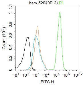
Blank control: Hela.
Primary Antibody (green line): Rabbit Anti-Cyclin E2 antibody (bsm-52049R)
Dilution: 1:50;
Secondary Antibody : Goat anti-rabbit IgG-AF488
Dilution: 1:1000.
Protocol
The cells were fixed with 4% PFA (10min at room temperature)and then permeabilized with 90% ice-cold methanol for 20 min at-20℃. The cells were then incubated in 5%BSA to block non-specific protein-protein interactions for 30 min at room temperature .Cells stained with Primary Antibody for 30 min at room temperature. The secondary antibody used for 40 min at room temperature. Acquisition of 20,000 events was performed.
-
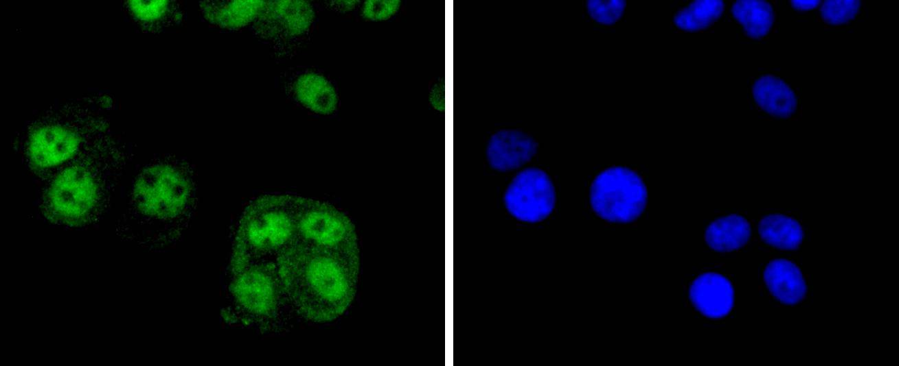
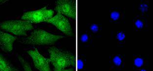
MCF-7 cell; 4% Paraformaldehyde-fixed; Triton X-100 at room temperature for 20 min; Blocking buffer (normal goat serum, C-0005) at 37°C for 20 min; Antibody incubation with (Cyclin E2) monoclonal Antibody, Unconjugated (bsm-52049R) 1:500, 90 minutes at 37°C; followed by a conjugated Goat Anti-Rabbit IgG antibody at 37°C for 90 minutes, DAPI (blue, C02-04002) was used to stain the cell nuclei.
-
NIH/3T3 cell; 4% Paraformaldehyde-fixed; Triton X-100 at room temperature for 20 min; Blocking buffer (normal goat serum, C-0005) at 37°C for 20 min; Antibody incubation with (Cyclin E2) monoclonal Antibody, Unconjugated (bsm-52049R) 1:500, 90 minutes at 37°C; followed by a conjugated Goat Anti-Rabbit IgG antibody at 37°C for 90 minutes, DAPI (blue, C02-04002) was used to stain the cell nuclei.
-
Hela cell; 4% Paraformaldehyde-fixed; Triton X-100 at room temperature for 20 min; Blocking buffer (normal goat serum, C-0005) at 37°C for 20 min; Antibody incubation with (Cyclin E2) monoclonal Antibody, Unconjugated (bsm-52049R) 1:500, 90 minutes at 37°C; followed by a conjugated Goat Anti-Rabbit IgG antibody at 37°C for 90 minutes, DAPI (blue, C02-04002) was used to stain the cell nuclei.
-
Paraformaldehyde-fixed, paraffin embedded (human breast carcinoma); Antigen retrieval by boiling in sodium citrate buffer (pH6.0) for 15min; Block endogenous peroxidase by 3% hydrogen peroxide for 20 minutes; Blocking buffer (normal goat serum) at 37°C for 30min; Antibody incubation with (Cyclin E2) Monoclonal Antibody, Unconjugated (bsm-52049R) at 1:50 overnight at 4°C, followed by operating according to SP Kit(Rabbit) (sp-0023) instructionsand DAB staining.
-
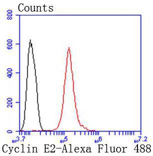
Blank control:MCF7.
Primary Antibody (green line): Rabbit Anti-Cyclin E2 antibody (bsm-52049R)
Dilution: 2μg /10^6 cells;
Isotype Control Antibody (orange line): Rabbit IgG .
Secondary Antibody : Goat anti-rabbit IgG-AF488
Dilution: 1μg /test.
Protocol
The cells were fixed with 4% PFA (10min at room temperature)and then permeabilized with 90% ice-cold methanol for 20 min at-20℃. The cells were then incubated in 5%BSA to block non-specific protein-protein interactions for 30 min at room temperature .Cells stained with Primary Antibody for 30 min at room temperature. The secondary antibody used for 40 min at room temperature. Acquisition of 20,000 events was performed.
RRID:RRID
产品名称:Rabbit Anti-Cyclin E2 antibody
别名: CCN E2; CCNE 2; CCNE2; CCNE2 protein; CYC E2; CYCE 2; CYCE2; CyclinE2; G1/S-specific cyclin E2; G1/S-specific cyclin E2; CCNE2_HUMAN.
中文名称:周期素E2重组兔单克隆抗体
英文名称:Rabbit Anti-Cyclin E2 antibody
抗体来源: Rabbit
克隆类型:单克隆
细胞定位:细胞核
性 状:Liquid
亚 型:IgG
纯化方法:affinity purified by Protein A
保存条件:Shipped at 4℃. Store at -20 °C for one year. Avoid repeated freeze/thaw cycles.
免 疫 原:Recombinant human Cyclin E2 protein, around C-terminal 100aa
SWISS:O96020
Gene ID :9134
Human Gene ID:9134
The human Cyclin E2 gene encodes a 404 amino acid protein that is most closely related to Cyclin E. Cyclin E2 mRNA levels peaks at the G1 / S transition. Cyclin E2 associates with Cdk2 in a functional kinase complex that is inhibited by both p27 (Kip1) and p21 (Cip1). Cyclin E2 / Cdk2 phosphorylates histone H1 in vitro. G1 cyclin E controls the initiation of DNA synthesis by activating CDK2. Abnormally high levels of cyclin E expression have frequently been observed in human cancers. Unlike Cyclin E1, which is expressed in great majority of proliferating normal and neoplastically transformed cells, Cyclin E2 levels are low to undetectable in non transformed cells and increase significantly in neoplasm derived cells.
Subunit:"Interacts with the CDK2 (in vivo) and CDK3 (in vitro) protein kinases to form a serine/threonine kinase holoenzyme complex. The cyclin subunit imparts substrate specificity to the complex.
Subcellular Location:Nucleus.
Tissue Specificity:According to PubMed:9858585, highest levels of expression in adult testis, thymus and brain. Lower levels in placenta, spleen and colon. Consistently elevated levels in tumor-derived cells compared to non-transformed proliferating cells. According to PubM
Post-translational modifications:Phosphorylation by CDK2 triggers its release from CDK2 and degradation via the ubiquitin proteasome pathway.
Similarity:Belongs to the cyclin family. Cyclin E subfamily.
Important Note:This product as supplied is intended for research use only, not for use in human, therapeutic or diagnostic applications.
 400-901-9800


 说明书
说明书 联系我们
联系我们 打印此页面
打印此页面 收藏
收藏