| Rabbit Anti-alpha smooth muscle Actin antibody |
| 反应物种(预测) |
Rat |
| 产品应用(已验证) |
WB,IHC,ICC,FCM |
| 产品应用(可尝试) |
IF |
| 推荐稀释比例 |
WB=1:1000-2000,IHC-P=1:50-200,IHC-F=1:50-200,Flow Cyt=1:50,IF=1:50-200,ICC=1:50, |
| 研究领域 |
肿瘤,细胞生物,免疫学,细胞骨架, |
| 标签 |
Array |
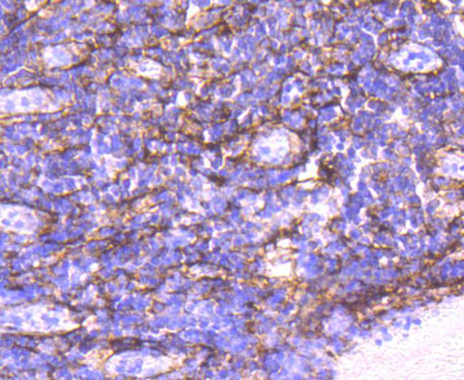
-
Paraformaldehyde-fixed, paraffin embedded (human tonsil tissue); Antigen retrieval by boiling in sodium citrate buffer (pH6.0) for 15min; Block endogenous peroxidase by 3% hydrogen peroxide for 20 minutes; Blocking buffer (normal goat serum) at 37°C for 30min; Antibody incubation with (alpha smooth muscle Actin) Monoclonal Antibody, Unconjugated (bsm-52396R ) at 1:50 overnight at 4°C, followed by operating according to SP Kit(Rabbit) (sp-0023) instructionsand DAB staining.
-
HepG2 cell; 4% Paraformaldehyde-fixed; Triton X-100 at room temperature for 20 min; Blocking buffer (normal goat serum) at 37°C for 20 min; Antibody incubation with (alpha smooth muscle Actin) Monoclonal Antibody, Unconjugated (bsm-52396R) 1:50, 90 minutes at 37°C; followed by a conjugated Goat Anti-Rabbit IgG antibody at 37°C for 90 minutes, DAPI (blue) was used to stain the cell nuclei.
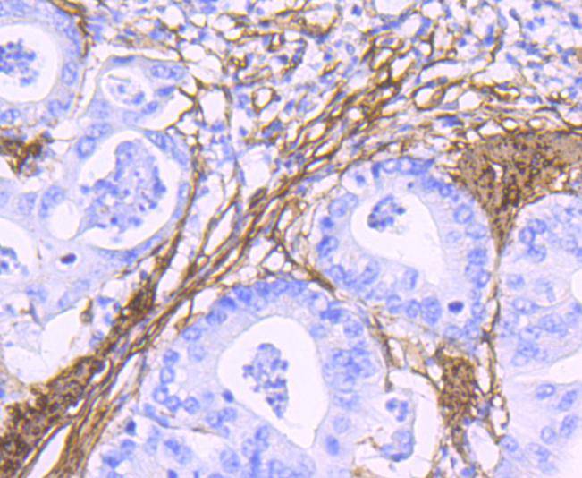
-
Paraformaldehyde-fixed, paraffin embedded (human stomach carcinoma tissue); Antigen retrieval by boiling in sodium citrate buffer (pH6.0) for 15min; Block endogenous peroxidase by 3% hydrogen peroxide for 20 minutes; Blocking buffer (normal goat serum) at 37°C for 30min; Antibody incubation with (alpha smooth muscle Actin) Monoclonal Antibody, Unconjugated (bsm-52396R ) at 1:50 overnight at 4°C, followed by operating according to SP Kit(Rabbit) (sp-0023) instructionsand DAB staining.
-
RH-35 cell; 4% Paraformaldehyde-fixed; Triton X-100 at room temperature for 20 min; Blocking buffer (normal goat serum) at 37°C for 20 min; Antibody incubation with (alpha smooth muscle Actin) Monoclonal Antibody, Unconjugated (bsm-52396R) 1:50, 90 minutes at 37°C; followed by a conjugated Goat Anti-Rabbit IgG antibody at 37°C for 90 minutes, DAPI (blue) was used to stain the cell nuclei.
-
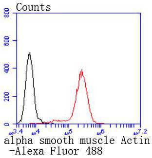
Blank control:Jurkat.
Primary Antibody (green line): Rabbit Anti-alpha smooth muscle Actin antibody (bsm-52396R)
Dilution: 1:50;
Isotype Control Antibody (orange line): Rabbit IgG .
Secondary Antibody : Goat anti-rabbit IgG-AF488
Dilution: 1:1000.
Protocol
The cells were fixed with 4% PFA (10min at room temperature)and then permeabilized with 0.1% PBST for 20 min at room temperature.The cells were then incubated in 5%BSA to block non-specific protein-protein interactions for 30 min at room temperature .Cells stained with Primary Antibody for 30 min at room temperature. The secondary antibody used for 40 min at room temperature. Acquisition of 20,000 events was performed.
-
A431 cell; 4% Paraformaldehyde-fixed; Triton X-100 at room temperature for 20 min; Blocking buffer (normal goat serum) at 37°C for 20 min; Antibody incubation with (alpha smooth muscle Actin) Monoclonal Antibody, Unconjugated (bsm-52396R) 1:50, 90 minutes at 37°C; followed by a conjugated Goat Anti-Rabbit IgG antibody at 37°C for 90 minutes, DAPI (blue) was used to stain the cell nuclei.
-
A549 cell; 4% Paraformaldehyde-fixed; Triton X-100 at room temperature for 20 min; Blocking buffer (normal goat serum) at 37°C for 20 min; Antibody incubation with (alpha smooth muscle Actin) Monoclonal Antibody, Unconjugated (bsm-52396R) 1:50, 90 minutes at 37°C; followed by a conjugated Goat Anti-Rabbit IgG antibody at 37°C for 90 minutes, DAPI (blue) was used to stain the cell nuclei.
-
Paraformaldehyde-fixed, paraffin embedded (human liver tissue); Antigen retrieval by boiling in sodium citrate buffer (pH6.0) for 15min; Block endogenous peroxidase by 3% hydrogen peroxide for 20 minutes; Blocking buffer (normal goat serum) at 37°C for 30min; Antibody incubation with (alpha smooth muscle Actin) Monoclonal Antibody, Unconjugated (bsm-52396R ) at 1:50 overnight at 4°C, followed by operating according to SP Kit(Rabbit) (sp-0023) instructionsand DAB staining.
-
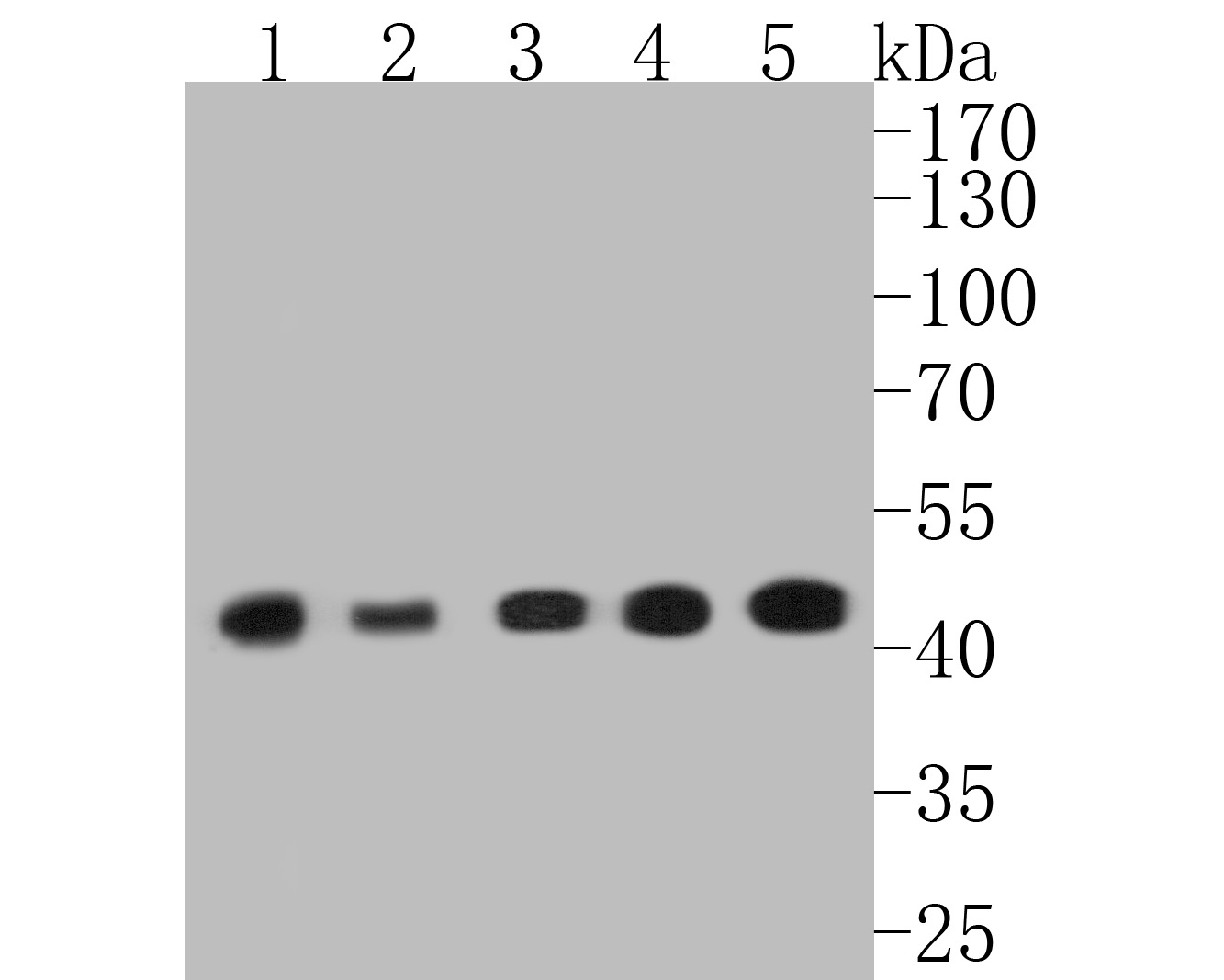
Sample:
Lane 1: A431 cell lysate
Lane 2: Hela cell lysate
Lane 3: NIH/3T3 cell lysate
Lane 4: mouse heart tissue lysate
Lane 5: mouse smooth muscle tissue lysate
Primary: Anti-alpha smooth muscle Actin (bsm-52396R) at 1:500 dilution
Secondary: Goat Anti-Rabbit IgG - HRP at 1:5000 dilution
Predicted band size: 42 kD
Observed band size: 42 kD
RRID:RRID
产品名称:Rabbit Anti-alpha smooth muscle Actin antibody
别名: alpha sarcomeric Actin; alpha smooth muscle Actin; Actin alpha; ASMA; ASM-A; alpha-SMA; alpha SMA; AAT6; ACTA2; Actin alpha 2 smooth muscle aorta; Actin aortic smooth muscle; ACTSA; ACTVS; Alpha 2 actin; Alpha cardiac actin; Alpha-actin 2; Cell growth inh
中文名称:α-SMA重组兔单克隆抗体
英文名称:Rabbit Anti-alpha smooth muscle Actin antibody
中文别名:肌动蛋白α/α-SMA/α Actin抗体
抗体来源: Rabbit
克隆类型:单克隆
细胞定位:细胞浆
性 状:Liquid
亚 型:IgG
纯化方法:affinity purified by Protein A
保存条件:Shipped at 4℃. Store at -20 °C for one year. Avoid repeated freeze/thaw cycles.
免 疫 原:KLH conjugated synthetic peptide derived from human Actin alpha
SWISS:P62736
Gene ID :59
Human Gene ID:59
All eukaryotic cells express Actin, which often constitutes as much as 50% of total cellular protein. Actin filaments can form both stable and labile structures and are crucial components of microvilli and the contractile apparatus of muscle cells. While lower eukaryotes, such as yeast, have only one Actin gene, higher eukaryotes have several isoforms encoded by a family of genes. At least six types of Actin are present in mammalian tissues and fall into three classes. alpha-Actin expression is limited to various types of muscle, whereas beta- and gamma-Actin are the principle constituents of filaments in other tissues. Members of the small GTPase family regulate the organization of the Actin cytoskeleton. Rho controls the assembly of Actin stress fibers and focal adhesion. Rac regulates Actin filament accumulation at the plasma membrane. Cdc42 stimulates formation of filopodia.
Function:Actins are highly conserved proteins that are involved in various types of cell motility and are ubiquitously expressed in all eukaryotic cells.
Subunit:Polymerization of globular actin (G-actin) leads to a structural filament (F-actin) in the form of a two-stranded helix. Each actin can bind to 4 others.
Subcellular Location:Cytoplasm, cytoskeleton.
Post-translational modifications:Oxidation of Met-46 by MICALs (MICAL1, MICAL2 or MICAL3) to form methionine sulfoxide promotes actin filament depolymerization. Methionine sulfoxide is produced stereospecifically, but it is not known whether the (S)-S-oxide or the (R)-S-oxide is produced
DISEASE:Defects in ACTA2 are the cause of aortic aneurysm familial thoracic type 6 (AAT6) [MIM:611788]. AATs are characterized by permanent dilation of the thoracic aorta usually due to degenerative changes in the aortic wall. They are primarily associated with a
Similarity:Belongs to the actin family.
Important Note:This product as supplied is intended for research use only, not for use in human, therapeutic or diagnostic applications.
 400-901-9800


 说明书
说明书 联系我们
联系我们 打印此页面
打印此页面 收藏
收藏