| Rabbit Anti-EGFR antibody |
| 产品应用(已验证) |
WB,ICC |
| 产品应用(可尝试) |
IF,ELISA,IP |
| 推荐稀释比例 |
WB=1:500-2000,Elisa=1:5000-10000,IP=1:50-100,IF=1:50-100,ICC=1:100, |
| 研究领域 |
肿瘤,细胞生物,信号转导,生长因子和激素,激酶和磷酸酶,细胞膜受体,新陈代谢,肿瘤细胞生物标志物, |
| 标签 |
Array |
-
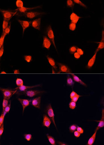
NIH/3T3 cell; 4% Paraformaldehyde-fixed; Triton X-100 at room temperature for 20 min; Blocking buffer (normal goat serum, C-0005) at 37°C for 20 min; Antibody incubation with (KO Validated)EGFR polyclonal Antibody, Unconjugated (bs-55061R) 1:100, 90 minutes at 37°C; followed by a conjugated Goat Anti-Rabbit IgG antibody at 37°C for 90 minutes, DAPI (blue, C02-04002) was used to stain the cell nuclei.
-
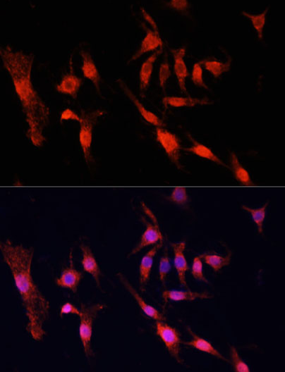
PC12 cell; 4% Paraformaldehyde-fixed; Triton X-100 at room temperature for 20 min; Blocking buffer (normal goat serum, C-0005) at 37°C for 20 min; Antibody incubation with (KO Validated)EGFR polyclonal Antibody, Unconjugated (bs-55061R) 1:100, 90 minutes at 37°C; followed by a conjugated Goat Anti-Rabbit IgG antibody at 37°C for 90 minutes, DAPI (blue, C02-04002) was used to stain the cell nuclei.
-
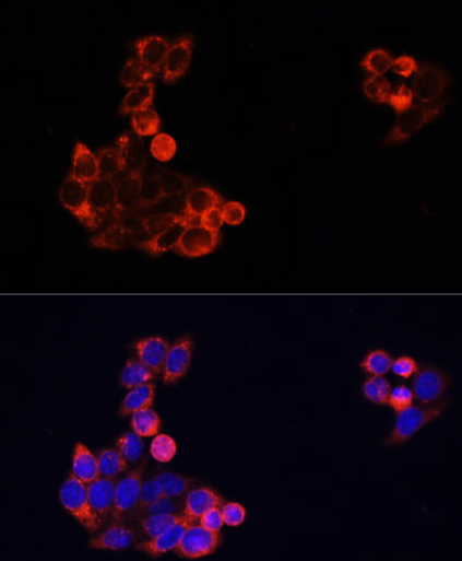
HeLa cell; 4% Paraformaldehyde-fixed; Triton X-100 at room temperature for 20 min; Blocking buffer (normal goat serum, C-0005) at 37°C for 20 min; Antibody incubation with (KO Validated)EGFR polyclonal Antibody, Unconjugated (bs-55061R) 1:100, 90 minutes at 37°C; followed by a conjugated Goat Anti-Rabbit IgG antibody at 37°C for 90 minutes, DAPI (blue, C02-04002) was used to stain the cell nuclei.
-
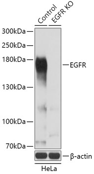
Sample:
Lane 1: Hela (Human) Cell Lysate at 25 ug
Lane 2: EGFR knockout (KO) Hela (Human) Cell Lysate at 25 ug
Primary: Anti-EGFR (bs-55061R) at 1/3000 dilution
Secondary: HRP Goat Anti-Rabbit IgG (H+L) at 1:10000 dilution
Predicted band size: 180 kD
Observed band size: 175 kD
-
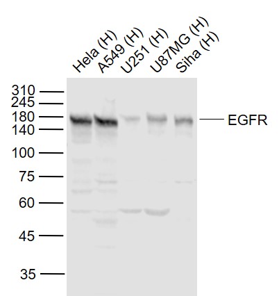
Sample:
Lane 1: Hela (Human) Cell Lysate at 30 ug
Lane 2: A549 (Human) Cell Lysate at 30 ug
Lane 3: U251 (Human) Cell Lysate at 30 ug
Lane 4: U87MG (Human) Cell Lysate at 30 ug
Lane 5: Siha (Human) Cell Lysate at 30 ug
Primary: Anti-EGFR (bs-55061R) at 1/1000 dilution
Secondary: IRDye800CW Goat Anti-Rabbit IgG at 1/20000 dilution
Predicted band size: 170 kD
Observed band size: 170 kD
RRID:RRID
产品名称:Rabbit Anti-EGFR antibody
别名: Avian erythroblastic leukemia viral (v erb b) oncogene homolog; Avian erythroblastic leukemia viral (verbb) oncogene homolog; Cell growth inhibiting protein 40; Cell proliferation inducing protein 61; EGF R; EGFR; Epidermal growth factor receptor (avian e
中文名称:表皮生长因子受体抗体
英文名称:Rabbit Anti-EGFR antibody
抗体来源: Rabbit
克隆类型:多克隆
细胞定位:细胞核,细胞浆,细胞膜,分泌型蛋白
性 状:Liquid
亚 型:IgG
纯化方法:affinity purified by Protein A
保存条件:Store at -20 °C for one year. Avoid repeated freeze/thaw cycles. The lyophilized antibody is stable at room temperature for at least one month and for greater than a year when kept at -20°C. When reconstituted in sterile pH 7.4 0.01M PBS or diluent of ant
免 疫 原:Recombinant human EGFR
抗原表位:1021-1210/1210
SWISS:P00533
Gene ID :1956
Human Gene ID:1956
The protein encoded by this gene is a transmembrane glycoprotein that is a member of the protein kinase superfamily. This protein is a receptor for members of the epidermal growth factor family. EGFR is a cell surface protein that binds to epidermal growth factor. Binding of the protein to a ligand induces receptor dimerization and tyrosine autophosphorylation and leads to cell proliferation. Mutations in this gene are associated with lung cancer. Multiple alternatively spliced transcript variants that encode different protein isoforms have been found for this gene. [provided by RefSeq, Jul 2010]
Function:Receptor tyrosine kinase binding ligands of the EGF family and activating several signaling cascades to convert extracellular cues into appropriate cellular responses. Known ligands include EGF, TGFA/TGF-alpha, amphiregulin, epigen/EPGN, BTC/betacellulin,
Subunit:Binding of the ligand triggers homo- and/or heterodimerization of the receptor triggering its autophosphorylation. Heterodimer with ERBB2. Interacts with ERRFI1; inhibits dimerization of the kinase domain and autophosphorylation. Part of a complex with ER
Subcellular Location:Cell membrane; Single-pass type I membrane protein. Endoplasmic reticulum membrane; Single-pass type I membrane protein. Golgi apparatus membrane; Single-pass type I membrane protein. Nucleus membrane; Single-pass type I membrane protein. Endosome. Endoso
Tissue Specificity:Ubiquitously expressed. Isoform 2 is also expressed in ovarian cancers.
Post-translational modifications:Phosphorylation at Ser-695 is partial and occurs only if Thr-693 is phosphorylated. Phosphorylation at Thr-678 and Thr-693 by PRKD1 inhibits EGF-induced MAPK8/JNK1 activation. Dephosphorylation by PTPRJ prevents endocytosis and stabilizes the receptor at
DISEASE:Defects in EGFR are associated with lung cancer (LNCR) [MIM:211980]. LNCR is a common malignancy affecting tissues of the lung. The most common form of lung cancer is non-small cell lung cancer (NSCLC) that can be divided into 3 major histologic subtypes:
Similarity:Belongs to the protein kinase superfamily. Tyr protein kinase family. EGF receptor subfamily.
Contains 1 protein kinase domain.
Important Note:This product as supplied is intended for research use only, not for use in human, therapeutic or diagnostic applications.
 400-901-9800


 说明书
说明书 联系我们
联系我们 打印此页面
打印此页面 收藏
收藏