| Rabbit Anti-CD59 antibody |
| 产品应用(已验证) |
WB,IHC,FCM |
| 推荐稀释比例 |
WB=1:500-2000,IHC-P=1:50-1:100,Flow Cyt=1:100, |
| 研究领域 |
心血管,免疫学,信号转导,干细胞 |
| 标签 |
Array |
-
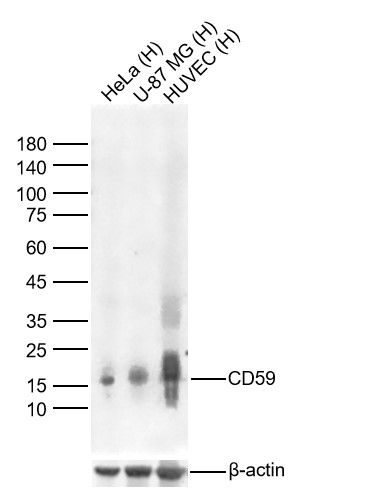
Sample:
Lane 1: Human Hela cell Lysates
Lane 2: Human U-87 MG cell Lysates
Lane 3: Human HUVEC cell Lysates
Primary: Anti-CD59 (bsm-60603R) at 1/1000 dilution
Secondary: IRDye800CW Goat Anti-Rabbit IgG at 1/20000 dilution
Predicted band size: 9kDa
Observed band size: 15kDa
-
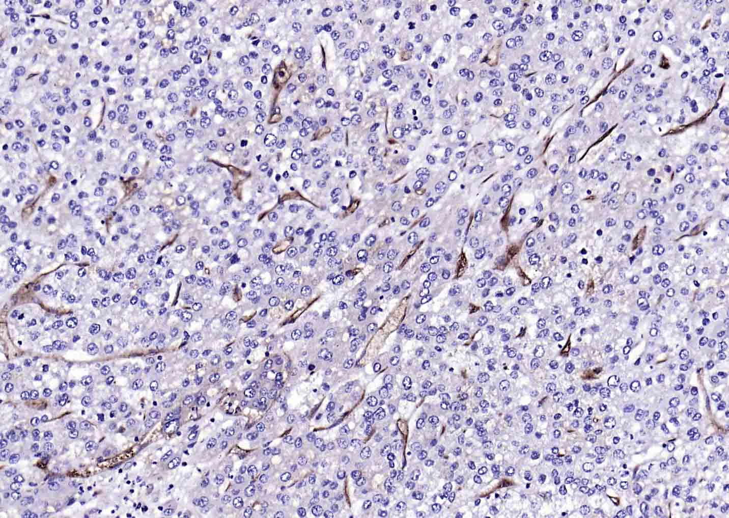
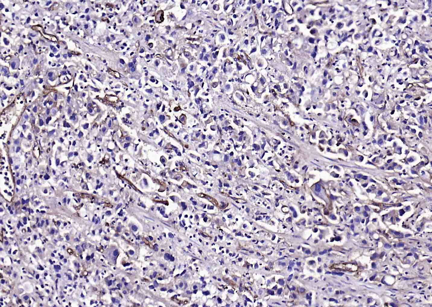
Paraformaldehyde-fixed, paraffin embedded (human liver carcinoma); Antigen retrieval by boiling in sodium citrate buffer (pH6.0) for 15min; Block endogenous peroxidase by 3% hydrogen peroxide for 20 minutes; Blocking buffer (normal goat serum) at 37°C for 30min; Antibody incubation with (CD59) Monoclonal Antibody, Unconjugated (bsm-60603R) at 1:200 overnight at 4°C, followed by operating according to SP Kit(Rabbit) (sp-0023) instructionsand DAB staining.
-
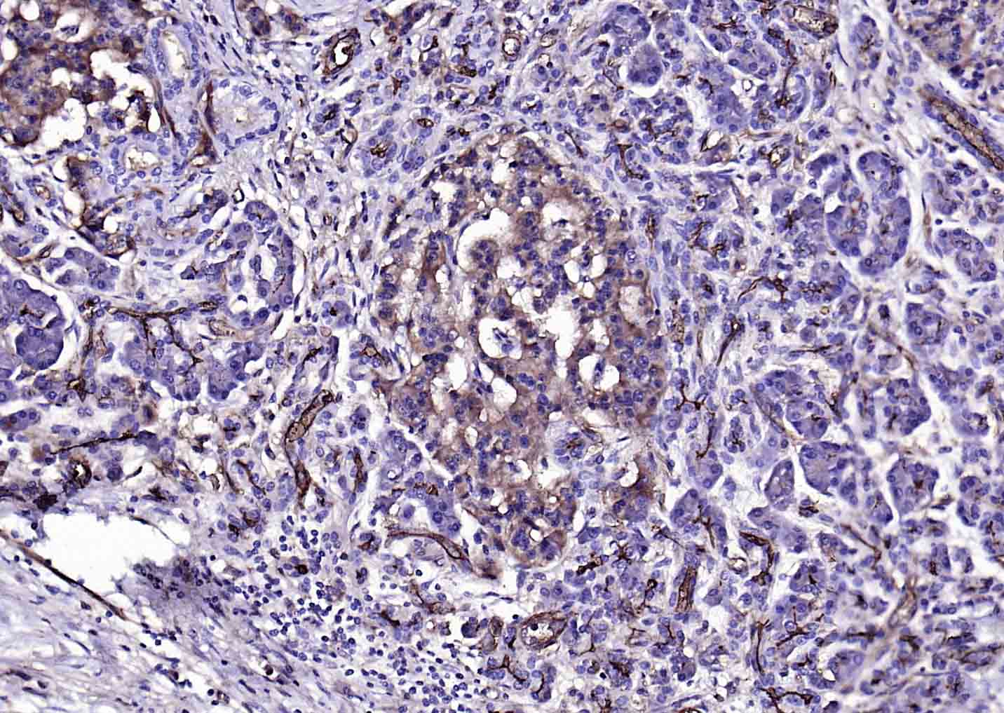
Paraformaldehyde-fixed, paraffin embedded (human pancreatic cancer); Antigen retrieval by boiling in sodium citrate buffer (pH6.0) for 15min; Block endogenous peroxidase by 3% hydrogen peroxide for 20 minutes; Blocking buffer (normal goat serum) at 37°C for 30min; Antibody incubation with (CD59) Monoclonal Antibody, Unconjugated (bsm-60603R) at 1:200 overnight at 4°C, followed by operating according to SP Kit(Rabbit) (sp-0023) instructionsand DAB staining.
-
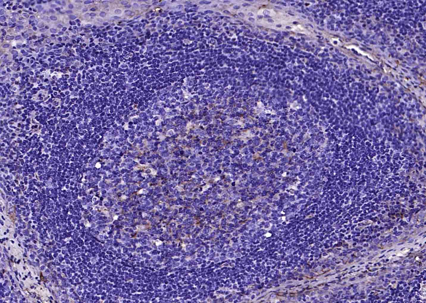
Paraformaldehyde-fixed, paraffin embedded (human tonsil); Antigen retrieval by boiling in sodium citrate buffer (pH6.0) for 15min; Block endogenous peroxidase by 3% hydrogen peroxide for 20 minutes; Blocking buffer (normal goat serum) at 37°C for 30min; Antibody incubation with (CD59) Monoclonal Antibody, Unconjugated (bsm-60603R) at 1:200 overnight at 4°C, followed by operating according to SP Kit(Rabbit) (sp-0023) instructionsand DAB staining.
-
Paraformaldehyde-fixed, paraffin embedded (human gastric carcinoma); Antigen retrieval by boiling in sodium citrate buffer (pH6.0) for 15min; Block endogenous peroxidase by 3% hydrogen peroxide for 20 minutes; Blocking buffer (normal goat serum) at 37°C for 30min; Antibody incubation with (CD59) Monoclonal Antibody, Unconjugated (bsm-60603R) at 1:200 overnight at 4°C, followed by operating according to SP Kit(Rabbit) (sp-0023) instructionsand DAB staining.
-
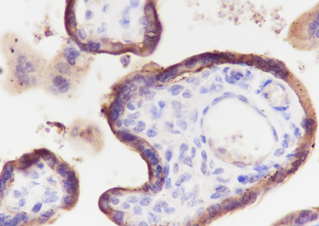
Tissue: Human placenta
Section type: Formalin fixed & Paraffin -embedded section
Retrieval method: High temperature and high pressure
Retrieval buffer: Tris/EDTA buffer, pH 9.0 Primary ab dilution: 1:100
Primary ab incubation condition: 1 hour at room temperature
Secondary ab: SP Kit(Rabbit) (sp-0023)
Counter stain: Hematoxylin (Blue)
Comment: Color brown is the positive signal for bsm-60603R
-
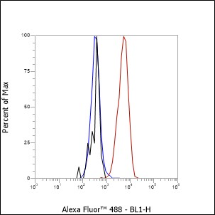
Cell line: PBMC
Fixative: 4% Paraformaldehyde
Permeabilization: 90% Methanol
Primary ab dilution: 1:100
Secondary ab: Goat anti Rabbit IgG
Unlabelled control: The cell without incubation with primary antibody and secondary antibody (Black line).
Isotype control: Rabbit monoclonal IgG (Blue line).
Comment: Line red is the positive signal for bsm-60603R
RRID:RRID
产品名称:Rabbit Anti-CD59 antibody
别名: CD59 glycoprotein; CD59 molecule (CD59 blood group); 1F5 antigen; 20 kDa homologous restriction factor; MAC-inhibitory protein; Membrane attack complex inhibition factor; Membrane inhibitor of reactive lysis; MEM43 antigen; MACIF;
1F5; EJ16; EJ30; EL32;
中文名称:CD59重组兔单克隆抗体
英文名称:Rabbit Anti-CD59 antibody
抗体来源: Rabbit
克隆类型:单克隆
细胞定位:细胞膜
性 状:Liquid
亚 型:IgG
纯化方法:affinity purified by Protein A
保存条件:Shipped at 4℃. Store at -20 °C for one year. Avoid repeated freeze/thaw cycles.
免 疫 原:KLH conjugated synthetic peptide derived from human CD59
SWISS:P13987
Gene ID :966
Human Gene ID:966
This gene encodes a cell surface glycoprotein that regulates complement-mediated cell lysis, and it is involved in lymphocyte signal transduction. This protein is a potent inhibitor of the complement membrane attack complex, whereby it binds complement C8 and/or C9 during the assembly of this complex, thereby inhibiting the incorporation of multiple copies of C9 into the complex, which is necessary for osmolytic pore formation. This protein also plays a role in signal transduction pathways in the activation of T cells. Mutations in this gene cause CD59 deficiency, a disease resulting in hemolytic anemia and thrombosis, and which causes cerebral infarction. Multiple alternatively spliced transcript variants, which encode the same protein, have been identified for this gene. [provided by RefSeq, Jul 2008]
Function:Potent inhibitor of the complement membrane attack complex (MAC) action. Acts by binding to the C8 and/or C9 complements of the assembling MAC, thereby preventing incorporation of the multiple copies of C9 required for complete formation of the osmolytic
Subunit:Interacts with T-cell surface antigen CD2.
Subcellular Location:Cell membrane; Lipid-anchor, GPI-anchor. Secreted. Note=Soluble form found in a number of tissues.
Post-translational modifications:N- and O-glycosylated. The N-glycosylation mainly consists of a family of biantennary complex-type structures with and without lactosamine extensions and outer arm fucose residues. Also significant amounts of triantennary complexes (22%). Variable sialyla
DISEASE:CD59 deficiency (CD59D) [MIM:612300]: A deficiency resulting in chronic complement-mediated intravascular hemolysis, anemia, hemoglobinuria and thrombosis. Note=The disease is caused by mutations affecting the gene represented in this entry.
Similarity:Contains 1 UPAR/Ly6 domain.
Important Note:This product as supplied is intended for research use only, not for use in human, therapeutic or diagnostic applications.
 400-901-9800


 说明书
说明书 联系我们
联系我们 打印此页面
打印此页面 收藏
收藏