| Rabbit Anti-beta-Actin (Loading Control) antibody |
| 反应物种(预测) |
Chicken,Dog,Pig,Rabbit,Sheep,Bee,Fish,GuineaPig,Hamster,Cat |
| 产品应用(已验证) |
WB,ICC,FCM,ELISA |
| 产品应用(可尝试) |
IHC |
| 推荐稀释比例 |
WB=1:5000-50000,Elisa=1:5000-20000,IHC-P=1:500-1000,Flow Cyt=1μg/Test,ICC=1:100, |
| 研究领域 |
肿瘤,细胞生物,信号转导,细胞骨架, |
| 标签 |
Array |
-
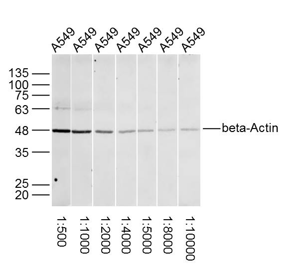
Sample:
A549 Cell (Human) Lysate at 30 ug
Primary:
Lane1: Anti-beta-Actin (bs-0061R) at 1/500 dilution
Lane2: Anti-beta-Actin (bs-0061R) at 1/1000 dilution
Lane3: Anti-beta-Actin (bs-0061R) at 1/2000 dilution
Lane4: Anti-beta-Actin (bs-0061R) at 1/4000 dilution
Lane5: Anti-beta-Actin (bs-0061R) at 1/5000 dilution
Lane6: Anti-beta-Actin (bs-0061R) at 1/8000 dilution
Lane7: Anti-beta-Actin (bs-0061R) at 1/10000 dilution
Secondary: IRDye800CW Goat Anti-Rabbit IgG at 1/20000 dilution
Predicted band size: 42 kD
Observed band size: 42 kD
-
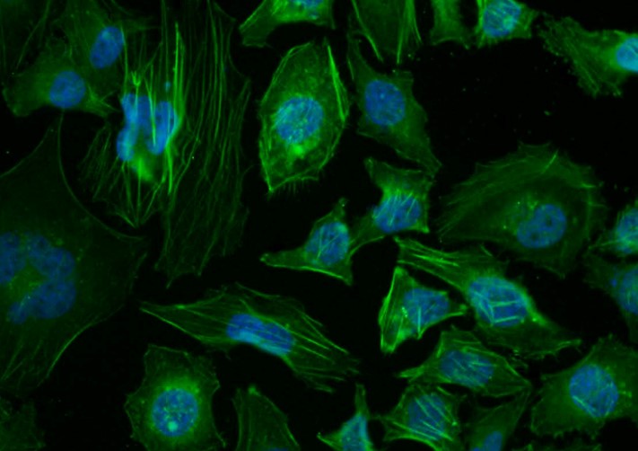
Tissue/cell: Hela cell; 4% Paraformaldehyde-fixed; Triton X-100 at room temperature for 20 min; Blocking buffer (normal goat serum, C-0005) at 37°C for 20 min; Antibody incubation with (beta-Actin) polyclonal Antibody, Unconjugated (bs-0061R) 1:100, 90 minutes at 37°C; followed by a conjugated Goat Anti-Rabbit IgG-FITC antibody at 37°C for 90 minutes, DAPI (blue, C02-04002) was used to stain the cell nuclei.
-
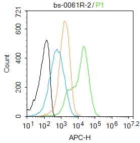
Blank control:Mouse spleen.
Primary Antibody (green line): Rabbit Anti-beta-Actin (Loading Control) antibody (bs-0061R)
Dilution: 2μg /10^6 cells;
Isotype Control Antibody (orange line): Rabbit IgG .
Secondary Antibody : Goat anti-rabbit IgG-AF647
Dilution: 1μg /test.
Protocol
The cells were fixed with 4% PFA (10min at room temperature)and then permeabilized with 90% ice-cold methanol for 20 min at-20℃. The cells were then incubated in 5%BSA to block non-specific protein-protein interactions for 30 min at room temperature .Cells stained with Primary Antibody for 30 min at room temperature. The secondary antibody used for 40 min at room temperature. Acquisition of 20,000 events was performed.
-
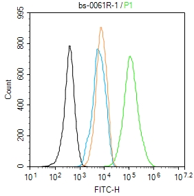
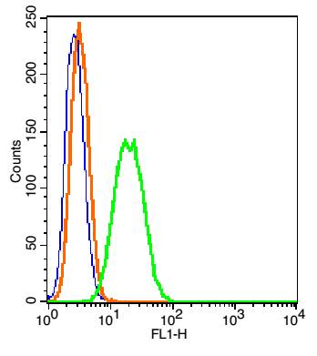
Blank control: NIH/3T3.
Primary Antibody (green line): Rabbit Anti-beta-Actin (Loading Control) antibody (bs-0061R)
Dilution: 1μg /10^6 cells;
Isotype Control Antibody (orange line): Rabbit IgG .
Secondary Antibody : Goat anti-rabbit IgG-AF488
Dilution: 1μg /test.
Protocol
The cells were fixed with 4% PFA (10min at room temperature)and then permeabilized with 90% ice-cold methanol for 20 min at -20℃. The cells were then incubated in 5%BSA to block non-specific protein-protein interactions for 30 min at room temperature .Cells stained with Primary Antibody for 30 min at room temperature. The secondary antibody used for 40 min at room temperature. Acquisition of 20,000 events was performed.
-
Sample:
Thymus (Mouse) Lysate at 40 ug
Primary:
Anti-beta-Actin (bs-0061R) at 1/1000~1/20000 dilution
Secondary: IRDye800CW Goat Anti-Rabbit IgG at 1/20000 dilution
Predicted band size: 42 kD
Observed band size: 42 kD
-
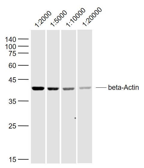
Sample:
SH-SY5Y (Human) Lysate at 40 ug
Primary:
Anti-beta-Actin (bs-0061R) at 1/2000~1/20000 dilution
Secondary: IRDye800CW Goat Anti-Rabbit IgG at 1/20000 dilution
Predicted band size: 42 kD
Observed band size: 42 kD
-
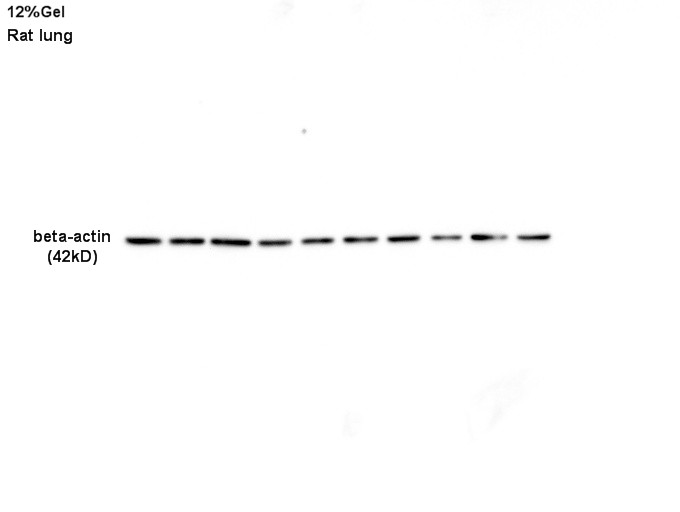
Sample: Lung lysate at 30ug;
Primary: Anti-beta-actin (bs-0061R) at 1:1000 dilution
Secondary: HRP conjugated Goat-Anti-Rabbit IgG(bse-0295G) at 1:3000 dilution
Predicted band size : 42kD
Observed band size : 42kD
-
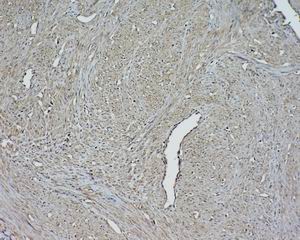
Tissue/cell: human cervical carcinoma; 4% Paraformaldehyde-fixed and paraffin-embedded;
Antigen retrieval: citrate buffer ( 0.01M, pH 6.0 ), Boiling bathing for 15min; Block endogenous peroxidase by 3% Hydrogen peroxide for 30min; Blocking buffer (normal goat serum,C-0005) at 37℃ for 20 min;
Incubation: Anti-Beta-actin Polyclonal Antibody, Unconjugated(bs-0061R) 1:1500, overnight at 4°C, followed by conjugation to the secondary antibody(SP-0023) and DAB(C-0010) staining
-
Blank control: RSC96(blue).
Primary Antibody: Rabbit Anti-beta-Actin /FITC Conjugated antibody (bs-0061R/FITC), Dilution: 1μg in 100 μL 1X PBS containing 0.5% BSA;
Isotype Control Antibody: Rabbit IgG/FITC(orange) ,used under the same conditions.
Protocol
The cells were fixed with 2% paraformaldehyde (10 min) and then permeabilized with ice-cold 90% methanol for 30 min on ice. The cells were washed twice with 1 X PBS. The cells were incubated in 1 X PBS containing 0.5% BSA + 1 0% goat serum (15 min) to block non-specific protein-protein interactions followed by the incubated with antibody (bs-0061R/FITC, 1μg /1x10^6 cells) for 30 min on ice. Acquisition of 20,000 events was performed.
-
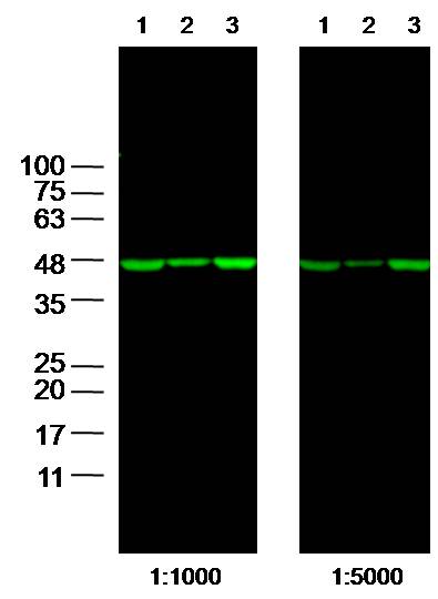
Sample:
Lane1: 293T Cell Lysate at 25 ug
Lane2: A549 Cell Lysate at 25 ug
Lane3: A431 Cell Lysate at 25 ug
Primary: Anti- beta-Actin (bs-0061R) at 1/1000 and 1/5000 dilution
Secondary: IRDye800CW Goat Anti-Rabbit IgG at 1/20000 dilution
Predicted band size: 42kD
Observed band size: 42 kD
-
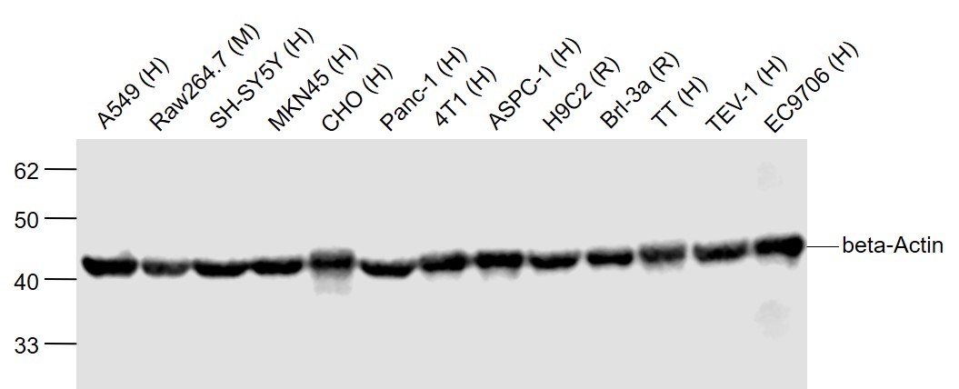
Sample:
A549 (Human) Cell Lysate at 40 ug
Raw264.7 (Mouse) Cell Lysate at 40 ug
SH-SY5Y (Human) Cell Lysate at 40 ug
MKN45 (Human) Cell Lysate at 40 ug
CHO (Human) Cell Lysate at 40 ug
Panc-1 (Human) Cell Lysate at 40 ug
4T1 (Human) Cell Lysate at 40 ug
ASPC-1 (Human) Cell Lysate at 40 ug
H9C2 (Rat) Cell Lysate at 40 ug
Brl-3a (Rat) Cell Lysate at 40 ug
TT (Human) Cell Lysate at 40 ug
TEV-1 (Human) Cell Lysate at 40 ug
EC9706 (Human) Cell Lysate at 40 ug
Primary: Anti-beta-Actin (bs-0061R) at 1/2000 dilution
Secondary: IRDye800CW Goat Anti-Rabbit IgG at 1/20000 dilution
Predicted band size: 42 kD
Observed band size: 42 kD
-
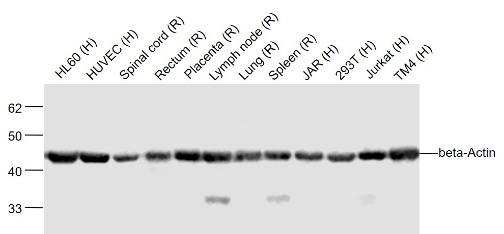
Sample:
HL60 (Human) Cell Lysate at 40 ug
HUVEC (Human) Cell Lysate at 40 ug
Spinal cord (Rat) Lysate at 40 ug
Rectum (Rat) Lysate at 40 ug
Placenta (Rat) Lysate at 40 ug
Lymph node (Rat) Lysate at 40 ug
Lung (Rat) Lysate at 40 ug
Spleen (Rat) Lysate at 40 ug
JAR (Human) Cell Lysate at 40 ug
293T (Human) Cell Lysate at 40 ug
Jurkat (Human) Cell Lysate at 40 ug
TM4 (Human) Cell Lysate at 40 ug
Primary: Anti-beta-Actin (bs-0061R) at 1/2000 dilution
Secondary: IRDye800CW Goat Anti-Rabbit IgG at 1/20000 dilution
Predicted band size: 42 kD
Observed band size: 42 kD
-
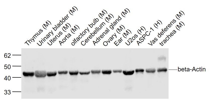
Sample:
Thymus (Mouse) Lysate at 40 ug
Urinary bladder (Mouse) Lysate at 40 ug
Uterus (Mouse) Cell Lysate at 40 ug
Aorta (Mouse) Lysate at 40 ug
olfactory bulb (Mouse) Lysate at 40 ug
Cerebellum (Mouse) Lysate at 40 ug
Adrenal gland (Mouse) Lysate at 40 ug
Ovary (Mouse) Lysate at 40 ug
Ear (Mouse) Lysate at 40 ug
U2os (Human) Cell Lysate at 40 ug
ASPC-1 (Human) Cell Lysate at 40 ug
Vas deferens (Mouse) Lysate at 40 ug
trachea (Mouse) Lysate at 40 ug
Primary: Anti-beta-Actin (bs-0061R) at 1/2000 dilution
Secondary: IRDye800CW Goat Anti-Rabbit IgG at 1/20000 dilution
Predicted band size: 42 kD
Observed band size: 42 kD
-
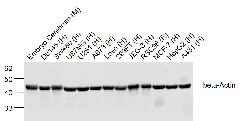
Sample:
Embryo Cerebrum (Mouse) Lysate at 40 ug
Du145 (Human) Lysate at 40 ug
SW480 (Human) Cell Lysate at 40 ug
U87MG (Human) Lysate at 40 ug
U251 (Human) Lysate at 40 ug
A673 (Human) Lysate at 40 ug
Lovo (Human) Lysate at 40 ug
293FT (Human) Lysate at 40 ug
JEG-3 (Human) Lysate at 40 ug
RSC96 (Rat) Cell Lysate at 40 ug
MCF-7 (Human) Cell Lysate at 40 ug
HepG2 (Human) Lysate at 40 ug
A431 (Human) Lysate at 40 ug
Primary: Anti-beta-Actin (bs-0061R) at 1/2000 dilution
Secondary: IRDye800CW Goat Anti-Rabbit IgG at 1/20000 dilution
Predicted band size: 42 kD
Observed band size: 42 kD
-
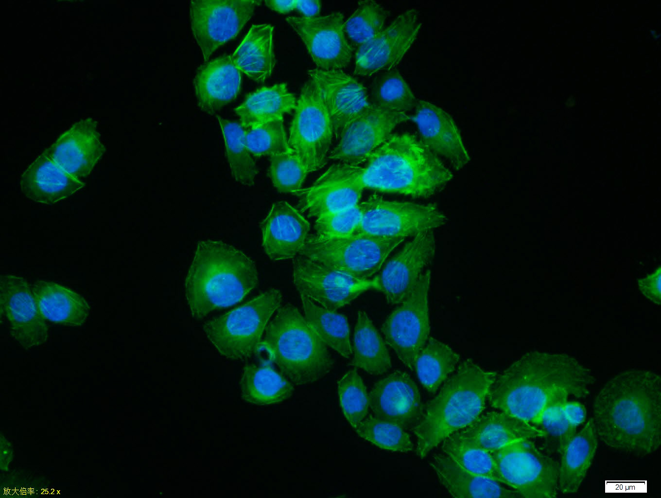
MCF7 cell; 4% Paraformaldehyde-fixed; Triton X-100 at room temperature for 20 min; Blocking buffer (normal goat serum, C-0005) at 37°C for 20 min; Antibody incubation with (beta-Actin) polyclonal Antibody, Unconjugated (bs-0061R) 1:100, 90 minutes at 37°C; followed by a conjugated Goat Anti-Rabbit IgG antibody at 37°C for 90 minutes, DAPI (blue, C02-04002) was used to stain the cell nuclei.
-
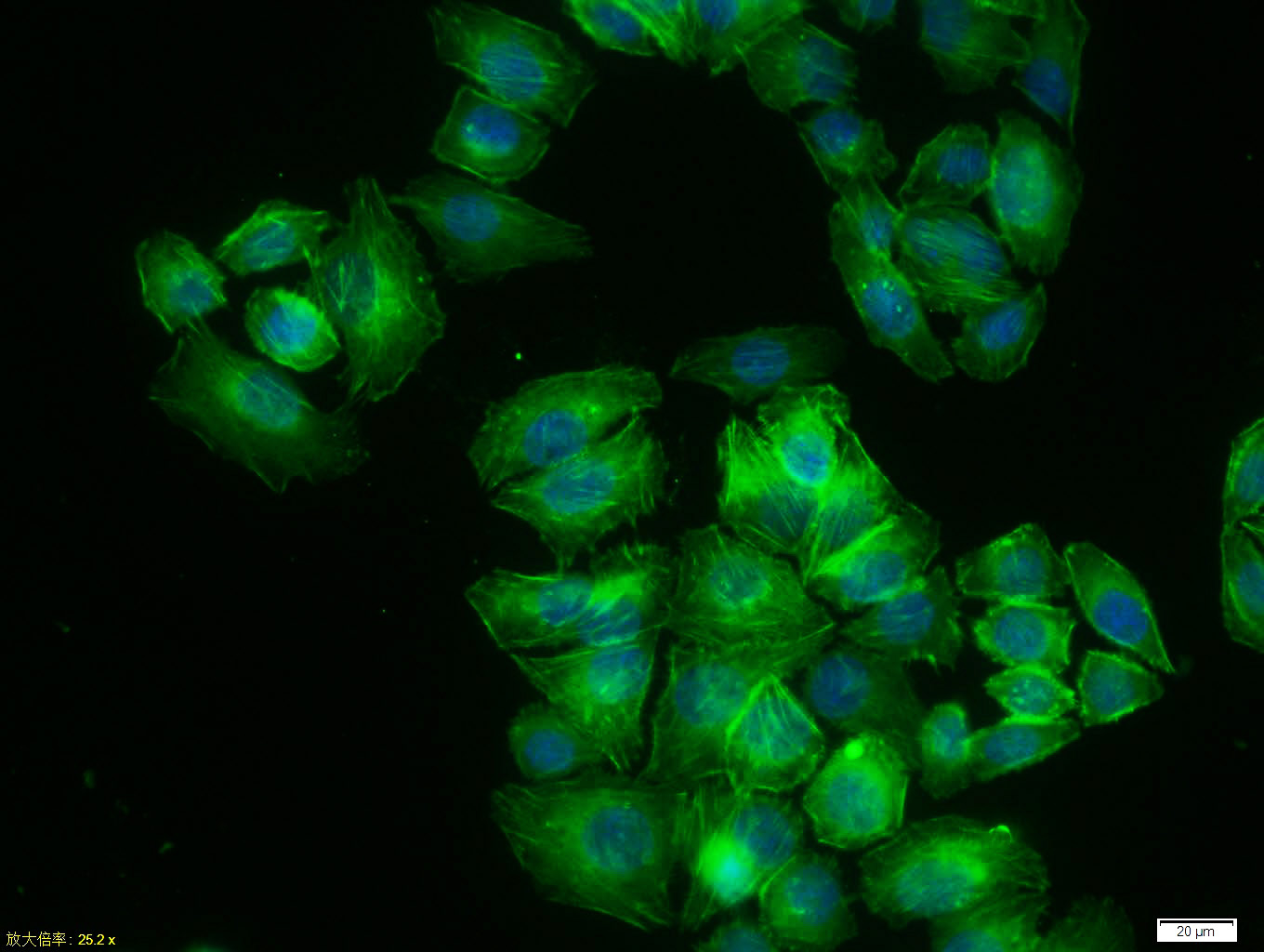
MCF7 cell; 4% Paraformaldehyde-fixed; Triton X-100 at room temperature for 20 min; Blocking buffer (normal goat serum, C-0005) at 37°C for 20 min; Antibody incubation with (beta-Actin) polyclonal Antibody, Unconjugated (bs-0061R) 1:100, 90 minutes at 37°C; followed by a conjugated Goat Anti-Rabbit IgG antibody at 37°C for 90 minutes, DAPI (blue, C02-04002) was used to stain the cell nuclei.
RRID:AB_10855480
产品名称:Rabbit Anti-beta-Actin (Loading Control) antibody
别名: Beta Actin; beta-Actin; ACTB; Actin cytoplasmic 1; Actin, beta; Beta actin; beta cytoskeletal actin; A X actin like protein; ACTB; Actin cytoplasmic 1; alpha sarcomeric Actin; Actx; Beta cytoskeletal actin; Melanoma X actin; PS1TP5BP1; ACTB_HUMAN.
中文名称:β-肌动蛋白/β-Actin(内参)抗体1
英文名称:Rabbit Anti-beta-Actin (Loading Control) antibody
中文别名:β actin; βactin;
抗体来源: Rabbit
克隆类型:多克隆
细胞定位:细胞浆
性 状:Liquid
亚 型:IgG
纯化方法:affinity purified by Protein A
保存条件:Shipped at 4℃. Store at -20 °C for one year. Avoid repeated freeze/thaw cycles.
免 疫 原:Synthetic MAP peptide derived from human beta-Actin
抗原表位:1-200/375
SWISS:P60709
Gene ID :60
Human Gene ID:60
Loading Control
This gene encodes one of six different actin proteins. Actins are highly conserved proteins that are involved in cell motility, structure, and integrity. This actin is a major constituent of the contractile apparatus and one of the two nonmuscle cytoskeletal actins. [provided by RefSeq, Jul 2008].
Function:Actins are highly conserved proteins that are involved in various types of cell motility and are ubiquitously expressed in all eukaryotic cells.
Subunit:Polymerization of globular actin (G-actin) leads to a structural filament (F-actin) in the form of a two-stranded helix. Each actin can bind to 4 others. Identified in a mRNP granule complex, at least composed of ACTB, ACTN4, DHX9, ERG, HNRNPA1, HNRNPA2B
Subcellular Location:Cytoplasm. cytoskeleton.
Tissue Specificity:Ubiquitously expressed in all eukaryotic cells.
Post-translational modifications:ISGylated.
Oxidation of Met-44 by MICALs (MICAL1, MICAL2 or MICAL3) to form methionine sulfoxide promotes actin filament depolymerization. Methionine sulfoxide is produced stereospecifically, but it is not known whether the (S)-S-oxide or the (R)-S-oxi
DISEASE:Defects in ACTA1 are the cause of nemaline myopathy type 3 (NEM3) [MIM:161800]. A form of nemaline myopathy. Nemaline myopathies are muscular disorders characterized by muscle weakness of varying severity and onset, and abnormal thread-or rod-like structu
Similarity:Belongs to the actin family.
Important Note:This product as supplied is intended for research use only, not for use in human, therapeutic or diagnostic applications.
 400-901-9800


 说明书
说明书 联系我们
联系我们 打印此页面
打印此页面 收藏
收藏