欢迎访问博奥森抗体公司官方网站!网站测试中
首页 > 产品 > 一抗 > 产品信息
Rabbit Anti-Bax antibody
多克隆  |   SKU:bs-0127R

说明书

联系我们

打印此页面

收藏

货号:bs-3351R
¥1280
订购号:bs-0127R
¥1098.00-2900.00
货期:现货
Rabbit Anti-Bax antibody
反应物种(预测)

Dog,Pig,Cow,Sheep

产品应用(已验证)

WB,IHC,ICC,FCM

产品应用(可尝试)

IF,ELISA

推荐稀释比例

WB=1:500-2000,Elisa=1:5000-10000,IHC-P=1:100-500,IHC-F=1:100-500,Flow Cyt=1μg /test,IF=1:100-500,ICC=1:100,

研究领域

细胞生物,信号转导,细胞凋亡,线抗体,

标签

Array

  • Paraformaldehyde-fixed, paraffin embedded (Rat brain); Antigen retrieval by boiling in sodium citrate buffer (pH6.0) for 15min; Block endogenous peroxidase by 3% hydrogen peroxide for 20 minutes; Blocking buffer (normal goat serum) at 37°C for 30min; Antibody incubation with (Bax) Polyclonal Antibody, Unconjugated (bs-0127R) at 1:400 overnight at 4°C, followed by operating according to SP Kit(Rabbit) (sp-0023) instructionsand DAB staining.
  • Tissue/cell: rat brain tissue; 4% Paraformaldehyde-fixed and paraffin-embedded;
    Antigen retrieval: citrate buffer ( 0.01M, pH 6.0 ), Boiling bathing for 15min; Block endogenous peroxidase by 3% Hydrogen peroxide for 30min; Blocking buffer (normal goat serum,C-0005) at 37℃ for 20 min;
    Incubation: Anti-Bax Polyclonal Antibody, Unconjugated(bs-0127R) 1:800, overnight at 4癈, followed by conjugation to the secondary antibody(SP-0023) and DAB(C-0010) staining
  • Sample: Brain(Rat)lysate 30ug;
    Liver(Mouse) lysates, 30ug;
    Primary: Anti-Bax (bs-0127R) at 1:200;
    Secondary: HRP conjugated Goat-Anti-Rabbit IgG(bse-0295G) at 1: 3000;
    ECL excitated the fluorescence;
    Predicted band size : 21kD
    Observed band size : 21kD
  • Overlay histogram showing HL 60 cells stained with bs-0127R (Green line).
    The cells were fixed with 90% methanol (5 min) and then permeabilized with 0.01M PBS-Tween for 20 min. The cells were then incubated in 1x PBS / 10% normal goat serum to block non-specific protein-protein interactions followed by the antibody (bs-0127R,1μg/1x10^6 cells) for 30 min at 22℃. The secondary antibody used was fluorescein isothiocyanate goat anti-rabbit IgG (H+L) (bs- 0295G-FITC , Brillant blue line) at 1/200 dilution for 30 min at 22℃. Isotype control antibody was rabbit IgG (polyclonal,bs-0295P,Orange line) (1μg/1x10^6 cells) used under the same conditions. Unlabelled sample (blue line) was also used as a control. Acquisition of 20,000 events were collected using a 20mW Argon ion laser (488nm) and 525/30 bandpass filter.
  • Tissue/cell: rat lung tissue; 4% Paraformaldehyde-fixed and paraffin-embedded;
    Antigen retrieval: citrate buffer ( 0.01M, pH 6.0 ), Boiling bathing for 15min; Block endogenous peroxidase by 3% Hydrogen peroxide for 30min; Blocking buffer (normal goat serum,C-0005) at 37℃ for 20 min;
    Incubation: Anti-Bax Polyclonal Antibody, Unconjugated(bs-0127R) 1:200, overnight at 4癈, followed by conjugation to the secondary antibody(SP-0023) and DAB(C-0010) staining
  • Tissue/cell: rat stomach tissue; 4% Paraformaldehyde-fixed and paraffin-embedded;
    Antigen retrieval: citrate buffer ( 0.01M, pH 6.0 ), Boiling bathing for 15min; Block endogenous peroxidase by 3% Hydrogen peroxide for 30min; Blocking buffer (normal goat serum,C-0005) at 37℃ for 20 min;
    Incubation: Anti-Bax Polyclonal Antibody, Unconjugated(bs-0127R) 1:200, overnight at 4癈, followed by conjugation to the secondary antibody(SP-0023) and DAB(C-0010) staining
  • Sample:
    Hela(Human) Cell Lysate at 30 ug
    Primary: Anti-Bax (bs-0127R) at 1/1000 dilution
    Secondary: IRDye800CW Goat Anti-Rabbit IgG at 1/20000 dilution
    Predicted band size: 21 kD
    Observed band size: 23 kD
  • Sample:
    Hela-UV(Human) Cell Lysate at 30 ug
    Primary: Anti-Bax (bs-0127R) at 1/1000 dilution
    Secondary: IRDye800CW Goat Anti-Rabbit IgG at 1/20000 dilution
    Predicted band size: 21 kD
    Observed band size: 23 kD
  • Paraformaldehyde-fixed, paraffin embedded (Human liver carcinoma); Antigen retrieval by boiling in sodium citrate buffer (pH6.0) for 15min; Block endogenous peroxidase by 3% hydrogen peroxide for 20 minutes; Blocking buffer (normal goat serum) at 37°C for 30min; Antibody incubation with (Bax) Polyclonal Antibody, Unconjugated (bs-0127R) at 1:400 overnight at 4°C, followed by operating according to SP Kit(Rabbit) (sp-0023) instructionsand DAB staining.
  • Paraformaldehyde-fixed, paraffin embedded (Human brain glioma); Antigen retrieval by boiling in sodium citrate buffer (pH6.0) for 15min; Block endogenous peroxidase by 3% hydrogen peroxide for 20 minutes; Blocking buffer (normal goat serum) at 37°C for 30min; Antibody incubation with (Bax) Polyclonal Antibody, Unconjugated (bs-0127R) at 1:400 overnight at 4°C, followed by operating according to SP Kit(Rabbit) (sp-0023) instructionsand DAB staining.
  • Tissue/cell:Sh-sy5y cell; 4% Paraformaldehyde-fixed; Triton X-100 at room temperature for 20 min; Blocking buffer (normal goat serum, C-0005) at 37°C for 20 min; Antibody incubation with (Bax) polyclonal Antibody, Unconjugated (bs-0127R) 1:100, 90 minutes at 37°C; followed by a FITC conjugated Goat Anti-Rabbit IgG antibody at 37°C for 90 minutes, DAPI (blue, C02-04002) was used to stain the cell nuclei.
  • Tissue/cell:Sh-sy5y cell; 4% Paraformaldehyde-fixed; Triton X-100 at room temperature for 20 min; Blocking buffer (normal goat serum, C-0005) at 37°C for 20 min; Antibody incubation with (Bax) polyclonal Antibody, Unconjugated (bs-0127R) 1:100, 90 minutes at 37°C; followed by a FITC conjugated Goat Anti-Rabbit IgG antibody at 37°C for 90 minutes, DAPI (blue, C02-04002) was used to stain the cell nuclei.
  • Tissue/cell:Sh-sy5y cell; 4% Paraformaldehyde-fixed; Triton X-100 at room temperature for 20 min; Blocking buffer (normal goat serum, C-0005) at 37°C for 20 min; Antibody incubation with (Bax) polyclonal Antibody, Unconjugated (bs-0127R) 1:100, 90 minutes at 37°C; followed by a FITC conjugated Goat Anti-Rabbit IgG antibody at 37°C for 90 minutes, DAPI (blue, C02-04002) was used to stain the cell nuclei.
  • Paraformaldehyde-fixed, paraffin embedded (Rat spinal cord); Antigen retrieval by boiling in sodium citrate buffer (pH6.0) for 15min; Block endogenous peroxidase by 3% hydrogen peroxide for 20 minutes; Blocking buffer (normal goat serum) at 37°C for 30min; Antibody incubation with (Bax) Polyclonal Antibody, Unconjugated (bs-0127R) at 1:400 overnight at 4°C, followed by operating according to SP Kit(Rabbit) (sp-0023) instructionsand DAB staining.
  • Tissue/cell:Sh-sy5y cell; 4% Paraformaldehyde-fixed; Triton X-100 at room temperature for 20 min; Blocking buffer (normal goat serum, C-0005) at 37°C for 20 min; Antibody incubation with (Bax) polyclonal Antibody, Unconjugated (bs-0127R) 1:100, 90 minutes at 37°C; followed by a FITC conjugated Goat Anti-Rabbit IgG antibody at 37°C for 90 minutes, DAPI (blue, C02-04002) was used to stain the cell nuclei.
  • Sample:
    Lane 1: Testis (Mouse) Lysate at 40 ug
    Lane 2: Kidney (Rat) Lysate at 40 ug
    Lane 3: Testis (Rat) Lysate at 40 ug
    Primary:
    Anti-Bax (bs-0127R) at 1/1000 dilution
    Secondary: IRDye800CW Goat Anti-Rabbit IgG at 1/20000 dilution
    Predicted band size: 21 kD
    Observed band size: 21 kD
  • Sample:
    Cerebral cortex (Rat) Lysate at 40 ug
    Primary: Anti-Bax (bs-0127R) at 1/1000 dilution
    Secondary: IRDye800CW Goat Anti-Rabbit IgG at 1/20000 dilution
    Predicted band size: 21 kD
    Observed band size: 21 kD

产品信息

RRID:AB_10856348
产品名称:Rabbit Anti-Bax antibody
别名: apoptosis regulator BAX; Apoptosis regulator BAX cytoplasmic isoform beta; Apoptosis regulator BAX membrane isoform alpha; Bax isoform psi; BAX protein cytoplasmic isoform delta; Bax protein cytoplasmic isoform delta. antibody Bax protein cytoplasmic isof
中文名称:Bax抗体
英文名称:Rabbit Anti-Bax antibody
抗体来源: Rabbit
克隆类型:多克隆
细胞定位:细胞浆,细胞膜
性 状:Liquid
亚 型:IgG
纯化方法:affinity purified by Protein A
保存条件:Shipped at 4℃. Store at -20 °C for one year. Avoid repeated freeze/thaw cycles.

免疫原信息

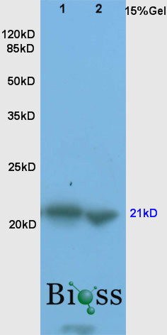
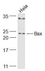
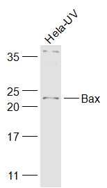
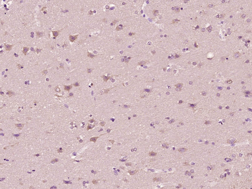
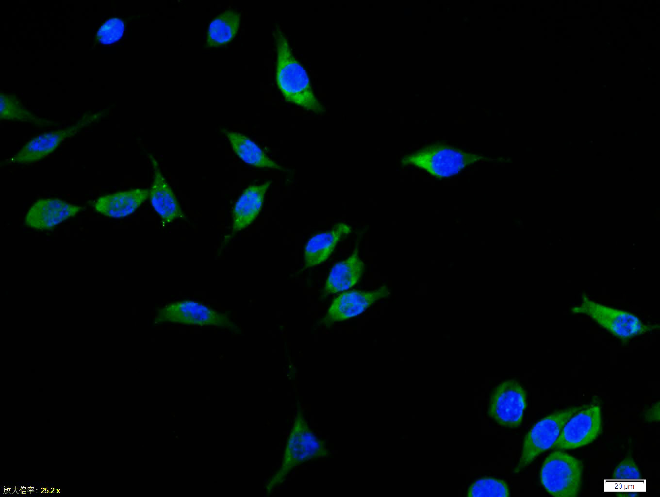
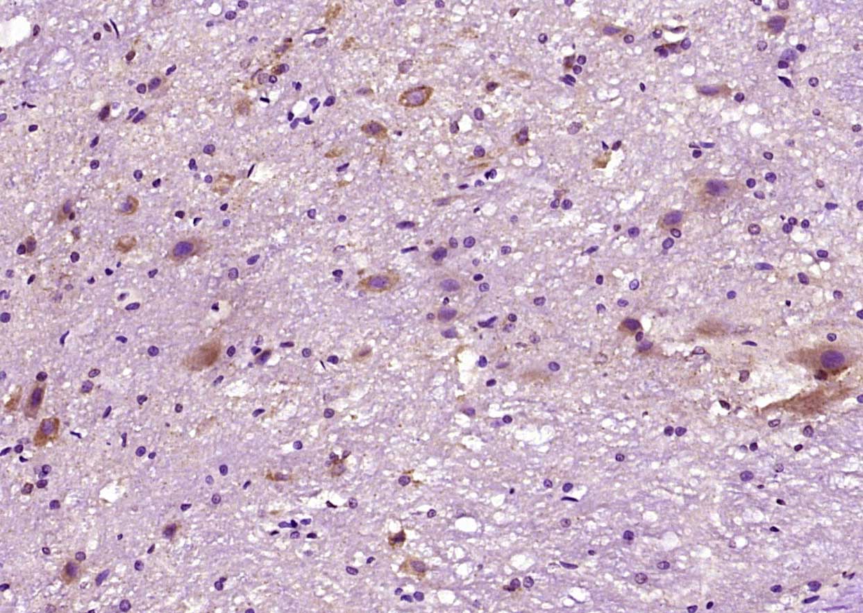
免 疫 原:KLH conjugated synthetic peptide derived from human Bax
抗原表位:84-175/192
SWISS:Q07812
Gene ID :581
Human Gene ID:581

产品介绍

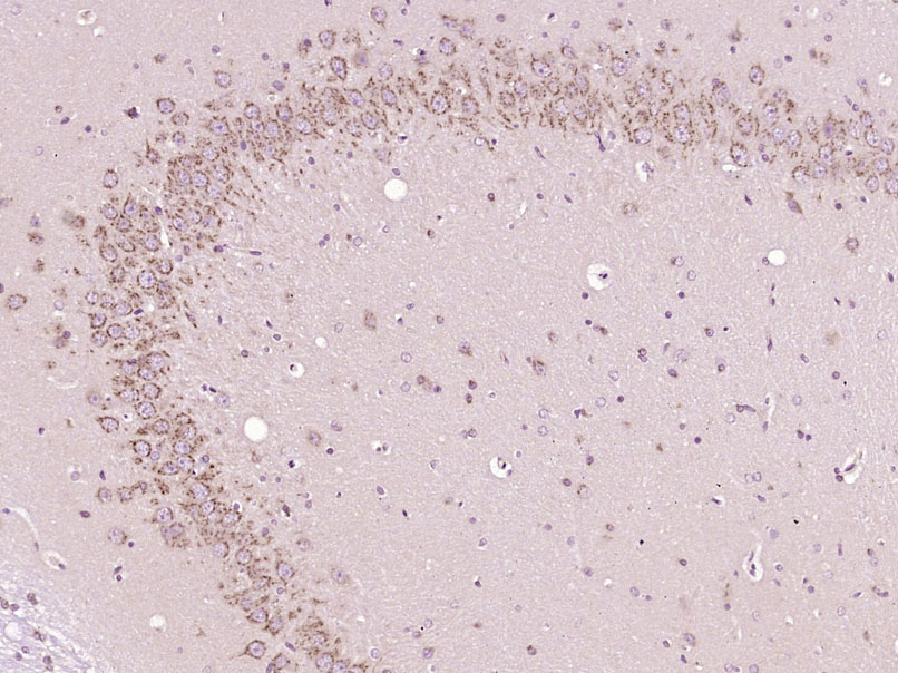
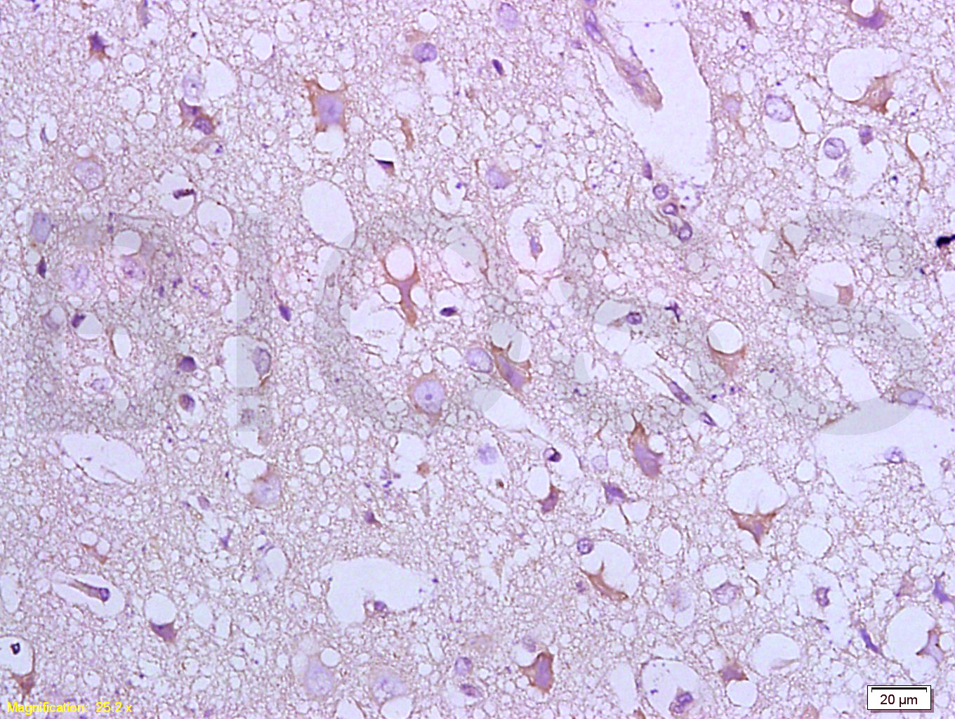
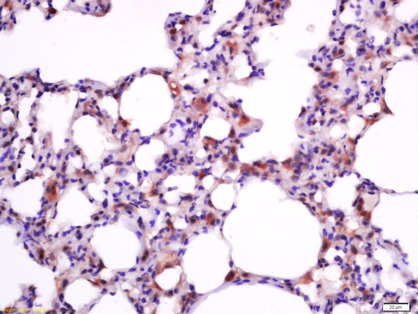
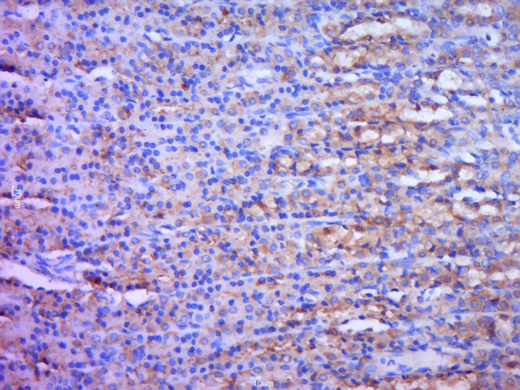
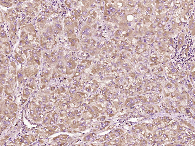
The protein encoded by this gene belongs to the BCL2 protein family. BCL2 family members form hetero- or homodimers and act as anti- or pro-apoptotic regulators that are involved in a wide variety of cellular activities. This protein forms a heterodimer with BCL2, and functions as an apoptotic activator. This protein is reported to interact with, and increase the opening of, the mitochondrial voltage-dependent anion channel (VDAC), which leads to the loss in membrane potential and the release of cytochrome c. The expression of this gene is regulated by the tumor suppressor P53 and has been shown to be involved in P53-mediated apoptosis. Multiple alternatively spliced transcript variants, which encode different isoforms, have been reported for this gene. [provided by RefSeq, Jul 2008].
Function:Accelerates programmed cell death by binding to, and antagonizing the apoptosis repressor BCL2 or its adenovirus homolog E1B 19k protein. Under stress conditions, undergoes a conformation change that causes translocation to the mitochondrion membrane, lea
Subunit:Homodimer. Forms higher oligomers under stress conditions. Interacts with BCL2L11. Interaction with BCL2L11 promotes BAX oligomerization and association with mitochondrial membranes, with subsequent release of cytochrome c. Forms heterodimers with BCL2, E
Subcellular Location:Isoform Alpha: Mitochondrion membrane; Single-pass membrane protein. Cytoplasm. Note=Colocalizes with 14-3-3 proteins in the cytoplasm. Under stress conditions, undergoes a conformation change that causes release from JNK-phosphorylated 14-3-3 proteins an
Tissue Specificity:Expressed in a wide variety of tissues. Isoform Psi is found in glial tumors. Isoform Alpha is expressed in spleen, breast, ovary, testis, colon and brain, and at low levels in skin and lung. Isoform Sigma is expressed in spleen, breast, ovary, testis, lu
Similarity:Belongs to the Bcl-2 family.
Important Note:This product as supplied is intended for research use only, not for use in human, therapeutic or diagnostic applications.

产品订购:sales@bioss.com.cn
邮政编码:101102
综合邮箱:bioss@bioss.com.cn
联系地址:北京市通州区马驹桥镇景盛南四街联东U谷西区2号院67号楼
版权所有 2019-2023 www.bioss.com.cn 北京博奥森生物技术有限公司 京ICP备05066980号-8 京公网安备110107000727号