| Rabbit Anti-Vimentin antibody |
| 反应物种(预测) |
Rat,Chicken,Pig,Cow,Goat |
| 产品应用(已验证) |
WB,IHC,ICC,FCM |
| 产品应用(可尝试) |
IF,ELISA |
| 推荐稀释比例 |
WB=1:500-2000,Elisa=1:5000-10000,IHC-P=1:100-500,IHC-F=1:100-500,Flow Cyt=1μg/Test,IF=1:100-500,ICC=1:100, |
| 研究领域 |
肿瘤,细胞生物,免疫学,发育生物学,神经生物学,信号转导,干细胞,细胞骨架,肿瘤细胞生物标志物, |
| 标签 |
Array |
-
Sample: Hela Cell (Human) Lysate at 40 ug
Primary: Anti-Vimentin (bs-0756R) at 1/300 dilution
Secondary: IRDye800CW Goat Anti-Rabbit IgG at 1/20000 dilution
Predicted band size: 53 kD
Observed band size: 53 kD
-
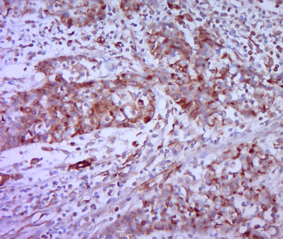
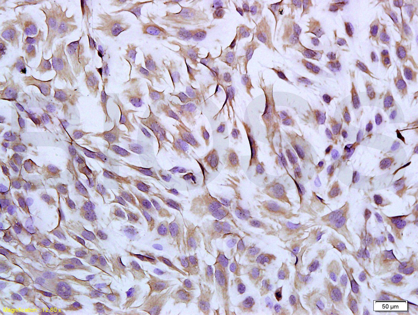
Paraformaldehyde-fixed, paraffin embedded (human cervical cancer); Antigen retrieval by boiling in sodium citrate buffer (pH6.0) for 15min; Block endogenous peroxidase by 3% hydrogen peroxide for 20 minutes; Blocking buffer (normal goat serum) at 37°C for 30min; Antibody incubation with (Vimentin) Polyclonal Antibody, Unconjugated (bs-0756R) at 1:400 overnight at 4°C, followed by a conjugated secondary (sp-0023) for 20 minutes and DAB staining.
-
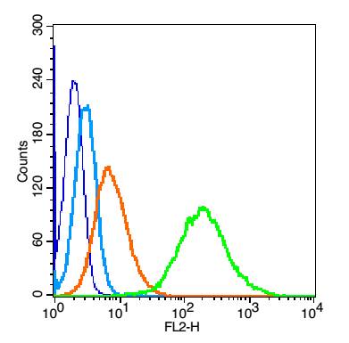
Blank control: Jurkat cells(blue).
Primary Antibody:Rabbit Anti-Vimentin antibody antibody(bs-0756R), Dilution: 1μg in 100 μL 1X PBS containing 0.5% BSA;
Isotype Control Antibody: Rabbit IgG(orange) ,used under the same conditions );
Secondary Antibody: Goat anti-rabbit IgG-PE(white blue), Dilution: 1:200 in 1 X PBS containing 0.5% BSA.
Protocol
The cells were fixed with 2% paraformaldehyde (10 min) , then permeabilized with 90% ice-cold methanol for 30 min on ice. Primary antibody (bs-0756R, 1μg /1x10^6 cells) were incubated for 30 min on the ice, followed by 1 X PBS containing 0.5% BSA + 1 0% goat serum (15 min) to block non-specific protein-protein interactions. Then the Goat Anti-rabbit IgG/PE antibody was added into the blocking buffer mentioned above to react with the primary antibody at 1/200 dilution for 30 min on ice. Acquisition of 20,000 events was performed.
-
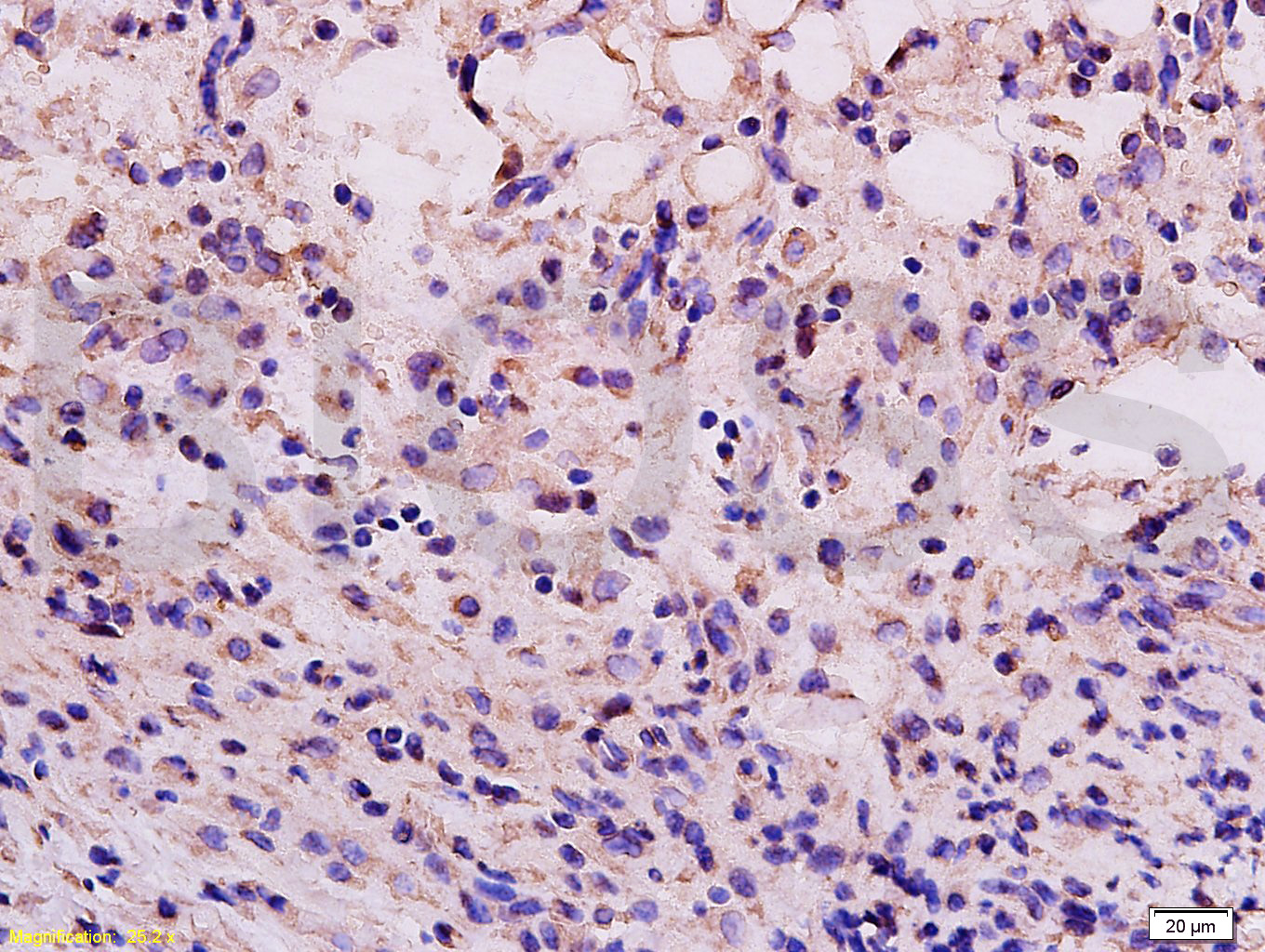
Tissue/cell: mouse kidney tissue; 4% Paraformaldehyde-fixed and paraffin-embedded;
Antigen retrieval: citrate buffer ( 0.01M, pH 6.0 ), Boiling bathing for 15min; Block endogenous peroxidase by 3% Hydrogen peroxide for 30min; Blocking buffer (normal goat serum,C-0005) at 37℃ for 20 min;
Incubation: Anti-Vimentin Polyclonal Antibody, Unconjugated(bs-0756R) 1:200, overnight at 4°C, followed by conjugation to the secondary antibody(SP-0023) and DAB(C-0010) staining
-
Paraformaldehyde-fixed, paraffin embedded (human cervical cancer); Antigen retrieval by boiling in sodium citrate buffer (pH6.0) for 15min; Block endogenous peroxidase by 3% hydrogen peroxide for 20 minutes; Blocking buffer (normal goat serum) at 37°C for 30min; Antibody incubation with (Vimentin) Polyclonal Antibody, Unconjugated (bs-0756R) at 1:400 overnight at 4°C, followed by a conjugated secondary (sp-0023) for 20 minutes and DAB staining.
-
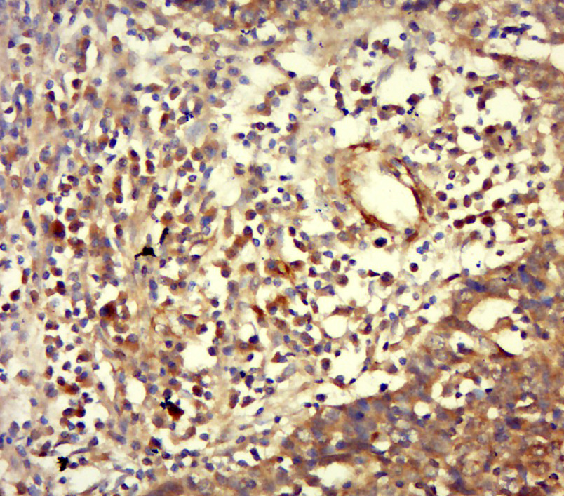
Tissue/cell: mouse mesenchymal stem cells;
Blocking buffer (normal goat serum,C-0005) at 37℃ for 20 min;
Incubation: Anti-Vimentin Polyclonal Antibody, Unconjugated(bs-0756R) 1:300, overnight at 4°C, followed by conjugation to the secondary antibody(SP-0023) and DAB(C-0010) staining
-
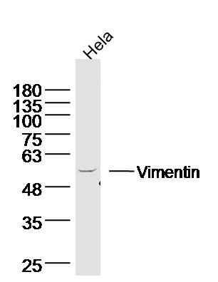
Sample: Hela (human)Cell Lysate at 40 ug
Primary: Anti- Vimentin (bs-0756R) at 1/300 dilution
Secondary: IRDye800CW Goat Anti-Rabbit IgG at 1/20000 dilution
Predicted band size: 53 kD
Observed band size: 53 kD
-
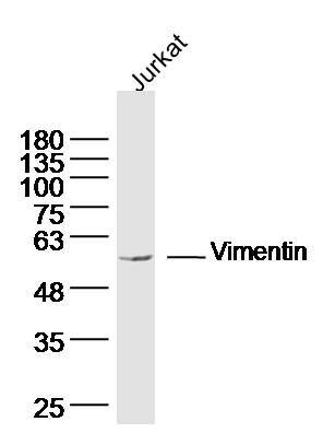
Sample: Jurkat (human)Cell Lysate at 40 ug
Primary: Anti- Vimentin (bs-0756R) at 1/300 dilution
Secondary: IRDye800CW Goat Anti-Rabbit IgG at 1/20000 dilution
Predicted band size: 53 kD
Observed band size: 53 kD
-
Tissue/cell: U-2OS cell; 4% Paraformaldehyde-fixed; Triton X-100 at room temperature for 20 min; Blocking buffer (normal goat serum, C-0005) at 37°C for 20 min; Antibody incubation with (Vimentin) Polyclonal Antibody, Unconjugated (bs-0756R) 1:50, 90 minutes at 37°C; followed by a conjugated Goat Anti-Rabbit IgG antibody (bs-0295G-FITC) at 37°C for 90 minutes, DAPI (5ug/ml, blue, C-0033) was used to stain the cell nuclei.
-
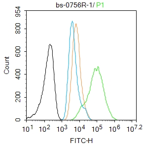
Blank control:A549.
Primary Antibody (green line): Rabbit Anti-Vimentin antibody (bs-0756R)
Dilution: 1μg /10^6 cells;
Isotype Control Antibody (orange line): Rabbit IgG .
Secondary Antibody : Goat anti-rabbit IgG-AF488
Dilution: 1μg /test.
Protocol
The cells were fixed with 4% PFA (10min at room temperature)and then permeabilized with 90% ice-cold methanol for 20 min at -20℃. The cells were then incubated in 5%BSA to block non-specific protein-protein interactions for 30 min at room temperature .Cells stained with Primary Antibody for 30 min at room temperature. The secondary antibody used for 40 min at room temperature. Acquisition of 20,000 events was performed.
-
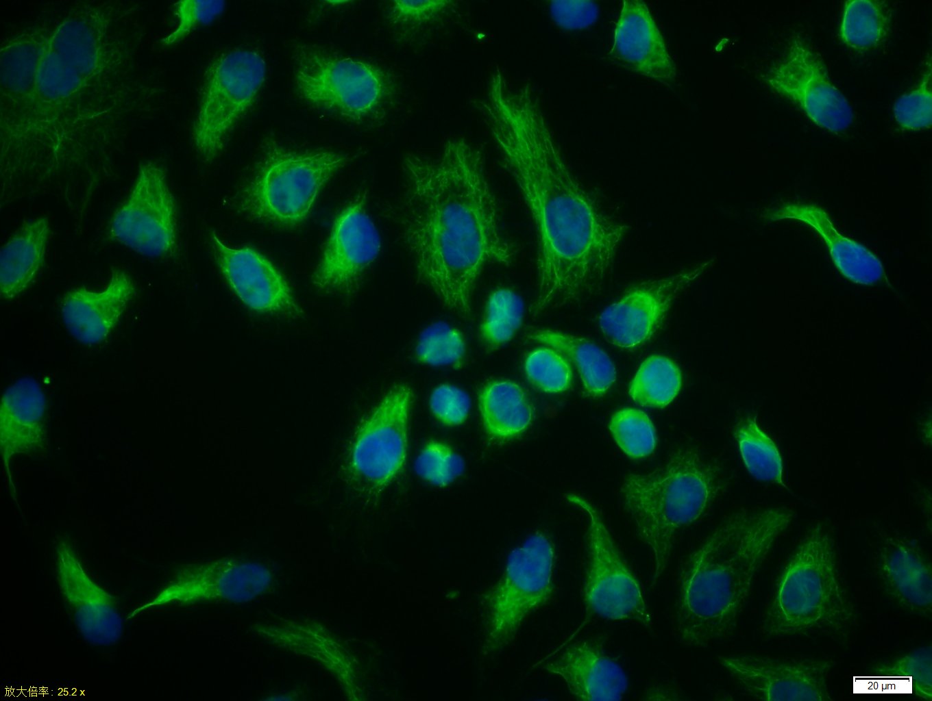
Tissue/cell: U251 cell; 4% Paraformaldehyde-fixed; Triton X-100 at room temperature for 20 min; Blocking buffer (normal goat serum, C-0005) at 37°C for 20 min; Antibody incubation with (Vimentin) Polyclonal Antibody, Unconjugated (bs-0756R) 1:50, 90 minutes at 37°C; followed by a conjugated Goat Anti-Rabbit IgG antibody (bs-0295G-FITC) at 37°C for 90 minutes, DAPI (blue, C02-04002) was used to stain the cell nuclei.
-
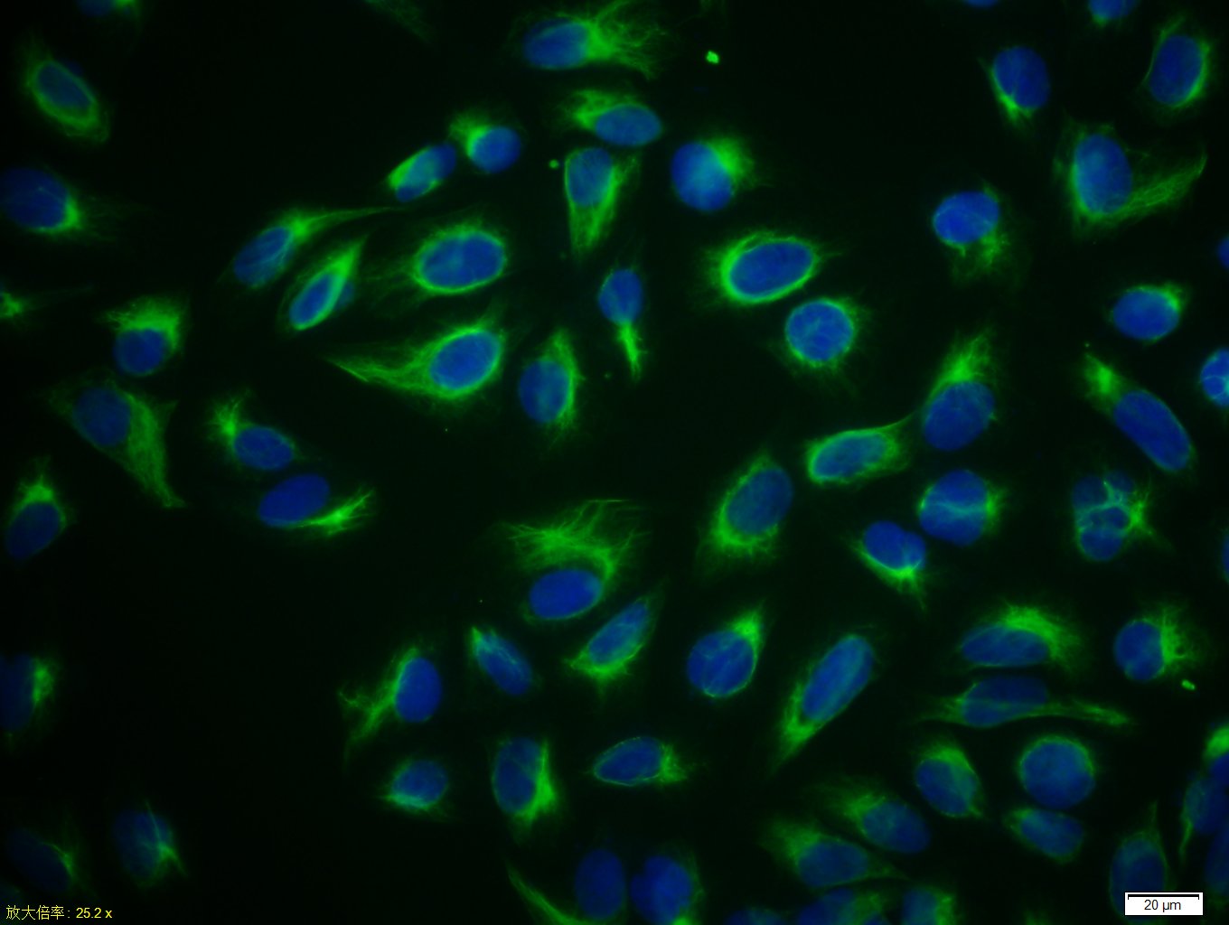
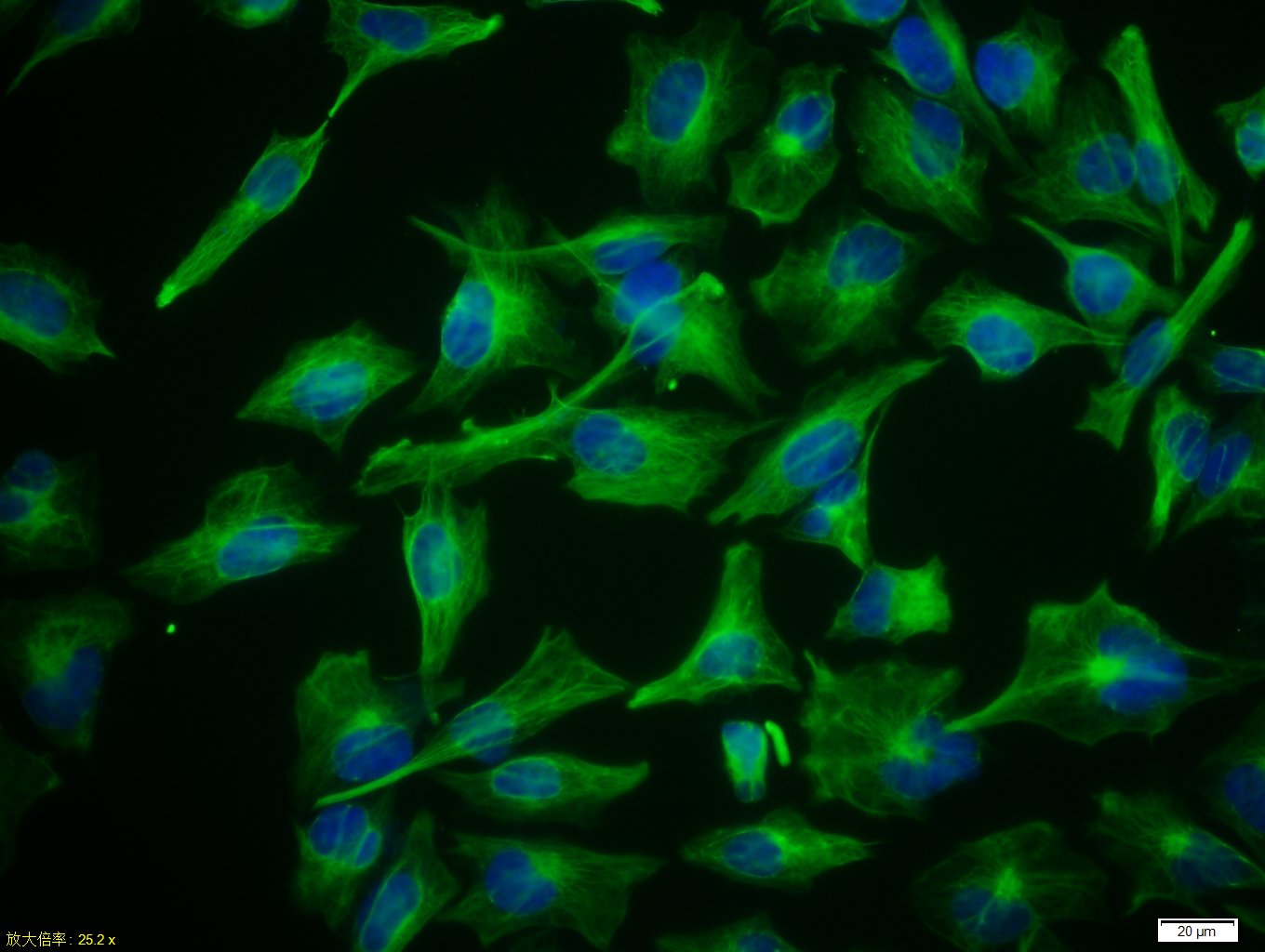
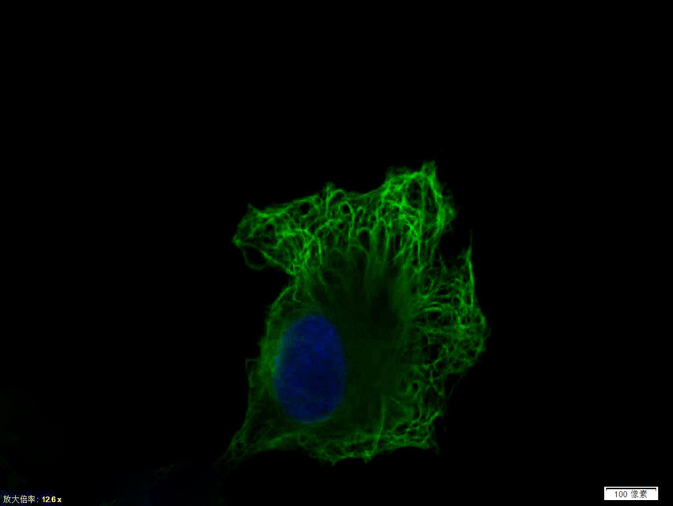
Tissue/cell: HeLa cell; 4% Paraformaldehyde-fixed; Triton X-100 at room temperature for 20 min; Blocking buffer (normal goat serum, C-0005) at 37°C for 20 min; Antibody incubation with (Vimentin) Polyclonal Antibody, Unconjugated (bs-0756R) 1:50, 90 minutes at 37°C; followed by a conjugated Goat Anti-Rabbit IgG antibody (bs-0295G-FITC) at 37°C for 90 minutes, DAPI (blue, C02-04002) was used to stain the cell nuclei.
-
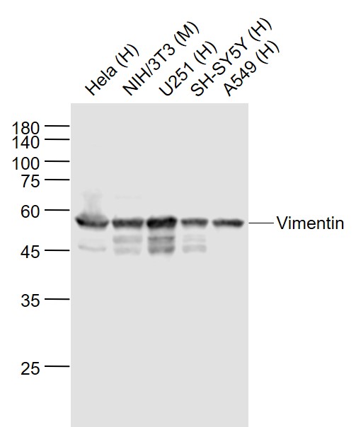
Sample:
Lane 1: Hela (Human) Cell Lysate at 30 ug
Lane 2: NIH/3T3 (Mouse) Cell Lysate at 30 ug
Lane 3: U251 (Human) Cell Lysate at 30 ug
Lane 4: SH-SY5Y (Human) Cell Lysate at 30 ug
Lane 5: A549 (Human) Cell Lysate at 30 ug
Primary: Anti-Vimentin (bs-0756R) at 1/1000 dilution
Secondary: IRDye800CW Goat Anti-Rabbit IgG at 1/20000 dilution
Predicted band size: 55 kD
Observed band size: 57 kD
-
Tissue/cell: U-87MG cell; 4% Paraformaldehyde-fixed; Triton X-100 at room temperature for 20 min; Blocking buffer (normal goat serum, C-0005) at 37°C for 20 min; Antibody incubation with (Vimentin) Polyclonal Antibody, Unconjugated (bs-0756R) 1:100, 90 minutes at 37°C; followed by a conjugated Goat Anti-Rabbit IgG antibody (bs-0295G-FITC) at 37°C for 90 minutes, DAPI (blue, C02-04002) was used to stain the cell nuclei.
-
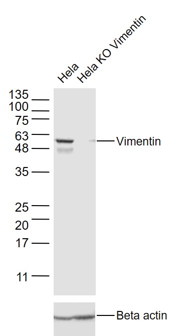
Sample:
Hela(Human) Cell Lysate at 30 ug
Hela KO Vimentin (Human) Cell Lysate at 30 ug
Primary: Anti- Vimentin (bs-0756R) at 1/1000 dilution
Secondary: IRDye800CW Goat Anti-Rabbit IgG at 1/20000 dilution
Predicted band size: 53 kD
Observed band size: 53 kD
-
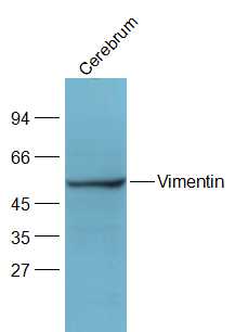
Sample:
Cerebrum (Mouse) Lysate at 40 ug
Primary: Anti-Vimentin (bs-0756R) at 1/2000 dilution
Secondary: IRDye800CW Goat Anti-Rabbit IgG at 1/20000 dilution
Predicted band size: 53 kD
Observed band size: 53 kD
RRID:AB_10855343
产品名称:Rabbit Anti-Vimentin antibody
别名: FLJ36605; OTTHUMP00000019224; VIM; VIME_HUMAN; Vimentin.
中文名称:波形蛋白抗体
英文名称:Rabbit Anti-Vimentin antibody
抗体来源: Rabbit
克隆类型:多克隆
细胞定位:细胞浆,细胞外基质
性 状:Liquid
亚 型:IgG
纯化方法:affinity purified by Protein A
保存条件:Shipped at 4℃. Store at -20 °C for one year. Avoid repeated freeze/thaw cycles.
免 疫 原:KLH conjugated synthetic peptide derived from human Vimentin
抗原表位:371-466/466
SWISS:P08670
Gene ID :7431
Human Gene ID:7431
This gene encodes a member of the intermediate filament family. Intermediate filamentents, along with microtubules and actin microfilaments, make up the cytoskeleton. The protein encoded by this gene is responsible for maintaining cell shape, integrity of the cytoplasm, and stabilizing cytoskeletal interactions. It is also involved in the immune response, and controls the transport of low-density lipoprotein (LDL)-derived cholesterol from a lysosome to the site of esterification. It functions as an organizer of a number of critical proteins involved in attachment, migration, and cell signaling. Mutations in this gene causes a dominant, pulverulent cataract.[provided by RefSeq, Jun 2009]
Function:Vimentins are class-III intermediate filaments found in various non-epithelial cells, especially mesenchymal cells. Vimentin is attached to the nucleus, endoplasmic reticulum, and mitochondria, either laterally or terminally.
Involved with LARP6 in the
Subunit:Homopolymer assembled from elementary dimers. Interacts with HCV core protein. Interacts with LGSN and SYNM. Interacts (via rod region) with PLEC (via CH 1 domain) (By similarity). Interacts with SLC6A4. Interacts with STK33. Interacts with LARP6. Interac
Subcellular Location:Cytoplasm.
Tissue Specificity:Highly expressed in fibroblasts, some expression in T- and B-lymphocytes, and little or no expression in Burkitt's lymphoma cell lines. Expressed in many hormone-independent mammary carcinoma cell lines.
Post-translational modifications:Filament disassembly during mitosis is promoted by phosphorylation at Ser-55 as well as by nestin (By similarity). One of the most prominent phosphoproteins in various cells of mesenchymal origin. Phosphorylation is enhanced during cell division, at which
Similarity:Belongs to the intermediate filament family.
Important Note:This product as supplied is intended for research use only, not for use in human, therapeutic or diagnostic applications.
 400-901-9800


 说明书
说明书 联系我们
联系我们 打印此页面
打印此页面 收藏
收藏