| Rabbit Anti-EGFR antibody |
| 反应物种(预测) |
Mouse,Dog,Pig |
| 产品应用(已验证) |
WB,IHC,IF,FCM |
| 产品应用(可尝试) |
ELISA |
| 推荐稀释比例 |
WB=1:500-2000,Elisa=1:5000-10000,IHC-P=1:100-500,IHC-F=1:100-500,Flow Cyt=1μg/Test,IF=1:100-500, |
| 研究领域 |
肿瘤,细胞生物,免疫学,生长因子和激素 |
| 标签 |
Array |
-
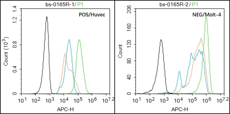
Black line : Positive blank control (HUVEC); Negative blank control (Molt-4)
Green line : Primary Antibody (Rabbit Anti-EGFR antibody (bs-0165R) )
Orange line:Isotype Control Antibody (Rabbit IgG) .
Blue line : Secondary Antibody (Goat anti-rabbit IgG-AF647)
HUVEC(Positive)and Molt-4(Negative control)cells (black) were fixed with 4% PFA for 10min at room temperature,permeabilized with 90% ice-cold methanol for 20 min at -20℃, and incubated in 5% BSA blocking buffer for 30 min at room temperature. Cells were then stained with EGFR Antibody(bs-0165R)at 1:50 dilution in blocking buffer and incubated for 30 min at room temperature, washed twice with 2% BSA in PBS, followed by secondary antibody(blue) incubation for 40 min at room temperature. Acquisitions of 20,000 events were performed. Cells stained with primary antibody (green), and isotype control (orange).
-
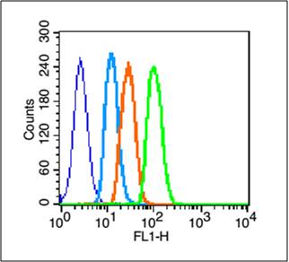
Blank control (blue line): A431 (blue).
Primary Antibody (green line): Rabbit Anti-EGFRantibody (bs-0165R)
Dilution: 3μg /10^6 cells;
Isotype Control Antibody (orange line): Rabbit IgG .
Secondary Antibody (white blue line): Goat anti-rabbit IgG-FITC
Dilution: 1μg /test.
Protocol
The cells were fixed with 2% paraformaldehyde (10 min) , then permeabilized with 90% ice-cold methanol for 30 min on ice. Cells stained with Primary Antibody for 30 min at room temperature. The cells were then incubated in 1 X PBS/2%BSA/10% goat serum to block non-specific protein-protein interactions followed by the antibody for 15 min at room temperature. The secondary antibody used for 40 min at room temperature. Acquisition of 20,000 events was performed.
-
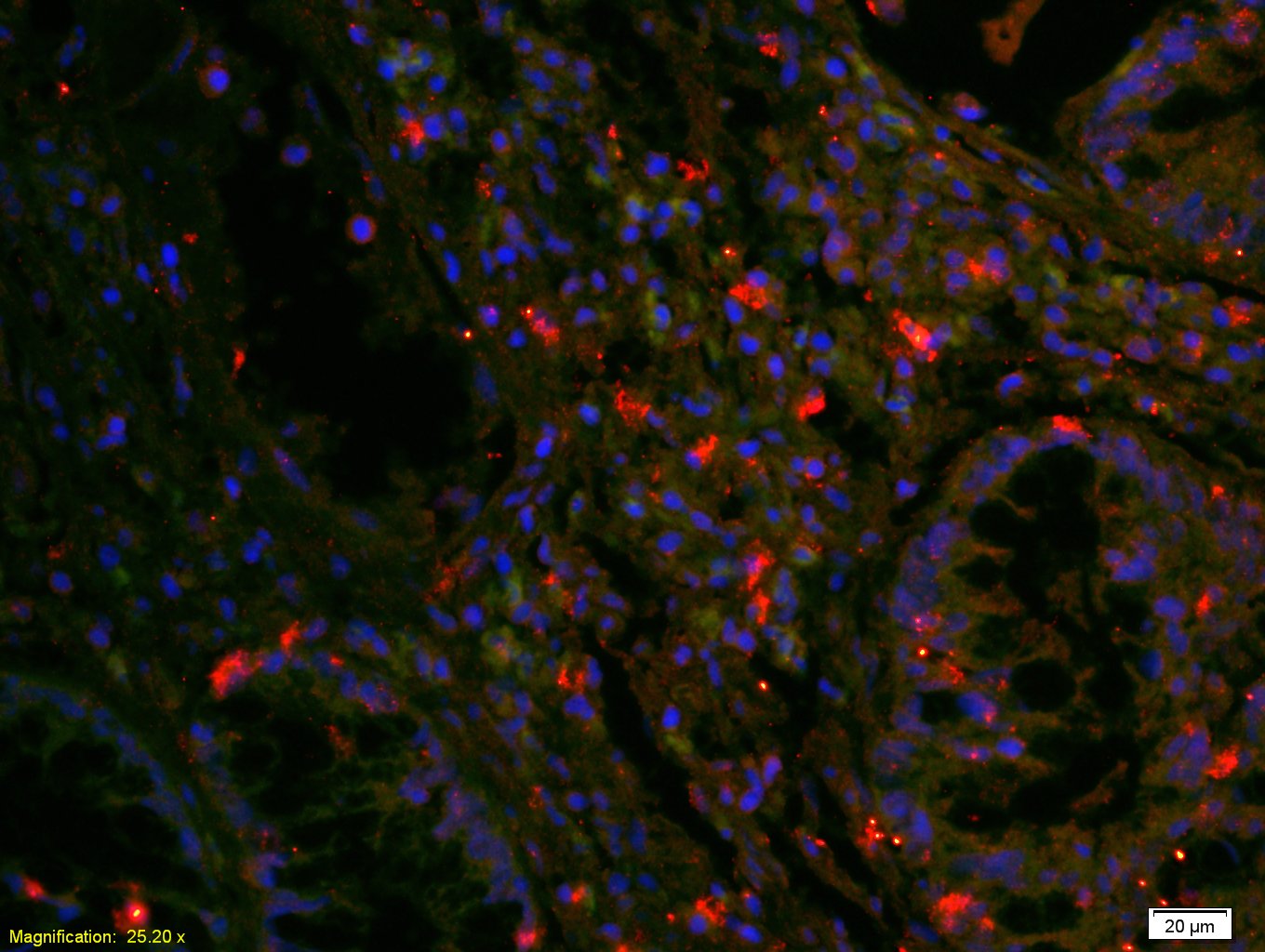
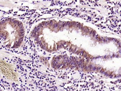
Tissue/cell: human rectal carcinoma;4% Paraformaldehyde-fixed and paraffin-embedded;
Antigen retrieval: citrate buffer ( 0.01M, pH 6.0 ), Boiling bathing for 15min; Blocking buffer (normal goat serum,C-0005) at 37℃ for 20 min;
Incubation: Anti-EGFR Polyclonal Antibody, Unconjugated(bs-0165R) 1:200, overnight at 4°C; The secondary antibody was Goat Anti-Rabbit IgG, Cy3 conjugated(bs-0295G-Cy3)used at 1:200 dilution for 40 minutes at 37°C. DAPI(5ug/ml,blue,C-0033) was used to stain the cell nuclei
-
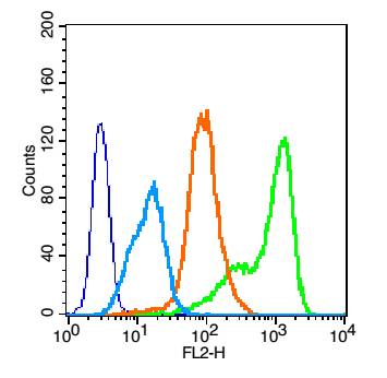
Blank control: HUVEC cells(blue).
Primary Antibody:Rabbit Anti-EGFR antibody(bs-0165R), Dilution: 1μg in 100 μL 1X PBS containing 0.5% BSA;
Isotype Control Antibody: Rabbit IgG(orange) ,used under the same conditions );
Secondary Antibody: Goat anti-rabbit IgG-PE(white blue), Dilution: 1:200 in 1 X PBS containing 0.5% BSA.
Protocol
The cells were fixed with 2% paraformaldehyde (10 min) , then permeabilized with 90% ice-cold methanol for 30 min on ice. Primary antibody (bs-0165R,1μg /1x10^6 cells) were incubated for 30 min on the ice, followed by 1 X PBS containing 0.5% BSA + 10% goat serum (15 min) to block non-specific protein-protein interactions. Then the Goat Anti-rabbit IgG/PE antibody was added into the blocking buffer mentioned above to react with the primary antibody at 1/200 dilution for 30 min on ice. Acquisition of 20,000 events was performed.
-
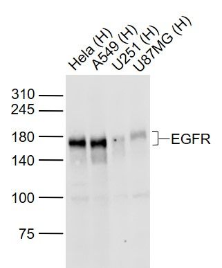
Sample:
Lane 1: Hela (Human) Cell Lysate at 30 ug
Lane 2: A549 (Human) Cell Lysate at 30 ug
Lane 3: U251 (Human) Cell Lysate at 30 ug
Lane 4: U87MG (Human) Cell Lysate at 30 ug
Primary: Anti-EGFR (bs-34018R) at 1/1000 dilution
Secondary: IRDye800CW Goat Anti-Rabbit IgG at 1/20000 dilution
Predicted band size: 170 kD
Observed band size: 170 kD
-
Paraformaldehyde-fixed, paraffin embedded (human gastric carcinoma); Antigen retrieval by boiling in sodium citrate buffer (pH6.0) for 15min; Block endogenous peroxidase by 3% hydrogen peroxide for 20 minutes; Blocking buffer (normal goat serum) at 37°C for 30min; Antibody incubation with (EGFR) Polyclonal Antibody, Unconjugated (bs-0165R) at 1:200 overnight at 4°C, followed by operating according to SP Kit(Rabbit) (sp-0023) instructionsand DAB staining.
-
Paraformaldehyde-fixed, paraffin embedded (rat placenta); Antigen retrieval by boiling in sodium citrate buffer (pH6.0) for 15min; Block endogenous peroxidase by 3% hydrogen peroxide for 20 minutes; Blocking buffer (normal goat serum) at 37°C for 30min; Antibody incubation with (EGFR) Polyclonal Antibody, Unconjugated (bs-0165R) at 1:200 overnight at 4°C, followed by operating according to SP Kit(Rabbit) (sp-0023) instructionsand DAB staining.
RRID:AB_10855278
产品名称:Rabbit Anti-EGFR antibody
别名: EGFR; Avian erythroblastic leukemia viral (v erb b) oncogene homolog; Avian erythroblastic leukemia viral (verbb) oncogene homolog; Cell growth inhibiting protein 40; Cell proliferation inducing protein 61; EGF R; EGFR; Epidermal growth factor receptor (a
中文名称:表皮生长因子受体抗体
英文名称:Rabbit Anti-EGFR antibody
抗体来源: Rabbit
克隆类型:多克隆
细胞定位:细胞核,细胞浆,细胞膜,分泌型蛋白
性 状:Liquid
亚 型:IgG
纯化方法:affinity purified by Protein A
保存条件:Shipped at 4℃. Store at -20 °C for one year. Avoid repeated freeze/thaw cycles.
免 疫 原:KLH conjugated synthetic peptide derived from human EGFR
抗原表位:951-1050/1210
抗原细胞定位:Cytoplasmic
SWISS:P00533
Gene ID :1956
Human Gene ID:1956
The protein encoded by this gene is a transmembrane glycoprotein that is a member of the protein kinase superfamily. This protein is a receptor for members of the epidermal growth factor family. EGFR is a cell surface protein that binds to epidermal growth factor. Binding of the protein to a ligand induces receptor dimerization and tyrosine autophosphorylation and leads to cell proliferation. Mutations in this gene are associated with lung cancer. Multiple alternatively spliced transcript variants that encode different protein isoforms have been found for this gene. [provided by RefSeq, Jul 2010]
Function:Receptor tyrosine kinase binding ligands of the EGF family and activating several signaling cascades to convert extracellular cues into appropriate cellular responses. Known ligands include EGF, TGFA/TGF-alpha, amphiregulin, epigen/EPGN, BTC/betacellulin,
Subunit:Binding of the ligand triggers homo- and/or heterodimerization of the receptor triggering its autophosphorylation. Heterodimer with ERBB2. Interacts with ERRFI1; inhibits dimerization of the kinase domain and autophosphorylation. Part of a complex with ER
Subcellular Location:Cell membrane; Single-pass type I membrane protein. Endoplasmic reticulum membrane; Single-pass type I membrane protein. Golgi apparatus membrane; Single-pass type I membrane protein. Nucleus membrane; Single-pass type I membrane protein. Endosome. Endoso
Tissue Specificity:Ubiquitously expressed. Isoform 2 is also expressed in ovarian cancers.
Post-translational modifications:Phosphorylation at Ser-695 is partial and occurs only if Thr-693 is phosphorylated. Phosphorylation at Thr-678 and Thr-693 by PRKD1 inhibits EGF-induced MAPK8/JNK1 activation. Dephosphorylation by PTPRJ prevents endocytosis and stabilizes the receptor at
DISEASE:Defects in EGFR are associated with lung cancer (LNCR) [MIM:211980]. LNCR is a common malignancy affecting tissues of the lung. The most common form of lung cancer is non-small cell lung cancer (NSCLC) that can be divided into 3 major histologic subtypes:
Similarity:Belongs to the protein kinase superfamily. Tyr protein kinase family. EGF receptor subfamily.
Contains 1 protein kinase domain.
Important Note:This product as supplied is intended for research use only, not for use in human, therapeutic or diagnostic applications.
 400-901-9800


 说明书
说明书 联系我们
联系我们 打印此页面
打印此页面 收藏
收藏