| Rabbit Anti-Caspase-9 antibody |
| 反应物种(预测) |
Mouse,Cow |
| 产品应用(已验证) |
WB,IHC,ICC,FCM |
| 产品应用(可尝试) |
IF,ELISA |
| 推荐稀释比例 |
WB=1:500-2000,Elisa=1:5000-10000,IHC-P=1:100-500,IHC-F=1:100-500,Flow Cyt=1μg/test,IF=1:100-500,ICC=1:100, |
| 研究领域 |
肿瘤,细胞生物,神经生物学,信号转导,细胞凋亡 |
| 标签 |
Array |
-
Tissue/cell: rat brain tissue; 4% Paraformaldehyde-fixed and paraffin-embedded;
Antigen retrieval: citrate buffer ( 0.01M, pH 6.0 ), Boiling bathing for 15min; Block endogenous peroxidase by 3% Hydrogen peroxide for 30min; Blocking buffer (normal goat serum,C-0005) at 37℃ for 20 min;
Incubation: Anti-Caspase-9 Polyclonal Antibody, Unconjugated(bs-0049R) 1:200, overnight at 4°C, followed by conjugation to the secondary antibody(SP-0023) and DAB(C-0010) staining
-
Tissue/cell: human colon carcinoma; 4% Paraformaldehyde-fixed and paraffin-embedded;
Antigen retrieval: citrate buffer ( 0.01M, pH 6.0 ), Boiling bathing for 15min; Block endogenous peroxidase by 3% Hydrogen peroxide for 30min; Blocking buffer (normal goat serum,C-0005) at 37℃ for 20 min;
Incubation: Anti-Caspase-9 Polyclonal Antibody, Unconjugated(bs-0049R) 1:200, overnight at 4°C, followed by conjugation to the secondary antibody(SP-0023) and DAB(C-0010) staining
-
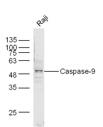
Sample: Raji Cell lysate at 30ug;
Primary: Anti-Caspase-9 (bs-0049R) at 1:300 dilution;
Secondary: IRDye800CW Goat Anti-Rabbit IgG at 1/20000 dilution
Predicted band size: 35/50 kD
Observed band size: 50 kD
-
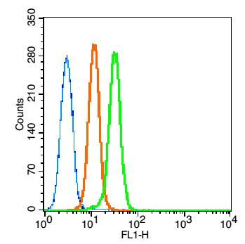
Blank control: RSC96(blue), the cells were fixed with 2% paraformaldehyde (10 min) and then permeabilized with ice-cold 90% methanol for 30 min on ice.
Isotype Control Antibody: Rabbit IgG(orange) ;
Secondary Antibody: Goat anti-rabbit IgG-PE(white blue),
Dilution: 1:200 in 1 X PBS containing 0.5% BSA ;
Primary Antibody Dilution: 1μg in 100 μL1X PBS containing 0.5% BSA(green).
-
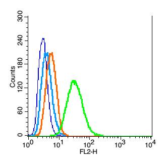
Blank control: K562 (blue).
Primary Antibody:Rabbit Anti-caspase-9 antibody (bs-0049R,Green); Dilution: 1μg in 100 μL 1X PBS containing 0.5% BSA;
Isotype Control Antibody: Rabbit IgG(orange) ,used under the same conditions;
Secondary Antibody: Goat anti-rabbit IgG-FITC(white blue), Dilution: 1:200 in 1 X PBS containing 0.5% BSA.
Protocol
The cells were fixed with 80% methanol (5 min) and and then permeabilized with 0.01M PBS-Tween for 20 min . Primary antibody (bs-0049R, 1μg /1x10^6 cells) were incubated for 30 min at room temperature, followed by 1 X PBS containing 0.5% BSA + 10% goat serum (30min) to block non-specific protein-protein interactions. Then the Goat Anti-rabbit IgG/FITC antibody was added into the blocking buffer mentioned above to react with the primary antibody at 1/200 dilution for 30 min at room temperature. Acquisition of 20,000 events was performed.
-
Paraformaldehyde-fixed, paraffin embedded (rat lung); Antigen retrieval by boiling in sodium citrate buffer (pH6.0) for 15min; Block endogenous peroxidase by 3% hydrogen peroxide for 20 minutes; Blocking buffer (normal goat serum) at 37°C for 30min; Antibody incubation with (Insulin like growth factor 1 ) Polyclonal Antibody, Unconjugated (bs-0014R) at 1:400 overnight at 4°C, followed by a conjugated secondary antibody (sp-0023) for 20 minutes and DAB staining.
-
HepG2 cell; 4% Paraformaldehyde-fixed; Triton X-100 at room temperature for 20 min; Blocking buffer (normal goat serum, C-0005) at 37°C for 20 min; Antibody incubation with (Caspase-9) polyclonal Antibody, Unconjugated (bs-0049R) 1:100, 90 minutes at 37°C; followed by a conjugated Goat Anti-Rabbit IgG antibody at 37°C for 90 minutes, DAPI (blue, C02-04002) was used to stain the cell nuclei.
-
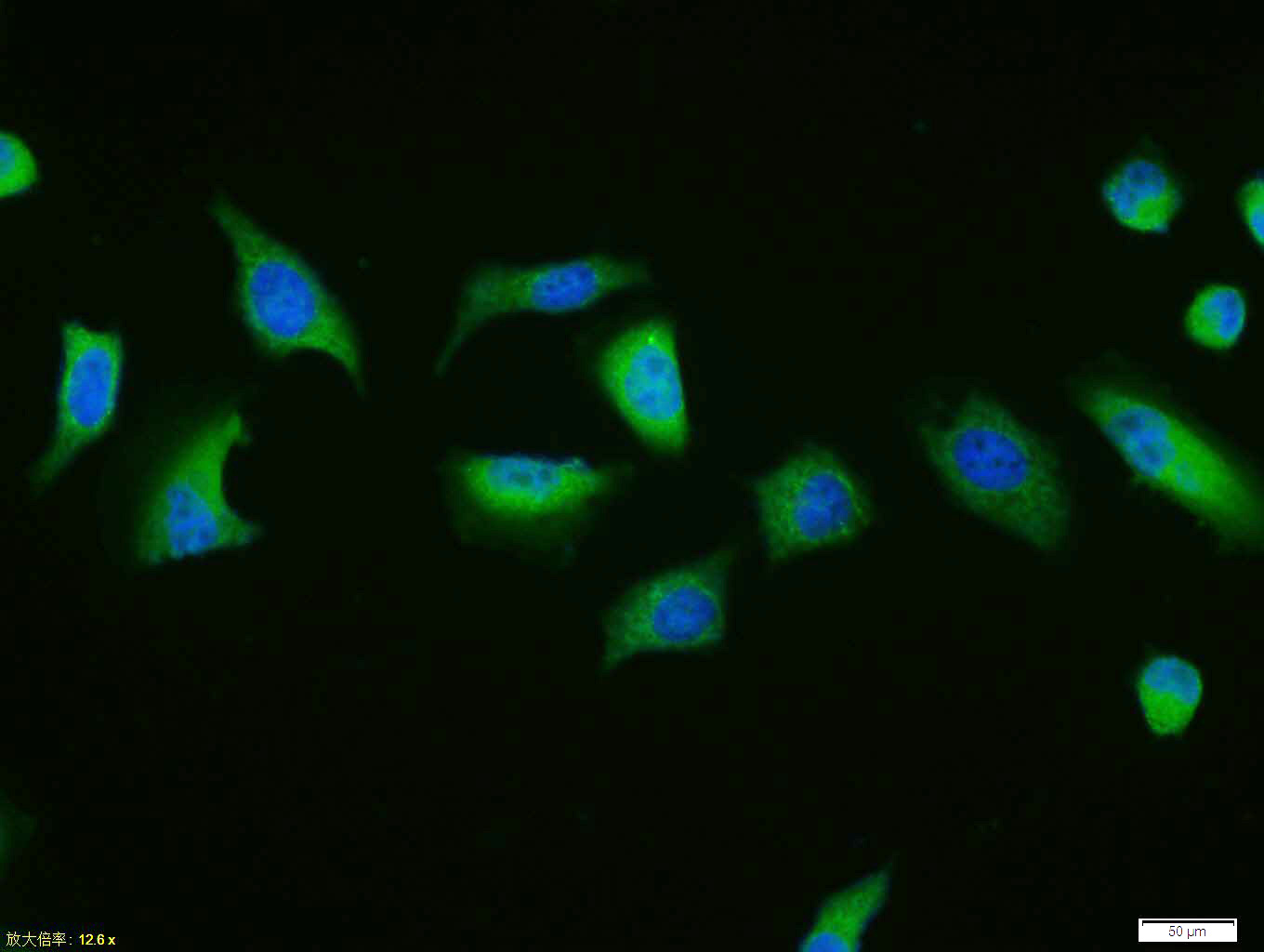
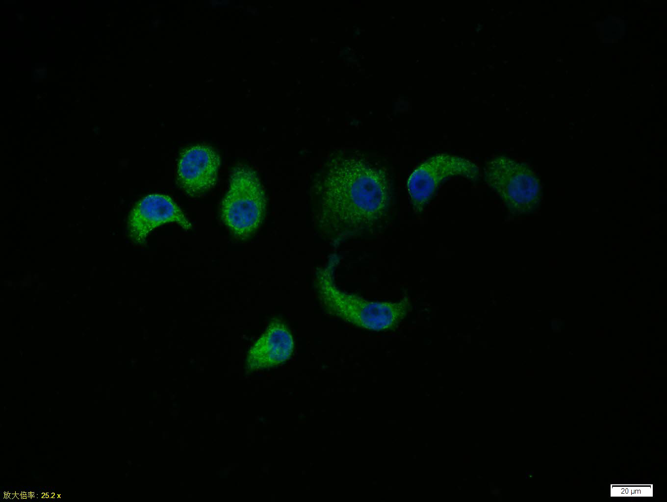
Tissue/cell: Hela cell; 4% Paraformaldehyde-fixed; Triton X-100 at room temperature for 20 min; Blocking buffer (normal goat serum, C-0005) at 37°C for 20 min; Antibody incubation with (Caspase-9) polyclonal Antibody, Unconjugated (bs-0049R) 1:100, 90 minutes at 37°C; followed by a FITC conjugated Goat Anti-Rabbit IgG antibody at 37°C for 90 minutes, DAPI (blue, C02-04002) was used to stain the cell nuclei.
-
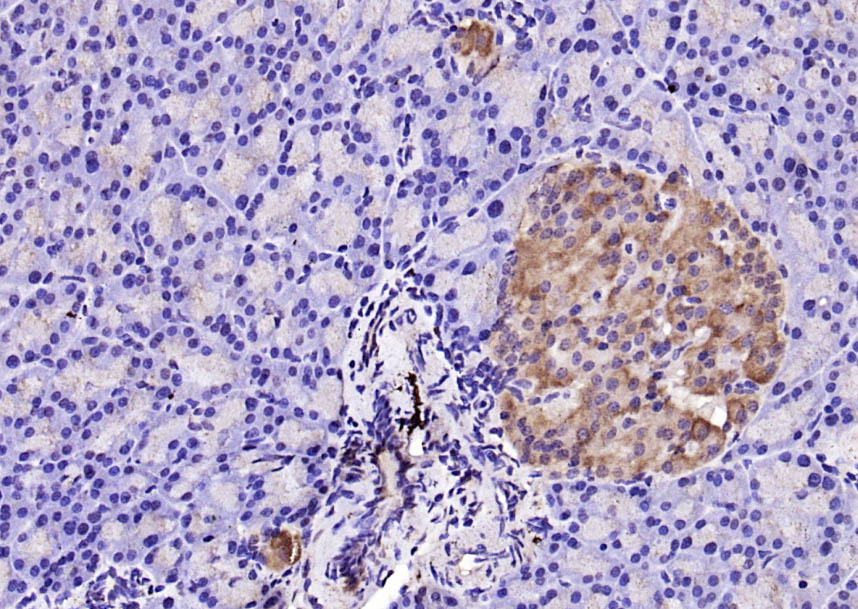
Paraformaldehyde-fixed, paraffin embedded (rat pancreas); Antigen retrieval by boiling in sodium citrate buffer (pH6.0) for 15min; Block endogenous peroxidase by 3% hydrogen peroxide for 20 minutes; Blocking buffer (normal goat serum) at 37°C for 30min; Antibody incubation with (Caspase-9) Polyclonal Antibody, Unconjugated (bs-0049R) at 1:200 overnight at 4°C, followed by operating according to SP Kit(Rabbit) (sp-0023) instructionsand DAB staining.
-
Tissue/cell: HepG2 cell; 4% Paraformaldehyde-fixed; Triton X-100 at room temperature for 20 min; Blocking buffer (normal goat serum, C-0005) at 37°C for 20 min; Antibody incubation with (Caspase-9) polyclonal Antibody, Unconjugated (bs-0049R) 1:100, 90 minutes at 37°C; followed by a FITC conjugated Goat Anti-Rabbit IgG antibody at 37°C for 90 minutes, DAPI (blue, C02-04002) was used to stain the cell nuclei.
-
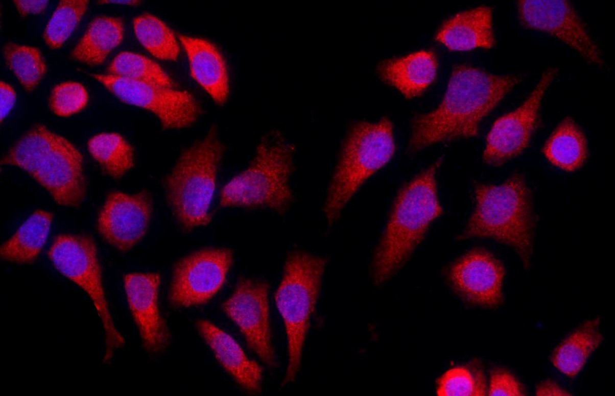
Tissue/cell: MCF-7 cell; 4% Paraformaldehyde-fixed; Triton X-100 at room temperature for 20 min; Blocking buffer (normal goat serum, C-0005) at 37°C for 20 min; Antibody incubation with (Caspase-9) Polyclonal Antibody, Unconjugated (bs-0049R) 1:50, 90 minutes at 37°C; followed by a conjugated Goat Anti-Rabbit IgG antibody (bs-0295G-Cy3) at 37°C for 90 minutes, DAPI (blue, C02-04002) was used to stain the cell nuclei.
-
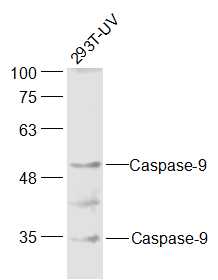
Sample:
293T-UV Cell (Human) Lysate at 30 ug
Primary: Anti-Caspase-9 (Bs- 0049R) at 1/300 dilution
Secondary: IRDye800CW Goat Anti-Rabbit IgG at 1/20000 dilution
Predicted band size: 35/50 kD
Observed band size: 35/50 kD
RRID:AB_10855484
产品名称:Rabbit Anti-Caspase-9 antibody
别名: Caspase-9 subunit p35; Apaf-3; APAF 3; APAF3; Apoptosis related cysteine peptidase; Apoptotic protease activating factor 3; Apoptotic protease MCH 6; Apoptotic protease MCH6; CASP 9; CASP9; Caspase 9; Caspase 9 apoptosis related cysteine protease; Caspase
中文名称:活化半胱胺酸蛋白酶蛋白-9抗体
英文名称:Rabbit Anti-Caspase-9 antibody
抗体来源: Rabbit
克隆类型:多克隆
细胞定位:细胞浆
性 状:Liquid
亚 型:IgG
纯化方法:affinity purified by Protein A
保存条件:Shipped at 4℃. Store at -20 °C for one year. Avoid repeated freeze/thaw cycles.
免 疫 原:KLH conjugated synthetic peptide derived from human Caspase-9 subunit p35
抗原表位:11-120/416
SWISS:P55211
Gene ID :842
Human Gene ID:842
Caspase 9 (also known as ICE like apoptotic protease 6 (ICE LAP6), apoptotic protease Mch6, and apoptotic protease activating factor 3 (Apaf3)) is a member of the peptidase family C14 that contains a CARD domain. This caspase is active as a heterotetramer and has been reported to have two isoforms. ProCaspase 9 has been reported to be approximately 47 kD. This caspase is present in the cytosol and, upon activation, translocates to the mitochondria. Caspase 9 is involved in the caspase activation cascade responsible for apoptosis execution and cleaves/activates Caspase 3 and Caspase 6. Caspase 9 is inhibited by the dominant negative isoform, BclXL, cIAP1, cIAP2, XIAP, and Livin. This caspase becomes activated when recruited to Apaf1/cytochrome c complex, and following cleavage by Apaf1, granzyme B, Caspase 3, possibly Caspase 8 and Caspase 10 into large p37 and small p10 subunits. Caspase 9 intereacts with BIRC7 and has been shown to cleave PARP and vimentin.
Function:Involved in the activation cascade of caspases responsible for apoptosis execution. Binding of caspase-9 to Apaf-1 leads to activation of the protease which then cleaves and activates caspase-3. Proteolytically cleaves poly(ADP-ribose) polymerase (PARP).<
Subunit:Heterotetramer that consists of two anti-parallel arranged heterodimers, each one formed by a 35 kDa (p35) and a 10 kDa (p10) subunit. Caspase-9 and APAF1 bind to each other via their respective NH2-terminal CED-3 homologous domains in the presence of cyt
Tissue Specificity:Ubiquitous, with highest expression in the heart, moderate expression in liver, skeletal muscle, and pancreas. Low levels in all other tissues. Within the heart, specifically expressed in myocytes.
Post-translational modifications:Cleavages at Asp-315 by granzyme B and at Asp-330 by caspase-3 generate the two active subunits. Caspase-8 and -10 can also be involved in these processing events.
Phosphorylated at Thr-125 by MAPK1/ERK2. Phosphorylation at Thr-125 is sufficient to blo
Similarity:Belongs to the peptidase C14A family.
Contains 1 CARD domain.
Important Note:This product as supplied is intended for research use only, not for use in human, therapeutic or diagnostic applications.
 400-901-9800


 说明书
说明书 联系我们
联系我们 打印此页面
打印此页面 收藏
收藏