| Rabbit Anti-IL-1 Beta antibody |
| 反应物种(预测) |
Dog,Horse,Rabbit |
| 产品应用(已验证) |
WB,IHC,FCM |
| 产品应用(可尝试) |
IF,ELISA |
| 推荐稀释比例 |
WB=1:500-2000,Elisa=1:5000-10000,IHC-P=1:100-500,IHC-F=1:100-500,Flow Cyt=2ug/Test,IF=1:100-500, |
| 研究领域 |
细胞生物,免疫学,生长因子和激素,糖蛋白, |
| 标签 |
Array |
-
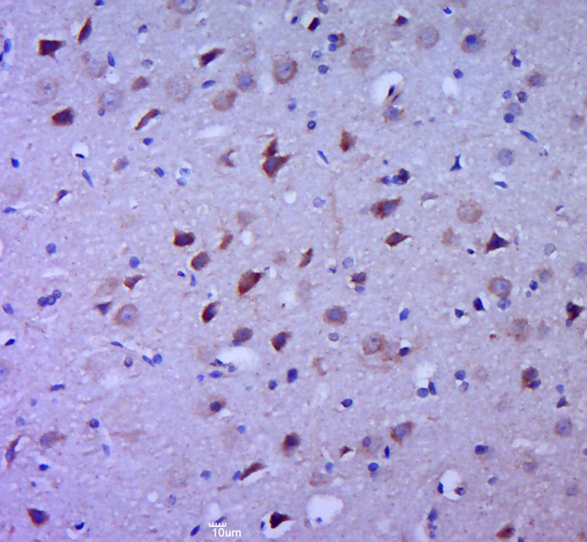
Paraformaldehyde-fixed, paraffin embedded (rat brain tissue); Antigen retrieval by boiling in sodium citrate buffer (pH6.0) for 15min; Block endogenous peroxidase by 3% hydrogen peroxide for 20 minutes; Blocking buffer (normal goat serum) at 37°C for 30min; Antibody incubation with (IL-1 Beta) Polyclonal Antibody, Unconjugated (bs-6319R) at 1:400 overnight at 4°C, followed by a conjugated secondary (sp-0023) for 20 minutes and DAB staining.
-
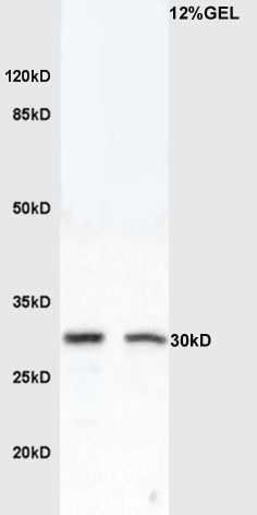
Sample:
Brain (Mouse) Lysate at 40 ug
Intestine (Mouse) Lysate at 40 ug
Primary: Anti-IL-1 Beta (bs-6319R) at 1/300 dilution
Secondary: HRP conjugated Goat-Anti-rabbit IgG (bs-0295G-HRP) at 1/5000 dilution
Predicted band size: 17/30 kD
Observed band size: 30 kD
-
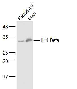
Sample:
Raw264.7(Mouse) Cell Lysate at 30 ug
Liver (Mouse) Lysate at 40 ug
Primary: Anti-IL-1 Beta (bs-6319R) at 1/500 dilution
Secondary: IRDye800CW Goat Anti-Rabbit IgG at 1/20000 dilution
Predicted band size: 17/30 kD
Observed band size: 30 kD
-
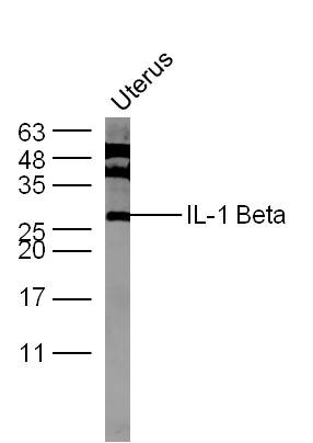
Sample:
uterus (Mouse) Lysate at 40 ug
Primary: Anti-IL-1 Beta (Bs- 6319R) at 1/300 dilution
Secondary: IRDye800CW Goat Anti-Rabbit IgG at 1/20000 dilution
Predicted band size: 17/30 kD
Observed band size: 27 kD
-
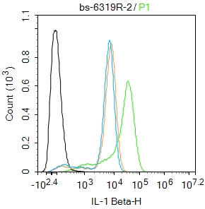
Blank control: THP-1.
Primary Antibody (green line): Rabbit Anti-IL-1 Beta antibody (bs-6319R)
Dilution: 2μg /10^6 cells;
Isotype Control Antibody (orange line): Rabbit IgG .
Secondary Antibody : Goat anti-rabbit IgG-FITC
Dilution: 0.5μg /test.
Protocol
The cells were fixed with 4% PFA (10min at room temperature)and then permeabilized with 0.1% PBST for 20 min at room temperature. The cells were then incubated in 5%BSA to block non-specific protein-protein interactions for 30 min at room temperature .Cells stained with Primary Antibody for 30 min at room temperature. The secondary antibody used for 40 min at room temperature. Acquisition of 20,000 events was performed.
-
Sample:
Raw264.7(Mouse) Cell Lysate at 30 ug
Primary: Anti- IL-1 Beta (bs-6319R) at 1/1000 dilution
Secondary: IRDye800CW Goat Anti-Rabbit IgG at 1/20000 dilution
Predicted band size: 17/30 kD
Observed band size: 30 kD
RRID:RRID
产品名称:Rabbit Anti-IL-1 Beta antibody
别名: Catabolin; H1; IL-1β; Hematopoietin 1; IFN beta inducing factor; IL 1; IL 1 beta; IL 1B; IL1B; IL1F2; Interleukin 1 beta; Interleukin 1 beta precursor; LAF; OAF; Osteoclast activating factor; Preinterleukin beta; Pro interleukin 1 beta; IL1B_HUMAN; Interl
中文名称:白介素1β抗体
英文名称:Rabbit Anti-IL-1 Beta antibody
抗体来源: Rabbit
克隆类型:多克隆
细胞定位:分泌型蛋白
性 状:Liquid
亚 型:IgG
纯化方法:affinity purified by Protein A
保存条件:Shipped at 4℃. Store at -20 °C for one year. Avoid repeated freeze/thaw cycles.
免 疫 原:KLH conjugated synthetic peptide derived from human IL-1 Beta
抗原表位:161-269/269
SWISS:P01584
Gene ID :3553
Human Gene ID:3553
The protein encoded by this gene is a member of the interleukin 1 cytokine family. This cytokine is produced by activated macrophages as a proprotein, which is proteolytically processed to its active form by caspase 1 (CASP1/ICE). This cytokine is an important mediator of the inflammatory response, and is involved in a variety of cellular activities, including cell proliferation, differentiation, and apoptosis. The induction of cyclooxygenase-2 (PTGS2/COX2) by this cytokine in the central nervous system (CNS) is found to contribute to inflammatory pain hypersensitivity. This gene and eight other interleukin 1 family genes form a cytokine gene cluster on chromosome 2. [provided by RefSeq, Jul 2008].
Function:Produced by activated macrophages, IL-1 stimulates thymocyte proliferation by inducing IL-2 release, B-cell maturation and proliferation, and fibroblast growth factor activity. IL-1 proteins are involved in the inflammatory response, being identified as e
Subunit:Monomer.
Subcellular Location:Secreted. Note=The lack of a specific hydrophobic segment in the precursor sequence suggests that IL-1 is released by damaged cells or is secreted by a mechanism differing from that used for other secretory proteins.
Similarity:Belongs to the IL-1 family.
Important Note:This product as supplied is intended for research use only, not for use in human, therapeutic or diagnostic applications.
 400-901-9800


 说明书
说明书 联系我们
联系我们 打印此页面
打印此页面 收藏
收藏