| Rabbit Anti-ZO-1 antibody |
| 反应物种(预测) |
Mouse,Rat,Chicken,Dog,Cow,Rabbit,GuineaPig |
| 产品应用(已验证) |
WB,IHC,FCM |
| 产品应用(可尝试) |
IF,ELISA |
| 推荐稀释比例 |
WB=1:500-2000,Elisa=1:5000-10000,IHC-P=1:100-500,IHC-F=1:100-500,Flow Cyt=1μg/Test,IF=1:100-500, |
| 研究领域 |
细胞生物,免疫学,信号转导,细胞粘附分子,细胞表面分子, |
| 标签 |
Array |
-
Black line : Positive blank control (293T); Negative blank control (HL60)
Green line : Primary Antibody (Rabbit Anti-ZO-1 antibody (bs-11320R) )
Orange line:Isotype Control Antibody (Rabbit IgG) .
Blue line : Secondary Antibody (Goat anti-rabbit IgG-AF647)
293T(Positive)and HL60(Negative control)cells (black) were fixed with 4% PFA for 10min at room temperature, permeabilized with PBST for 20 min at room temperature, and incubated in 5% BSA blocking buffer for 30 min at room temperature. Cells were then stained with ZO-1 Antibody(bs-1329R)at 1:50 dilution in blocking buffer and incubated for 30 min at room temperature, washed twice with 2% BSA in PBS, followed by secondary antibody(blue) incubation for 40 min at room temperature. Acquisitions of 20,000 events were performed. Cells stained with primary antibody (green), and isotype control (orange).
-
Independently Validated Antibody, image provided by Science Direct, badge number 029577:Formalin-fixed and paraffin embedded human testis labeled with Anti-ZO-1 Polyclonal Antibody, Unconjugated (bs-1329R) at 1:250 followed by conjugation to the secondary antibody.
-
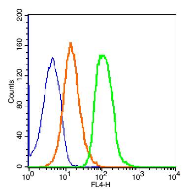
Blank control: 293T Cells(blue).
Primary Antibody: Rabbit Anti-ZO-1/AF647 Conjugated antibody (bs-1329R-AF647), Dilution: 1μg in 100 μL 1X PBS containing 0.5% BSA;
Isotype Control Antibody: Rabbit IgG/AF647(orange) ,used under the same conditions.
Protocol
The cells were washed twice with phosphate-buffered saline (PBS). The cells were incubated in 1 X PBS containing 0.5% BSA + 1 0% goat serum (15 min) to block non-specific protein-protein interactions followed by the antibody (bs-1329R-AF647, 1μg /1x10^6 cells) for 30 min on ice. Acquisition of 20,000 events was performed.
-
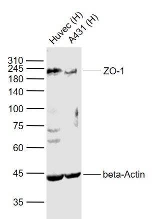
Sample:
Lane 1: Huvec (Human) Cell Lysate at 30 ug
Lane 2: A431 (Human) Cell Lysate at 30 ug
Primary:
Anti-ZO-1 (bs-1329R) at 1/1000 dilution
Anti-beta-Actin (bs-0061R) at 1/2000 dilution
Secondary: IRDye800CW Goat Anti-Rabbit IgG at 1/20000 dilution
Predicted band size: 220 kD
Observed band size: 220 kD
RRID:AB_10855948
产品名称:Rabbit Anti-ZO-1 antibody
别名: ZO1 tight junction protein;
Tight junction protein 1;
Tight junction protein ZO-1;
Tight junction protein ZO1;
TJP1;
zo-1;
Zo1;
ZO1_HUMAN;
Zona occludens 1;
Zona occludens 1 protein;
Zona occludens protein 1;
Zonula occludens 1 protein;
Zonula
中文名称:胞质紧密粘连蛋白1/闭锁小带蛋白1抗体
英文名称:Rabbit Anti-ZO-1 antibody
抗体来源: Rabbit
克隆类型:多克隆
细胞定位:细胞浆,细胞膜
性 状:Liquid
亚 型:IgG
纯化方法:affinity purified by Protein A
保存条件:Shipped at 4℃. Store at -20 °C for one year. Avoid repeated freeze/thaw cycles.
免 疫 原:KLH conjugated synthetic peptide derived from human ZO-1
抗原表位:1551-1702/1733
SWISS:Q07157
Gene ID :7082
Human Gene ID:7082
This gene encodes a protein located on a cytoplasmic membrane surface of intercellular tight junctions. The encoded protein may be involved in signal transduction at cell-cell junctions. Two transcript variants encoding distinct isoforms have been identified for this gene. The N-terminal may be involved in transducing a signal required for tight junction assembly, while the C-terminal may have specific properties of tight junctions. The alpha domain might be involved in stabilizing junctions.
Function:The N-terminal may be involved in transducing a signal required for tight junction assembly, while the C-terminal may have specific properties of tight junctions. The alpha domain might be involved in stabilizing junctions. Plays a role in the regulation
Subunit:Interacts with BVES (via the C-terminus cytoplasmic tail). Interacts with HSPA4 and KIRREL1. Homodimer, and heterodimer with TJP2/ZO-2 and TJP3/ZO-3. Interacts with OCLN, CALM, claudins, CGN/cingulin, CXADR, GJA12, GJD3 and UBN1. Interacts (via ZU5 domain
Subcellular Location:Cell membrane; Peripheral membrane protein; Cytoplasmic side. Cell junction, tight junction. Cell junction. Cell junction, gap junction. Note=Moves from the cytoplasm to the cell membrane concurrently with cell-cell contact. Detected at the leading edge o
Tissue Specificity:The alpha-containing isoform is found in most epithelial cell junctions. The short isoform is found both in endothelial cells and the highly specialized epithelial junctions of renal glomeruli and Sertoli cells of the seminiferous tubules.
Post-translational modifications:Phosphorylated. Dephosphorylated by PTPRJ.
Similarity:Belongs to the MAGUK family.
Contains 1 guanylate kinase-like domain.
Contains 3 PDZ (DHR) domains.
Contains 1 SH3 domain.
Contains 1 ZU5 domain.
Important Note:This product as supplied is intended for research use only, not for use in human, therapeutic or diagnostic applications.
 400-901-9800


 说明书
说明书 联系我们
联系我们 打印此页面
打印此页面 收藏
收藏