| Rabbit Anti-phospho-STAT3 (Tyr705) antibody |
| 反应物种(预测) |
Dog,Pig,Cow,Rabbit,Sheep,GuineaPig |
| 产品应用(已验证) |
WB,IHC,FCM |
| 产品应用(可尝试) |
IF,ELISA |
| 推荐稀释比例 |
WB=1:500-2000,Elisa=1:5000-10000,IHC-P=1:100-500,IHC-F=1:100-500,Flow Cyt=1μg /test,IF=1:100-500, |
| 研究领域 |
肿瘤,心血管,细胞生物,免疫学,信号转导,干细胞,细胞凋亡,转录调节因子,表观遗传学, |
| 标签 |
Array |
-
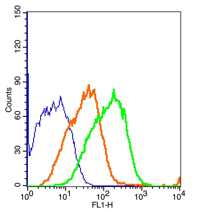
The blue histogram is unstained cells (mouse kidney).
The Orange histogram is cells stained with Rabbit IgG/FITC (bs-0295P-FITC)isotype control antibody.
The green histogram is cells stained with Rabbit Anti-phospho-STAT3 (Tyr705)/FITC Conjugated antibody (bs-1658R-FITC).
Concentration: 2μg/10^6 cells or 5μg/10^6 cells.
-
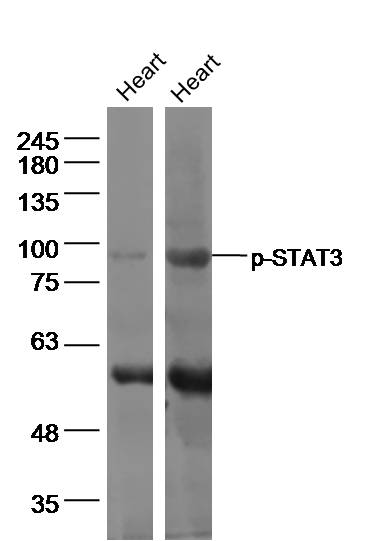
Sample:
Heart (Mouse) Lysate at 40 ug
Heart (Rat) Lysate at 40 ug
Primary: Anti-p-STAT3 (bs-1658R) at 1/500 dilution
Secondary: IRDye800CW Goat Anti-Rabbit IgG at 1/20000 dilution
Predicted band size: 85 kD
Observed band size: 85 kD
-
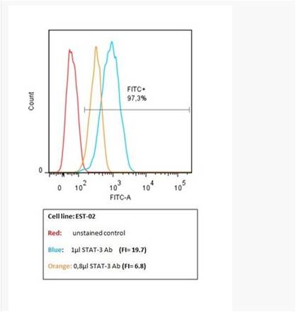
This image has been kindly submitted by Tübingen Ageing and Tumour Immunology Group (TATI), Centre for Medical Research (ZMF), University of Tübingen Medical School www.tati-group.de via our distributor Biozol.
Human Melanoma cell line, EST02, was stained with Rabbit Anti-phospho-STAT3 (Tyr705) Polyclonal Antibody, FITC Conjugated (bs-1658R-FITC)for 30 minutes, on ice.
-
Paraformaldehyde-fixed, paraffin embedded (Transplantation tumor); Antigen retrieval by boiling in sodium citrate buffer (pH6.0) for 15min; Block endogenous peroxidase by 3% hydrogen peroxide for 20 minutes; Blocking buffer (normal goat serum) at 37°C for 30min; Antibody incubation with (Phosphorylated signal Transductor and Activator of Transcription 3; p-STAT3) Polyclonal Antibody, Unconjugated (bs-1658R) at 1:400 overnight at 4°C, followed by a conjugated secondary antibody (sp-0023) for 20 minutes and DAB staining.
-
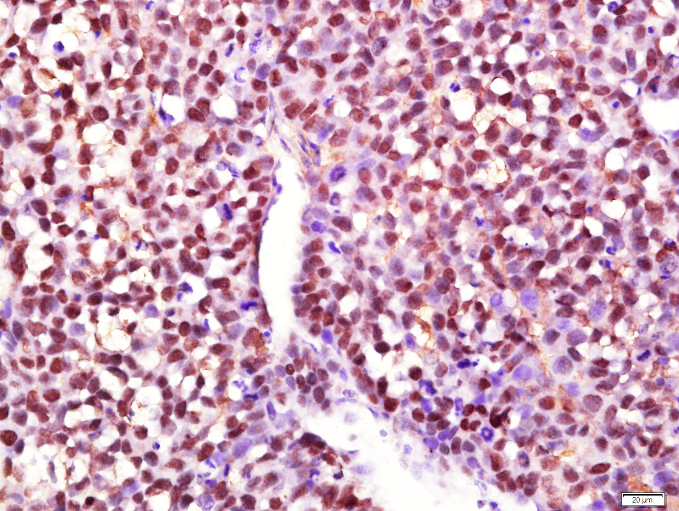
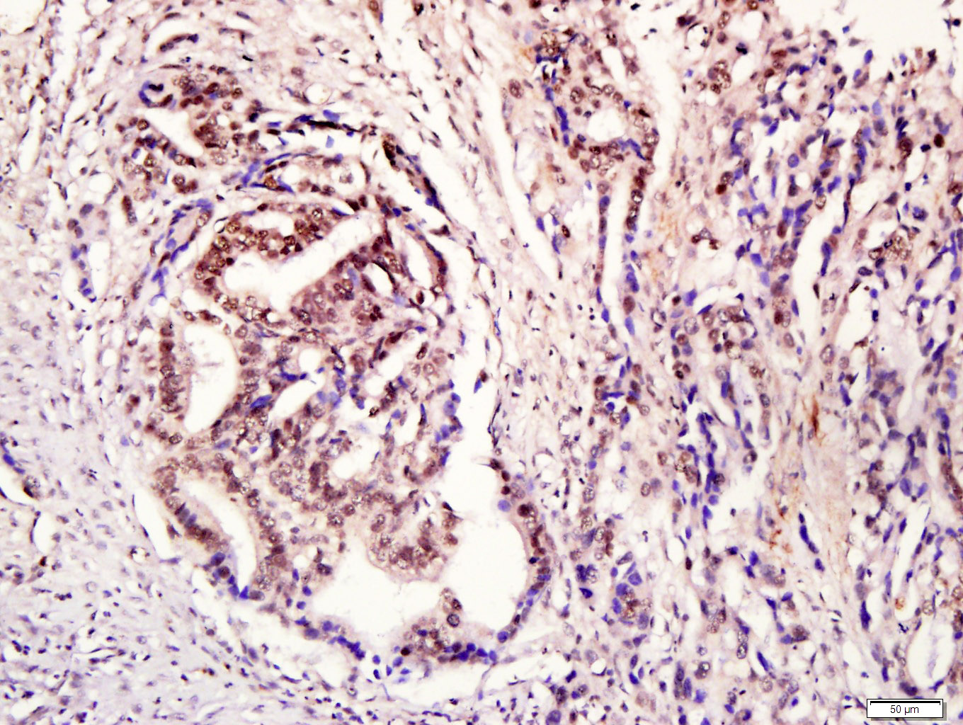
Tissue/cell: human gastric cancer; 4% Paraformaldehyde-fixed and paraffin-embedded;
Antigen retrieval: citrate buffer ( 0.01M, pH 6.0 ), Boiling bathing for 15min; Block endogenous peroxidase by 3% Hydrogen peroxide for 30min; Blocking buffer (normal goat serum,C-0005) at 37℃ for 20 min;
Incubation: Anti- phospho-STAT3 Polyclonal Antibody, Unconjugated(bs-1658R) 1:200, overnight at 4°C, followed by conjugation to the secondary antibody(SP-0023) and DAB(C-0010) staining
-
Blank control: A431.
Primary Antibody (green line): Rabbit Anti-phospho-STAT3 (Tyr705) antibody (bs-1658R)
Dilution: 1μg /10^6 cells;
Isotype Control Antibody (orange line): Rabbit IgG .
Secondary Antibody : Goat anti-rabbit IgG-AF647
Dilution: 1μg /test.
Protocol
The cells were fixed with 4% PFA (10min at room temperature)and then permeabilized with 90% ice-cold methanol for 20 min at-20℃. The cells were then incubated in 5%BSA to block non-specific protein-protein interactions for 30 min at room temperature .Cells stained with Primary Antibody for 30 min at room temperature. The secondary antibody used for 40 min at room temperature. Acquisition of 20,000 events was performed.
-
Paraformaldehyde-fixed, paraffin embedded (mouse heart); Antigen retrieval by boiling in sodium citrate buffer (pH6.0) for 15min; Block endogenous peroxidase by 3% hydrogen peroxide for 20 minutes; Blocking buffer (normal goat serum) at 37°C for 30min; Antibody incubation with (phospho-STAT3 (Tyr705)) Polyclonal Antibody, Unconjugated (bs-1658R) at 1:200 overnight at 4°C, followed by operating according to SP Kit(Rabbit) (sp-0023) instructionsand DAB staining.
-
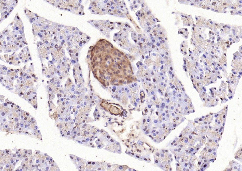
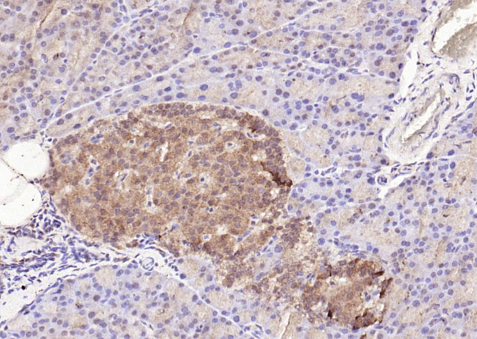
Paraformaldehyde-fixed, paraffin embedded (mouse pancreas); Antigen retrieval by boiling in sodium citrate buffer (pH6.0) for 15min; Block endogenous peroxidase by 3% hydrogen peroxide for 20 minutes; Blocking buffer (normal goat serum) at 37°C for 30min; Antibody incubation with (phospho-STAT3 (Tyr705)) Polyclonal Antibody, Unconjugated (bs-1658R) at 1:200 overnight at 4°C, followed by operating according to SP Kit(Rabbit) (sp-0023) instructionsand DAB staining.
-
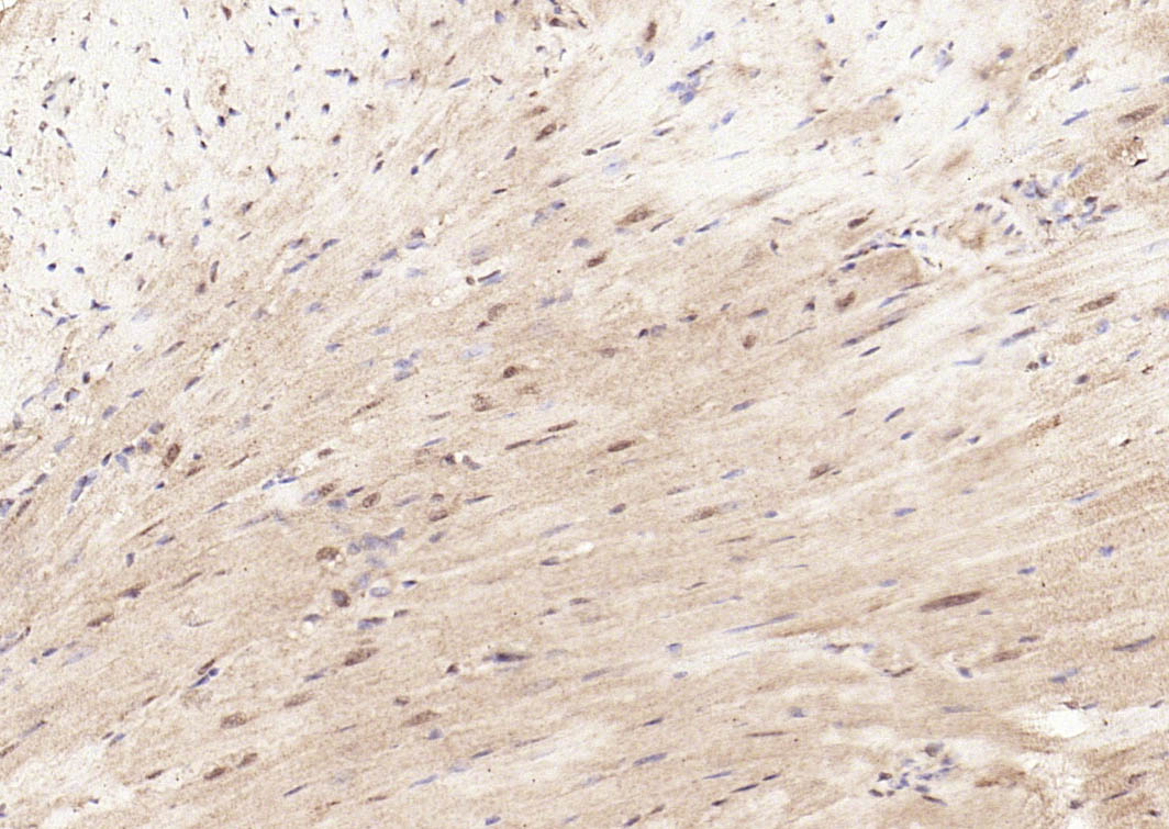
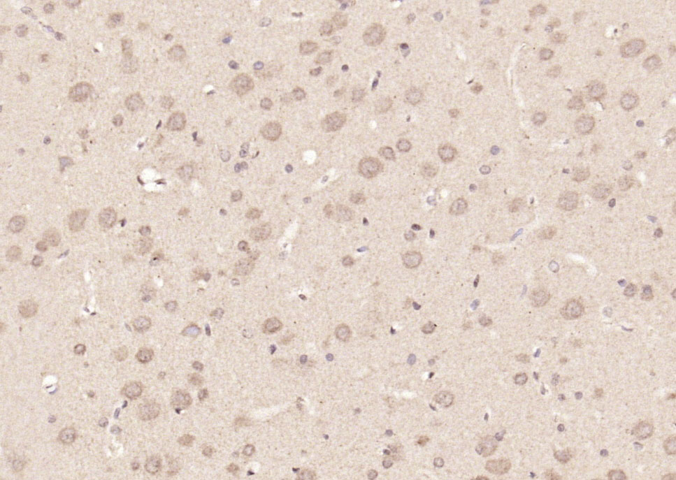
Paraformaldehyde-fixed, paraffin embedded (rat brain); Antigen retrieval by boiling in sodium citrate buffer (pH6.0) for 15min; Block endogenous peroxidase by 3% hydrogen peroxide for 20 minutes; Blocking buffer (normal goat serum) at 37°C for 30min; Antibody incubation with (phospho-STAT3 (Tyr705)) Polyclonal Antibody, Unconjugated (bs-1658R) at 1:200 overnight at 4°C, followed by operating according to SP Kit(Rabbit) (sp-0023) instructionsand DAB staining.
-
Paraformaldehyde-fixed, paraffin embedded (rat pancreas); Antigen retrieval by boiling in sodium citrate buffer (pH6.0) for 15min; Block endogenous peroxidase by 3% hydrogen peroxide for 20 minutes; Blocking buffer (normal goat serum) at 37°C for 30min; Antibody incubation with (phospho-STAT3 (Tyr705)) Polyclonal Antibody, Unconjugated (bs-1658R) at 1:200 overnight at 4°C, followed by operating according to SP Kit(Rabbit) (sp-0023) instructionsand DAB staining.
-
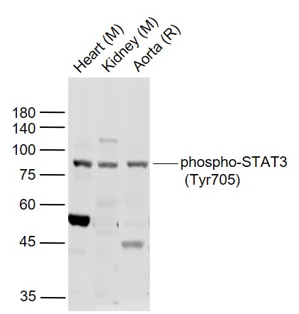
Sample:
Lane 1: Heart (Mouse) Lysate at 40 ug
Lane 2: Kidney (Mouse) Lysate at 40 ug
Lane 3: Aorta (Rat) Lysate at 40 ug
Primary:
Anti-phospho-STAT3 (Tyr705) (bs-1658R) at 1/1000 dilution
Secondary: IRDye800CW Goat Anti-Rabbit IgG at 1/20000 dilution
Predicted band size: 92/84 kD
Observed band size: 84 kD
-
Sample:
Kidney (Mouse) Lysate at 40 ug
Primary: Anti-phospho-STAT3 (Tyr705) (bs-1658R) at 1/1000 dilution
Secondary: IRDye800CW Goat Anti-Rabbit IgG at 1/20000 dilution
Predicted band size: 85 kD
Observed band size: 85 kD
RRID:AB_10855117
产品名称:Rabbit Anti-phospho-STAT3 (Tyr705) antibody
别名: STAT3 (phospho Y705); p-STAT3 (phospho Y705); Phospho-Stat3(pTyr705); STAT3(Phospho-Tyr705); p-STAT3(Tyr705); Phosphorylated Stat3(pTyr705); p-Stat3; Acute Phase Response Factor; APRF; DNA binding protein APRF; FLJ20882; MGC16063; Signal Transductor and
中文名称:磷酸化信号转导和转录激活因子3抗体
英文名称:Rabbit Anti-phospho-STAT3 (Tyr705) antibody
抗体来源: Rabbit
克隆类型:多克隆
细胞定位:细胞核,细胞浆
性 状:Liquid
亚 型:IgG
纯化方法:affinity purified by Protein A
保存条件:Shipped at 4℃. Store at -20 °C for one year. Avoid repeated freeze/thaw cycles.
免 疫 原:KLH conjugated Synthesised phosphopeptide derived from human STAT3 around the phosphorylation site of Tyr705
抗原表位:AP(p-Y)LK
SWISS:P40763
Gene ID :6774
Human Gene ID:6774
The protein encoded by this gene is a member of the STAT protein family. In response to cytokines and growth factors, STAT family members are phosphorylated by the receptor associated kinases, and then form homo- or heterodimers that translocate to the cell nucleus where they act as transcription activators. This protein is activated through phosphorylation in response to various cytokines and growth factors including IFNs, EGF, IL5, IL6, HGF, LIF and BMP2. This protein mediates the expression of a variety of genes in response to cell stimuli, and thus plays a key role in many cellular processes such as cell growth and apoptosis. The small GTPase Rac1 has been shown to bind and regulate the activity of this protein. PIAS3 protein is a specific inhibitor of this protein. Mutations in this gene are associated with infantile-onset multisystem autoimmune disease and hyper-immunoglobulin E syndrome. Alternative splicing results in multiple transcript variants encoding distinct isoforms. [provided by RefSeq, Sep 2015]
Function:Transcription factor that binds to the interleukin-6 (IL-6)-responsive elements identified in the promoters of various acute-phase protein genes. Activated by IL31 through IL31RA.
Subcellular Location:Cytoplasm. Nucleus. Shuttles between the nucleus and the cytoplasm. Constitutive nuclear presence is independent of tyrosine phosphorylation. Predominantly present in the cytoplasm without stimuli. Upon leukemia inhibitory factor (LIF) stimulation, accumu
Tissue Specificity:Heart, brain, placenta, lung, liver, skeletal muscle, kidney and pancreas.
Post-translational modifications:Tyrosine phosphorylated upon stimulation with EGF (By similarity). Tyrosine phosphorylated in response to IL-6, IL-11, CNTF, LIF, CSF-1, EGF, PDGF, IFN-alpha and OSM. Phosphorylated on serine upon DNA damage, probably by ATM or ATR. Serine phosphorylation
DISEASE:Defects in STAT3 are the cause of hyperimmunoglobulin E recurrent infection syndrome autosomal dominant (AD-HIES) [MIM:147060]; also known as hyper-IgE syndrome or Job syndrome. AD-HIES is a rare disorder of immunity and connective tissue characterized by
Important Note:This product as supplied is intended for research use only, not for use in human, therapeutic or diagnostic applications.
 400-901-9800


 说明书
说明书 联系我们
联系我们 打印此页面
打印此页面 收藏
收藏