| Rabbit Anti-MDR1/P Glycoprotein antibody |
| 产品应用(已验证) |
WB,IHC,ICC,FCM |
| 产品应用(可尝试) |
IF,ELISA |
| 推荐稀释比例 |
WB=1:500-2000,Elisa=1:5000-10000,IHC-P=1:100-500,IHC-F=1:100-500,Flow Cyt=1µg/Test,IF=1:100-500,ICC=1:100, |
| 研究领域 |
肿瘤,细胞生物,免疫学,信号转导,细胞凋亡,转录调节因子,糖蛋白, |
| 标签 |
Array |
-
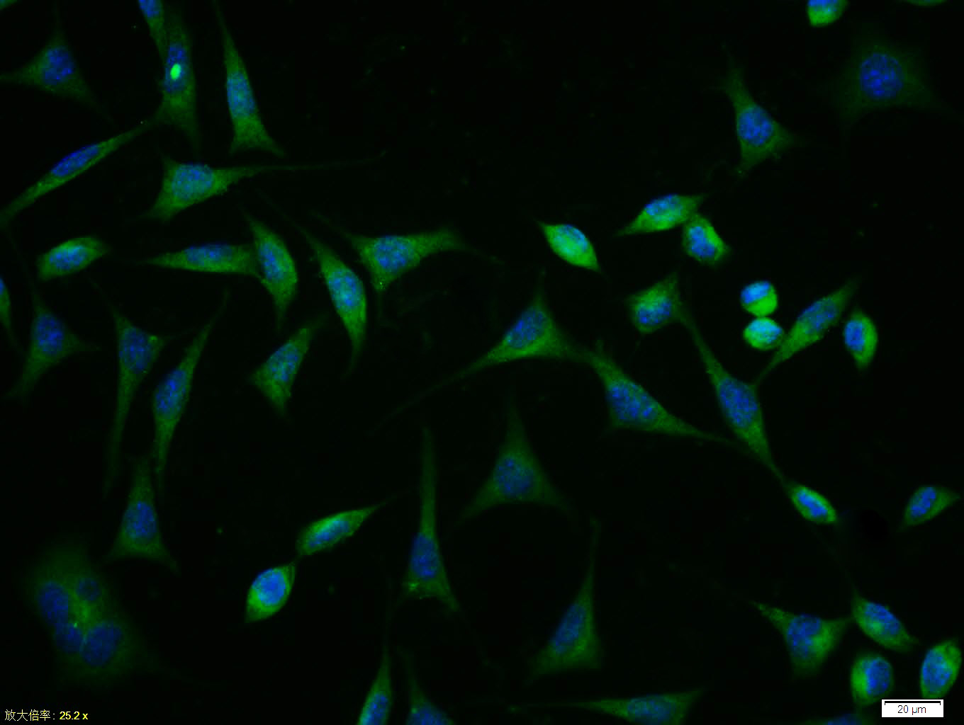
Tissue/cell:SH-SY5Y cell; 4% Paraformaldehyde-fixed; Triton X-100 at room temperature for 20 min; Blocking buffer (normal goat serum,C-0005) at 37°C for 20 min; Antibody incubation with (MDR1/P Glycoprotein) polyclonal Antibody, Unconjugated (bs-1468R) 1:100, 90 minutes at 37°C; followed by a FITC conjugated Goat Anti-Rabbit IgG antibody at 37°C for 90 minutes, DAPI (blue, C02-04002) was used to stain the cell nuclei.
-
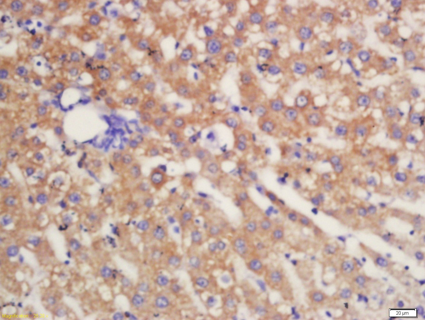
Tissue/cell: Rat liver tissue; 4% Paraformaldehyde-fixed and paraffin-embedded;
Antigen retrieval: citrate buffer ( 0.01M, pH 6.0 ), Boiling bathing for 15min; Block endogenous peroxidase by 3% Hydrogen peroxide for 30min; Blocking buffer (normal goat serum,C-0005) at 37℃ for 20 min;
Incubation: Anti-MDR1 / P Glycoprotein Polyclonal Antibody, Unconjugated(bs-1468R) 1:200, overnight at 4°C, followed by conjugation to the secondary antibody(SP-0023) and DAB(C-0010) staining
-
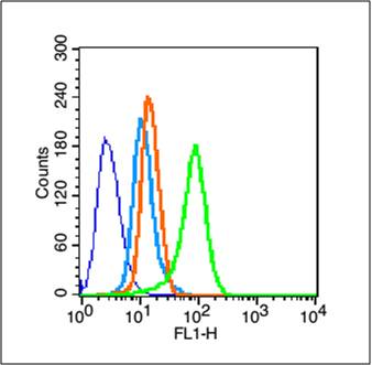
Blank control (blue line): Hela (blue).
Primary Antibody (green line): Rabbit Anti-MDR1 antibody (bs-1468R)
Dilution: 1μg /10^6 cells;
Isotype Control Antibody (orange line): Rabbit IgG .
Secondary Antibody (white blue line): Goat anti-rabbit IgG-FITC
Dilution: 1μg /test.
Protocol
The cells were fixed with 70% methanol (Overnight at -20℃) and then permeabilized with ice-cold 90% methanol for 30 min on ice. Cells stained with Primary Antibody for 30 min at room temperature. The cells were then incubated in 1 X PBS/2%BSA/10% goat serum to block non-specific protein-protein interactions followed by the antibody for 15 min at room temperature. The secondary antibody used for 40 min at room temperature. Acquisition of 20,000 events was performed.
-
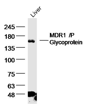
Sample: liver (Rat) Lysate at 40 ug
Primary: Anti-MDR1'P Glycoprotein (bs-1468R)at 1/300 dilution
Secondary: IRDye800CW Goat Anti-Rabbit IgG at 1/20000 dilution
Predicted band size: 141kD
Observed band size: 141 kD
RRID:AB_10854437
产品名称:Rabbit Anti-MDR1/P Glycoprotein antibody
别名: ABC20;
ABCB1;
ATP binding cassette, sub family B (MDR/TAP), member 1;
ATP-binding cassette sub-family B member 1;
CD243;
CLCS;
Colchicin sensitivity;
Doxorubicin resistance;
GP170;
MDR1;
MDR1_HUMAN;
Multidrug resistance 1;
Multidrug resistance
中文名称:多药耐药蛋白/P-糖蛋白(C端)抗体
英文名称:Rabbit Anti-MDR1/P Glycoprotein antibody
中文别名:p-gp; P糖蛋白
抗体来源: Rabbit
克隆类型:多克隆
细胞定位:细胞膜
性 状:Liquid
亚 型:IgG
纯化方法:affinity purified by Protein A
保存条件:Shipped at 4℃. Store at -20 °C for one year. Avoid repeated freeze/thaw cycles.
免 疫 原:KLH conjugated synthetic peptide derived from human MDR1
抗原表位:1051-1280/1280
抗原细胞定位:Cytoplasmic
SWISS:P08183
Gene ID :5243
Human Gene ID:5243
P Glycoprotein, the product of the MDR1 gene, is expressed in distinct non-malignant cells, typically cells with secretory and excretory functions. It is assumed to function as an ATP-dependent drug efflux pump with broad substrate specificity. The highest expression of P Glycoprotein has been observed in kidney (proximal tubules), liver (bile canaliculi), adrenal gland and intestine, suggesting that the primary role of P Glycoprotein is in the normal secretion of physiological metabolites and ingested chemicals into bile, urine and the lumen of the intestinal tract. Elevated levels of P Glycoprotein have also been reported in multidrug-resistant cell lines and in colon, endometrial, ovarian, and breast tumors, as well as in sarcomas and leukemias / lymphomas.
Function:Energy-dependent efflux pump responsible for decreased drug accumulation in multidrug-resistant cells.
Subunit:Interacts with PSMB5.
Subcellular Location:Cell membrane; Multi-pass membrane protein (By similarity).
Tissue Specificity:Expressed in liver, kidney, small intestine and brain.
DISEASE:Genetic variations in ABCB1 are associated with susceptibility to inflammatory bowel disease type 13 (IBD13) [MIM:612244]. Inflammatory bowel disease is characterized by a chronic relapsing intestinal inflammation. It is subdivided into Crohn disease and
Similarity:Belongs to the ABC transporter superfamily. ABCB family. Multidrug resistance exporter (TC 3.A.1.201) subfamily.
Contains 2 ABC transmembrane type-1 domains.
Contains 2 ABC transporter domains.
Important Note:This product as supplied is intended for research use only, not for use in human, therapeutic or diagnostic applications.
 400-901-9800


 说明书
说明书 联系我们
联系我们 打印此页面
打印此页面 收藏
收藏