| Rabbit Anti-C-jun antibody |
| 反应物种(预测) |
Chicken,Dog,Pig,Cow,Sheep |
| 产品应用(已验证) |
WB,IHC,FCM |
| 产品应用(可尝试) |
IF,ELISA |
| 推荐稀释比例 |
WB=1:500-2000,Elisa=1:5000-10000,IHC-P=1:100-500,IHC-F=1:100-500,Flow Cyt=1μg/Test,IF=1:100-500, |
| 研究领域 |
肿瘤,细胞生物,信号转导,转录调节因子,激酶和磷酸酶 |
| 标签 |
Array |
-
-
Blank control (blue line): HepG2 (blue).
Primary Antibody (green line): Rabbit Anti-C-jun antibody (bs-4601R)
Dilution: 1μg /10^6 cells;
Isotype Control Antibody (orange line): Rabbit IgG .
Secondary Antibody (white blue line): Goat anti-rabbit IgG-PE
Dilution: 1μg /test.
Protocol
The cells were fixed with 70% methanol (Overnight at 4℃) and then permeabilized with 90% ice-cold methanol for 20 min at -20℃. Cells stained with Primary Antibody for 30 min at room temperature. The cells were then incubated in 1 X PBS/2%BSA/10% goat serum to block non-specific protein-protein interactions followed by the antibody for 15 min at room temperature. The secondary antibody used for 40 min at room temperature. Acquisition of 20,000 events was performed.
-
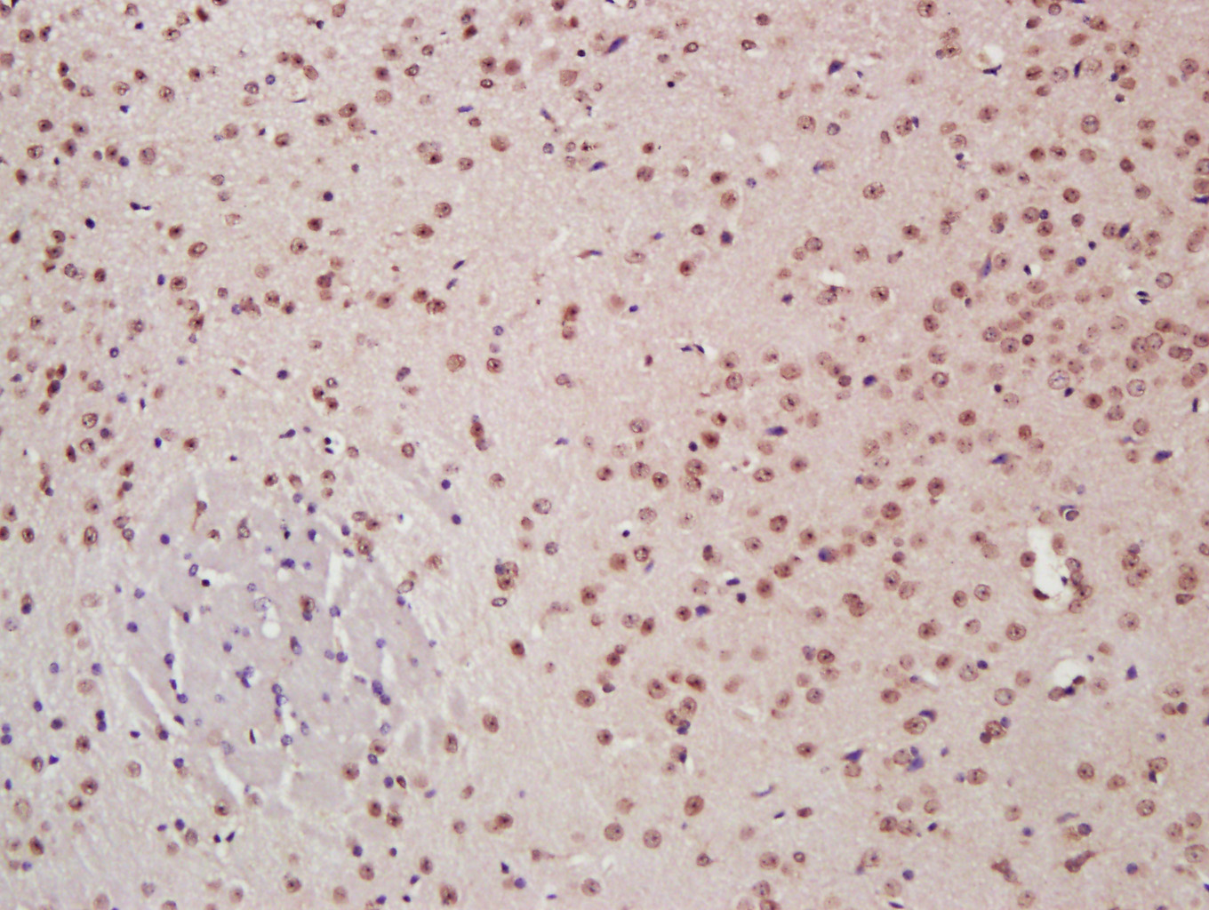
Paraformaldehyde-fixed, paraffin embedded (Mouse brain); Antigen retrieval by boiling in sodium citrate buffer (pH6.0) for 15min; Block endogenous peroxidase by 3% hydrogen peroxide for 20 minutes; Blocking buffer (normal goat serum) at 37°C for 30min; Antibody incubation with (C-jun) Polyclonal Antibody, Unconjugated (bs-0670R) at 1:400 overnight at 4°C, followed by a conjugated secondary antibody (sp-0023) for 20 minutes and DAB staining.
-
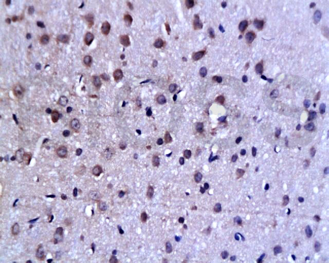
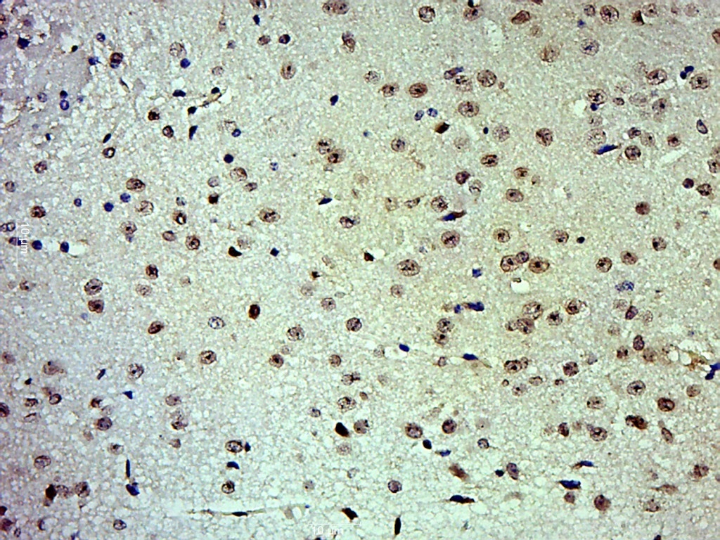
Tissue/cell: rat brain tissue; 4% Paraformaldehyde-fixed and paraffin-embedded;
Antigen retrieval: citrate buffer ( 0.01M, pH 6.0 ), Boiling bathing for 15min; Block endogenous peroxidase by 3% Hydrogen peroxide for 30min; Blocking buffer (normal goat serum,C-0005) at 37℃ for 20 min;
Incubation: Anti-C-Jun Polyclonal Antibody, Unconjugated(bs-0670R) 1:200, overnight at 4°C, followed by conjugation to the secondary antibody(SP-0023) and DAB(C-0010) staining
-
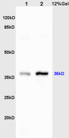
Sample:
Liver(Rat)lysate at 30ug;
Brain(Rat) lysates at 30ug;
Primary: Anti-C-jun/AP-1 (bs-0670R) at 1:200;
Secondary: HRP conjugated Goat-Anti-Rabbit IgG(bs-0295G-HRP) at 1: 3000;
Predicted band size : 36kD
Observed band size : 36kD
-
Paraformaldehyde-fixed, paraffin embedded (Mouse brain); Antigen retrieval by boiling in sodium citrate buffer (pH6.0) for 15min; Block endogenous peroxidase by 3% hydrogen peroxide for 20 minutes; Blocking buffer (normal goat serum) at 37°C for 30min; Antibody incubation with (C-jun) Polyclonal Antibody, Unconjugated (bs-0670R) at 1:500 overnight at 4°C, followed by a conjugated secondary (sp-0023) for 20 minutes and DAB staining.
-
Paraformaldehyde-fixed, paraffin embedded (Rat testis); Antigen retrieval by boiling in sodium citrate buffer (pH6.0) for 15min; Block endogenous peroxidase by 3% hydrogen peroxide for 20 minutes; Blocking buffer (normal goat serum) at 37°C for 30min; Antibody incubation with (C-jun) Polyclonal Antibody, Unconjugated (bs-0670R) at 1:400 overnight at 4°C, followed by a conjugated secondary (sp-0023) for 20 minutes and DAB staining.
-
Blank control: Hela.
Primary Antibody (green line): Rabbit Anti-C-jun antibody (bs-0670R)
Dilution: 1μg /10^6 cells;
Isotype Control Antibody (orange line): Rabbit IgG .
Secondary Antibody : Goat anti-rabbit IgG-AF647
Dilution: 1μg /test.
Protocol
The cells were fixed with 4% PFA (10min at room temperature)and then permeabilized with 90% ice-cold methanol for 20 min at -20℃. The cells were then incubated in 5%BSA to block non-specific protein-protein interactions for 30 min at room temperature .Cells stained with Primary Antibody for 30 min at room temperature. The secondary antibody used for 40 min at room temperature. Acquisition of 20,000 events was performed.
-
Sample:
Lane 1: Kidney (Mouse) Lysate at 40 ug
Lane 2: Lung (Mouse) Lysate at 40 ug
Lane 3: NIH/3T3 (Mouse) Cell Lysate at 30 ug
Lane 4: Hela (Human) Cell Lysate at 30 ug
Primary: Anti-C-jun (bs-0670R) at 1/1000 dilution
Secondary: IRDye800CW Goat Anti-Rabbit IgG at 1/20000 dilution
Predicted band size: 43/36 kD
Observed band size: 45 kD
RRID:AB_10857880
产品名称:Rabbit Anti-C-jun antibody
别名: Transcription factor AP-1; Jun oncogene; JUN; AP 1; AP1; AP-1; Enhancer Binding Protein AP1; Jun Activation Domain Binding Protein; JUN protein; JUNC; p39; Proto oncogene cJun; Transcription Factor AP1; V jun avian sarcoma virus 17 oncogene homolog; vJun
中文名称:原癌基因蛋白/活化蛋白1抗体
英文名称:Rabbit Anti-C-jun antibody
抗体来源: Rabbit
克隆类型:多克隆
细胞定位:细胞核
性 状:Liquid
亚 型:IgG
纯化方法:affinity purified by Protein A
保存条件:Shipped at 4℃. Store at -20 °C for one year. Avoid repeated freeze/thaw cycles.
免 疫 原:KLH conjugated synthetic peptide derived from human Transcription factor AP-1
抗原表位:31-331/331
SWISS:P05412
Gene ID :3725
Human Gene ID:3725
The human protooncogene JUN is the putative transforming gene of avian sarcoma virus 17, and it encodes a protein which is highly homologous to the viral protein. cJun (previously known as the Fos binding protein p39) and c Fos form a complex in the nucleus. AP 1 (activating protein 1) is a collective term referring to these dimeric transcription factors composed of Jun, Fos or ATF subunits that bind to a common DNA site, the AP1 binding site. AP 1 proteins, mostly the Jun group, regulate the expression and function of cell cycle regulators such as Cyclin D1, p53, p21 (cip1/waf1), p19 (ARF) and p16. Fos and Jun proto oncogene expression is induced transiently by a variety of extracellular stimuli associated with mitogenesis, differentiation processes or depolarization of neurons. JUN has been mapped to 1p32 to p31, a chromosomal region involved in both translocations and deletions in human malignancies.
Function:Transcription factor that recognizes and binds to the enhancer heptamer motif 5'-TGA[CG]TCA-3'. Promotes activity of NR5A1 when phosphorylated by HIPK3 leading to increased steroidogenic gene expression upon cAMP signaling pathway stimulation.
Subunit:Heterodimer with either FOS or BATF3 or ATF7. The ATF7/JUN heterodimer is essential for ATF7 transactivation activity. Interacts with DSIPI; the interaction inhibits the binding of active AP1 to its target DNA. Interacts with HIVEP3 and MYBBP1A. Interacts
Subcellular Location:Nucleus.
Post-translational modifications:Phosphorylated by CaMK4 and PRKDC; phosphorylation enhances the transcriptional activity. Phosphorylated by HIPK3.
[PTM] Phosphorylated at Thr-239, Ser-243 and Ser-249 by GSK3B; phosphorylation reduces its ability to bind DNA.
Phosphorylated by PAK2 a
Similarity:Belongs to the bZIP family. Jun subfamily.
Contains 1 bZIP domain.
Important Note:This product as supplied is intended for research use only, not for use in human, therapeutic or diagnostic applications.
 400-901-9800


 说明书
说明书 联系我们
联系我们 打印此页面
打印此页面 收藏
收藏