| Rabbit Anti-IKB alpha antibody |
| 产品应用(已验证) |
WB,IHC,ICC,FCM |
| 产品应用(可尝试) |
IF,ELISA |
| 推荐稀释比例 |
WB=1:500-2000,Elisa=1:5000-10000,IHC-P=1:100-500,IHC-F=1:100-500,Flow Cyt=1μg/Test,IF=1:100-500,ICC=1:100, |
| 研究领域 |
肿瘤,免疫学,信号转导,转录调节因子,激酶和磷酸酶 |
| 标签 |
Array |
-
Sample:
skin (Mouse) Lysate at 40 ug
Primary: Anti-IKB alpha (Bs- 1287R) at 1/300 dilution
Secondary: IRDye800CW Goat Anti-Rabbit IgG at 1/20000 dilution
Predicted band size: 36 kD
Observed band size: 35 kD
-
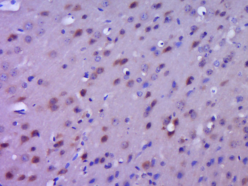
Paraformaldehyde-fixed, paraffin embedded (Mouse brain); Antigen retrieval by boiling in sodium citrate buffer (pH6.0) for 15min; Block endogenous peroxidase by 3% hydrogen peroxide for 20 minutes; Blocking buffer (normal goat serum) at 37°C for 30min; Antibody incubation with (IKB alpha) Polyclonal Antibody, Unconjugated (bs-1287R) at 1:400 overnight at 4°C, followed by operating according to SP Kit(Rabbit) (sp-0023) instructionsand DAB staining.
-
Paraformaldehyde-fixed, paraffin embedded (mouse lung); Antigen retrieval by boiling in sodium citrate buffer (pH6.0) for 15min; Block endogenous peroxidase by 3% hydrogen peroxide for 20 minutes; Blocking buffer (normal goat serum) at 37°C for 30min; Antibody incubation with (IKB alpha) Polyclonal Antibody, Unconjugated (bs-1287R) at 1:200 overnight at 4°C, followed by operating according to SP Kit(Rabbit) (sp-0023) instructionsand DAB staining.
-
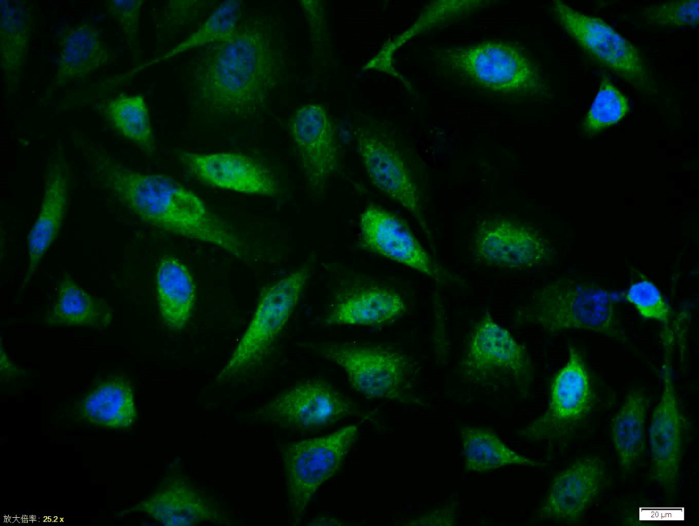
Tissue/cell:Hela cell; 4% Paraformaldehyde-fixed; Triton X-100 at room temperature for 20 min; Blocking buffer (normal goat serum,C-0005) at 37°C for 20 min; Antibody incubation with (IKB alpha) polyclonal Antibody, Unconjugated (bs-1287R) 1:100, 90 minutes at 37°C; followed by a FITC conjugated Goat Anti-Rabbit IgG antibody at 37°C for 90 minutes, DAPI (blue, C02-04002) was used to stain the cell nuclei.
-
Sample:
Lane 1: HepG2 (Human) Cell Lysate at 30 ug
Lane 2: Hela (Human) Cell Lysate at 30 ug
Lane 3: Siha (Human) Cell Lysate at 30 ug
Lane 4: U251 (Human) Cell Lysate at 30 ug
Lane 5: Spleen (Mouse) Lysate at 40 ug
Lane 6: Thymus (Mouse) Lysate at 40 ug
Lane 7: Thymus (Rat) Lysate at 40 ug
Lane 8: Lymph node (Mouse) Lysate at 40 ug
Lane 9: Lymph node (Rat) Lysate at 40 ug
Primary: Anti-IKB alpha (bs-1287R) at 1/1000 dilution
Secondary: IRDye800CW Goat Anti-Rabbit IgG at 1/20000 dilution
Predicted band size: 39 kD
Observed band size: 37 kD
-
Blank control: Hela.
Primary Antibody (green line): Rabbit Anti-IKB alpha antibody (bs-1287R)
Dilution: 1μg /10^6 cells;
Isotype Control Antibody (orange line): Rabbit IgG .
Secondary Antibody : Goat anti-rabbit IgG-AF647
Dilution: 1μg /test.
Protocol
The cells were fixed with 4% PFA (10min at room temperature)and then permeabilized with 90% ice-cold methanol for 20 min at -20℃. The cells were then incubated in 5%BSA to block non-specific protein-protein interactions for 30 min at room temperature .Cells stained with Primary Antibody for 30 min at room temperature. The secondary antibody used for 40 min at room temperature. Acquisition of 20,000 events was performed.
-
Tissue/cell:Hela cell; 4% Paraformaldehyde-fixed; Triton X-100 at room temperature for 20 min; Blocking buffer (normal goat serum,C-0005) at 37°C for 20 min; Antibody incubation with (IKB alpha) polyclonal Antibody, Unconjugated (bs-1287R) 1:100, 90 minutes at 37°C; followed by a FITC conjugated Goat Anti-Rabbit IgG antibody at 37°C for 90 minutes, DAPI (blue, C02-04002) was used to stain the cell nuclei.
-
Sample:
Lymph node (Mouse) Lysate at 40 ug
Thymus (Mouse) Lysate at 40 ug
Primary: Anti- IKB alpha (bs-1287R) at 1/1000 dilution
Secondary: IRDye800CW Goat Anti-Rabbit IgG at 1/20000 dilution
Predicted band size: 36 kD
Observed band size: 36 kD
-
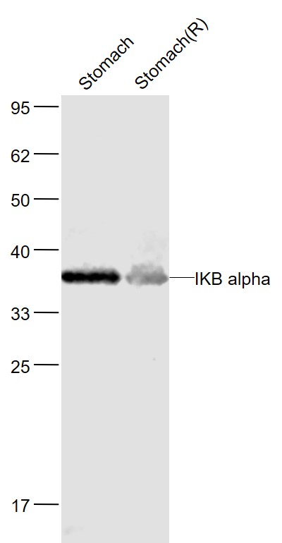
Sample:
Stomach(Mouse) Lysate at 40 ug
Stomach(Rat) Lysate at 40 ug
Primary: Anti-IKB alpha (bs-1287R) at 1/1000 dilution
Secondary: IRDye800CW Goat Anti-Rabbit IgG at 1/20000 dilution
Predicted band size: 35 kD
Observed band size: 37 kD
-
Sample:
Hela(Human) Cell Lysate at 30 ug
Primary: Anti-IKB alpha (bs-1287R) at 1/1000 dilution
Secondary: IRDye800CW Goat Anti-Rabbit IgG at 1/20000 dilution
Predicted band size: 36 kD
Observed band size: 36 kD
-
Sample:
HepG2 (Human) Cell Lysate at 30 ug
Primary: Anti-IKB alpha (bs-1287R) at 1/1000 dilution
Secondary: IRDye800CW Goat Anti-Rabbit IgG at 1/20000 dilution
Predicted band size: 36 kD
Observed band size: 36 kD
-
Sample:
Thymus gland (Mouse) Lysate at 40 ug
Primary: Anti-IKB alpha (bs-1287R) at 1/1000 dilution
Secondary: IRDye800CW Goat Anti-Rabbit IgG at 1/20000 dilution
Predicted band size: 36 kD
Observed band size: 36 kD
-
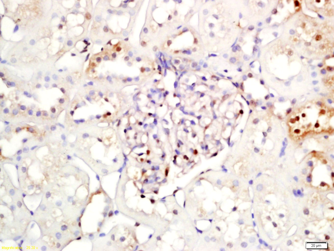
Tissue/cell: rat kidney tissue; 4% Paraformaldehyde-fixed and paraffin-embedded;
Antigen retrieval: citrate buffer ( 0.01M, pH 6.0 ), Boiling bathing for 15min; Block endogenous peroxidase by 3% Hydrogen peroxide for 30min; Blocking buffer (normal goat serum,C-0005) at 37℃ for 20 min;
Incubation: Anti-IKB alpha Polyclonal Antibody, Unconjugated(bs-1287R) 1:200, overnight at 4°C, followed by conjugation to the secondary antibody(SP-0023) and DAB(C-0010) staining
-
Sample: Lung (mouse) Lysate at 40 ug
Primary: Anti- IKB alpha (bs-1287R) at 1/300 dilution
Secondary: IRDye800CW Goat Anti-Rabbit IgG at 1/20000 dilution
Predicted band size: 36 kD
Observed band size: 36 kD
-
Blank control (blue line): Jurkat (blue).
Primary Antibody (green line): Rabbit Anti-IKB alpha antibody (bs-1287R)
Dilution: 1μg /10^6 cells;
Isotype Control Antibody (orange line): Rabbit IgG .
Secondary Antibody (white blue line): Goat anti-rabbit IgG-FITC
Dilution: 1μg /test.
Protocol
The cells were fixed with 2% paraformaldehyde (10 min) , then permeabilized with 90% ice-cold methanol for 30 min on ice. Cells stained with Primary Antibody for 30 min at room temperature. The cells were then incubated in 1 X PBS/2%BSA/10% goat serum to block non-specific protein-protein interactions followed by the antibody for 15 min at room temperature. The secondary antibody used for 40 min at room temperature. Acquisition of 20,000 events was performed.
-
Sample: Spleen (mouse) Lysate at 40 ug
Primary: Anti- IKB alpha (bs-1287R) at 1/300 dilution
Secondary: IRDye800CW Goat Anti-Rabbit IgG at 1/20000 dilution
Predicted band size: 36 kD
Observed band size: 35 kD
-
Sample:
Spleen (Mouse) Lysate at 40 ug
Primary: Anti-IKB alpha (bs-1287R) at 1/300 dilution
Secondary: IRDye800CW Goat Anti-Rabbit IgG at 1/20000 dilution
Predicted band size: 36 kD
Observed band size: 36 kD
RRID:AB_10857466
产品名称:Rabbit Anti-IKB alpha antibody
别名: NFKBIA; Inhibitor of KB alpha; I kappa B alpha; I(Kappa)B(alpha); IkappaBalpha; IKBA; IKBalpha; MAD 3; MAD3; Major histocompatibility complex enhancer binding protein MAD3; NF kappa B inhibitor alpha; NFKBI; Nuclear factor of kappa light chain gene enhanc
中文名称:核因子κB抑制蛋白α抗体
英文名称:Rabbit Anti-IKB alpha antibody
抗体来源: Rabbit
克隆类型:多克隆
细胞定位:细胞核,细胞浆
性 状:Liquid
亚 型:IgG
纯化方法:affinity purified by Protein A
保存条件:Shipped at 4℃. Store at -20 °C for one year. Avoid repeated freeze/thaw cycles.
免 疫 原:KLH conjugated synthetic peptide derived from human NFKBIA
抗原表位:1-120/314
SWISS:Q9Z1E3
Gene ID :4792
Human Gene ID:4792
This gene encodes a member of the NF-kappa-B inhibitor family, which contain multiple ankrin repeat domains. The encoded protein interacts with REL dimers to inhibit NF-kappa-B/REL complexes which are involved in inflammatory responses. The encoded protein moves between the cytoplasm and the nucleus via a nuclear localization signal and CRM1-mediated nuclear export. Mutations in this gene have been found in ectodermal dysplasia anhidrotic with T-cell immunodeficiency autosomal dominant disease. [provided by RefSeq, Aug 2011]
Function:Inhibits the activity of dimeric NF-kappa-B/REL complexes by trapping REL dimers in the cytoplasm through masking of their nuclear localization signals. On cellular stimulation by immune and proinflammatory responses, becomes phosphorylated promoting ubiq
Subunit:Interacts with RELA; the interaction requires the nuclear import signal. Interacts with NKIRAS1 and NKIRAS2. Part of a 70-90 kDa complex at least consisting of CHUK, IKBKB, NFKBIA, RELA, IKBKAP and MAP3K14. Interacts with HBV protein X. Interacts with RWD
Subcellular Location:Cytoplasm. Nucleus.
Tissue Specificity:Highly expressed in lymph node, thymus followed by liver, brain, muscle, kidney, gastrointestinal and reproductive tract.
Post-translational modifications:Phosphorylated; disables inhibition of NF-kappa-B DNA-binding activity. Phosphorylation at positions 32 and 36 is prerequisite to recognition by UBE2D3 leading to polyubiquitination and subsequent degradation.
Sumoylated; sumoylation requires the pre
DISEASE:Defects in NFKBIA are the cause of ectodermal dysplasia anhidrotic with T-cell immunodeficiency autosomal dominant (ADEDAID) [MIM:612132]. Ectodermal dysplasia defines a heterogeneous group of disorders due to abnormal development of two or more ectoderma
Similarity:Belongs to the NF-kappa-B inhibitor family.
Contains 5 ANK repeats.
Important Note:This product as supplied is intended for research use only, not for use in human, therapeutic or diagnostic applications.
 400-901-9800


 说明书
说明书 联系我们
联系我们 打印此页面
打印此页面 收藏
收藏