| Rabbit Anti-SMAD7 antibody |
| 反应物种(预测) |
Pig,Cow |
| 产品应用(已验证) |
WB,IHC,ICC,FCM |
| 产品应用(可尝试) |
IF,ELISA |
| 推荐稀释比例 |
WB=1:500-2000,Elisa=1:5000-10000,IHC-P=1:100-500,IHC-F=1:100-500,Flow Cyt=1ug/Test,IF=1:100-500,ICC=1:100, |
| 研究领域 |
肿瘤,细胞生物,信号转导,细胞凋亡,生长因子和激素,激酶和磷酸酶 |
| 标签 |
Array |
-
U-2OS cell; 4% Paraformaldehyde-fixed; Triton X-100 at room temperature for 20 min; Blocking buffer (normal goat serum, C-0005) at 37°C for 20 min; Antibody incubation with (MADH7/Smad7) polyclonal Antibody, Unconjugated (bs-0566R) 1:100, 90 minutes at 37°C; followed by a conjugated Goat Anti-Rabbit IgG antibody at 37°C for 90 minutes, DAPI (blue, C02-04002) was used to stain the cell nuclei.
-
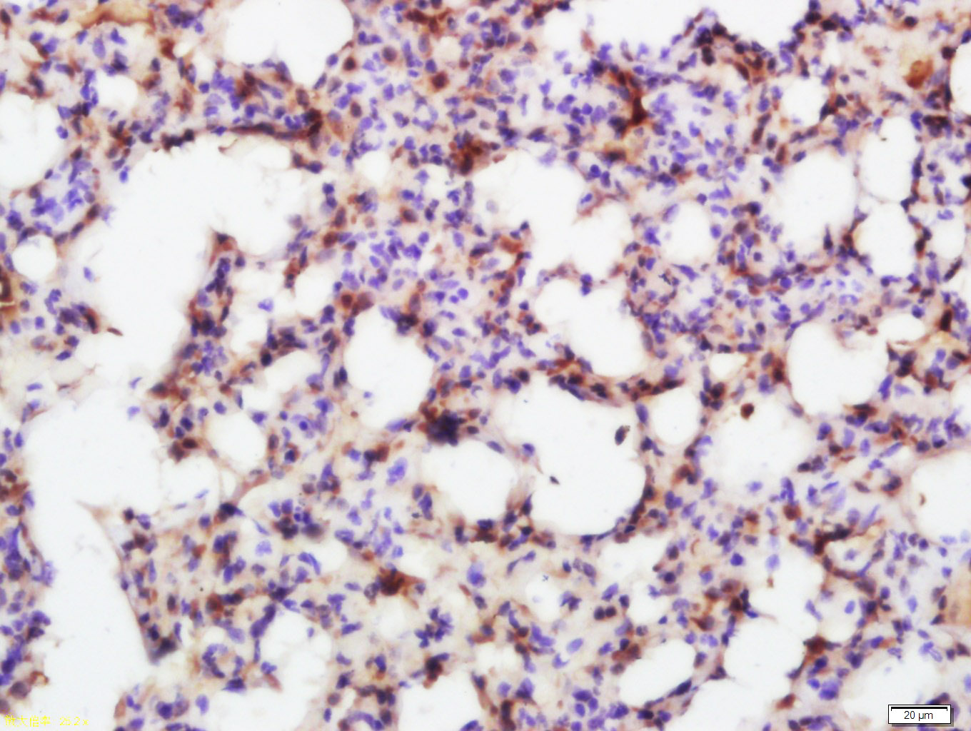
Paraformaldehyde-fixed, paraffin embedded (rat kidney); Antigen retrieval by boiling in sodium citrate buffer (pH6.0) for 15min; Block endogenous peroxidase by 3% hydrogen peroxide for 20 minutes; Blocking buffer (normal goat serum) at 37°C for 30min; Antibody incubation with (MADH7) Polyclonal Antibody, Unconjugated (bs-0566R) at 1:200 overnight at 4°C, followed by operating according to SP Kit(Rabbit) (sp-0023) instructionsand DAB staining.
-
Paraformaldehyde-fixed, paraffin embedded (mouse kidney); Antigen retrieval by boiling in sodium citrate buffer (pH6.0) for 15min; Block endogenous peroxidase by 3% hydrogen peroxide for 20 minutes; Blocking buffer (normal goat serum) at 37°C for 30min; Antibody incubation with (MADH7) Polyclonal Antibody, Unconjugated (bs-0566R) at 1:200 overnight at 4°C, followed by operating according to SP Kit(Rabbit) (sp-0023) instructionsand DAB staining.
-
Sample:
Lane 1: Stomach (Mouse) Lysate at 40 ug
Lane 2: Spleen (Mouse) Lysate at 40 ug
Lane 3: Lung (Mouse) Lysate at 40 ug
Lane 4: SH-SY5Y (Human) Cell Lysate at 30 ug
Lane 5: 293T (Human) Cell Lysate at 30 ug
Primary: Anti-MADH7/Smad7 (bs-0566R) at 1/1000 dilution
Secondary: IRDye800CW Goat Anti-Rabbit IgG at 1/20000 dilution
Predicted band size: 50 kD
Observed band size: 50 kD
-
Sample:
Lane 1: SH-SY5Y (Human) Cell Lysate at 30 ug
Lane 2: HepG2 (Human) Cell Lysate at 30 ug
Lane 3: Cerebrum (Mouse) Lysate at 40 ug
Lane 4: Cerebrum (Rat) Lysate at 40 ug
Lane 5: Stomach (Mouse) Lysate at 40 ug
Lane 6: Lung (Mouse) Lysate at 40 ug
Lane 7: Kidney (Mouse) Lysate at 40 ug
Primary: Anti-MADH7/Smad7 (bs-0566R) at 1/1000 dilution
Secondary: IRDye800CW Goat Anti-Rabbit IgG at 1/20000 dilution
Predicted band size: 50 kD
Observed band size: 50 kD
-
Paraformaldehyde-fixed, paraffin embedded (rat stomach); Antigen retrieval by boiling in sodium citrate buffer (pH6.0) for 15min; Block endogenous peroxidase by 3% hydrogen peroxide for 20 minutes; Blocking buffer (normal goat serum) at 37°C for 30min; Antibody incubation with (MADH7) Polyclonal Antibody, Unconjugated (bs-0566R) at 1:200 overnight at 4°C, followed by operating according to SP Kit(Rabbit) (sp-0023) instructionsand DAB staining.
-
U-2OS cell; 4% Paraformaldehyde-fixed; Triton X-100 at room temperature for 20 min; Blocking buffer (normal goat serum, C-0005) at 37°C for 20 min; Antibody incubation with (MADH7/Smad7) polyclonal Antibody, Unconjugated (bs-0566R) 1:100, 90 minutes at 37°C; followed by a conjugated Goat Anti-Rabbit IgG antibody at 37°C for 90 minutes, DAPI (blue, C02-04002) was used to stain the cell nuclei.
-
Paraformaldehyde-fixed, paraffin embedded (rat lung); Antigen retrieval by boiling in sodium citrate buffer (pH6.0) for 15min; Block endogenous peroxidase by 3% hydrogen peroxide for 20 minutes; Blocking buffer (normal goat serum) at 37°C for 30min; Antibody incubation with (Smad7) Polyclonal Antibody, Unconjugated (bs-0566R) at 1:600 overnight at 4°C, followed by a conjugated secondary (sp-0023) for 20 minutes and DAB staining.
-
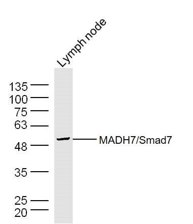
Sample:Lymph node (Mouse) Lysate at 30 ug
Primary: Anti- MADH7/Smad7 (bs-0566R) at 1/300 dilution
Secondary: IRDye800CW Goat Anti-Rabbit IgG at 1/20000 dilution
Predicted band size: 46 kD
Observed band size: 50 kD
-
Sample:Spleen(Mouse) Lysate at 30 ug
Primary: Anti- MADH7/Smad7 (bs-0566R) at 1/300 dilution
Secondary: IRDye800CW Goat Anti-Rabbit IgG at 1/20000 dilution
Predicted band size: 46 kD
Observed band size: 50 kD
-
Sample:Stomach (Mouse) Lysate at 30 ug
Primary: Anti- MADH7/Smad7 (bs-0566R) at 1/300 dilution
Secondary: IRDye800CW Goat Anti-Rabbit IgG at 1/20000 dilution
Predicted band size: 46 kD
Observed band size: 50 kD
-
Tissue/cell: rat kidney tissue; 4% Paraformaldehyde-fixed and paraffin-embedded;
Antigen retrieval: citrate buffer ( 0.01M, pH 6.0 ), Boiling bathing for 15min; Block endogenous peroxidase by 3% Hydrogen peroxide for 30min; Blocking buffer (normal goat serum,C-0005) at 37℃ for 20 min;
Incubation: Anti-Smad7/Smad6 Polyclonal Antibody, Unconjugated(bs-0071R) 1:200, overnight at 4°C, followed by conjugation to the secondary antibody(SP-0023) and DAB(C-0010) staining
-
Sample:
Lung(Mouse) Lysate at 30 ug
Placenta(Mouse) Lysate at 30 ug
Large intestine(Mouse) Lysate at 30 ug
Primary: Anti- MADH7/Smad7 (bs-0566R) at 1/300 dilution
Secondary: IRDye800CW Goat Anti-Rabbit IgG at 1/20000 dilution
Predicted band size: 46 kD
Observed band size: 50 kD
-
Blank control: SH-SY5Y.
Primary Antibody (green line): Rabbit Anti-MADH7/Smad7 antibody (bs-0566R)
Dilution: 1μg /10^6 cells;
Isotype Control Antibody (orange line): Rabbit IgG .
Secondary Antibody : Goat anti-rabbit IgG-AF647
Dilution: 1μg /test.
Protocol
The cells were fixed with 4% PFA (10min at room temperature)and then permeabilized with 90% ice-cold methanol for 20 min at-20℃.The cells were then incubated in 5%BSA to block non-specific protein-protein interactions for 30 min at room temperature .Cells stained with Primary Antibody for 30 min at room temperature. The secondary antibody used for 40 min at room temperature. Acquisition of 20,000 events was performed.
RRID:AB_10855651
产品名称:Rabbit Anti-SMAD7 antibody
别名: MAD (mothers against decapentaplegic Drosophila) homolog 7;
MAD;
Mad homolog 7;
MAD mothers against decapentaplegic homolog 7;
MADH 7;
MADH 8;
MADH8;
Mothers Against Decapentaplegic Drosophila Homolog of 7;
Mothers against decapentaplegic homolog
中文名称:Smad7抗体
英文名称:Rabbit Anti-SMAD7 antibody
抗体来源: Rabbit
克隆类型:多克隆
细胞定位:细胞核,细胞浆
性 状:Liquid
亚 型:IgG
纯化方法:affinity purified by Protein A
保存条件:Shipped at 4℃. Store at -20 °C for one year. Avoid repeated freeze/thaw cycles.
免 疫 原:KLH conjugated synthetic peptide derived from human Smad7
抗原表位:1-100/426
SWISS:O15105
Gene ID :4092
Human Gene ID:4092
The protein encoded by this gene is a nuclear protein that binds the E3 ubiquitin ligase SMURF2. Upon binding, this complex translocates to the cytoplasm, where it interacts with TGF-beta receptor type-1 (TGFBR1), leading to the degradation of both the encoded protein and TGFBR1. Expression of this gene is induced by TGFBR1. Variations in this gene are a cause of susceptibility to colorectal cancer type 3 (CRCS3). Several transcript variants encoding different isoforms have been found for this gene. [provided by RefSeq, Jun 2010]
Function:Antagonist of signaling by TGF-beta (transforming growth factor) type 1 receptor superfamily members; has been shown to inhibit TGF-beta (Transforming growth factor) and activin signaling by associating with their receptors thus preventing SMAD2 access. F
Subunit:Interacts with WWP1. Interacts with COPS5. Interacts with NEDD4L. Interacts with STAMBP. Interacts with RNF111, AXIN1 and AXIN2. Interacts with PPP1R15A. Interacts (via MH2 domain) with EP300. Interacts with ACVR1B, SMURF1, SMURF2 and TGFBR1; SMAD7 recrui
Subcellular Location:Nucleus. Cytoplasm. Note=Interaction with NEDD4L or RNF111 or induces translocation from the nucleus to the cytoplasm. TGF-beta stimulates its translocation from the nucleus to the cytoplasm. PDPK1 inhibits its translocation from the nucleus to the cytopl
Tissue Specificity:Ubiquitous with higher expression in the lung and vascular endothelium.
Post-translational modifications:Phosphorylation on Ser-249 does not affect its stability, nuclear localization or inhibitory function in TGFB signaling; however it affects its ability to regulate transcription. Phosphorylated by PDPK1.
Ubiquitinated by WWP1 (By similarity). Polyubiq
DISEASE:Genetic variations in SMAD7 influence susceptibility to colorectal cancer type 3 (CRCS3) [MIM:612229]. Colorectal cancer consists of tumors or cancer of either the colon or rectum or both. Cancers of the large intestine are the second most common form of
Similarity:Belongs to the dwarfin/SMAD family.
Contains 1 MH1 (MAD homology 1) domain.
Contains 1 MH2 (MAD homology 2) domain.
Important Note:This product as supplied is intended for research use only, not for use in human, therapeutic or diagnostic applications.
 400-901-9800


 说明书
说明书 联系我们
联系我们 打印此页面
打印此页面 收藏
收藏