| Rabbit Anti-Notch1 antibody |
| 产品应用(已验证) |
WB,IHC,FCM |
| 产品应用(可尝试) |
IF,ELISA |
| 推荐稀释比例 |
WB=1:500-2000,Elisa=1:5000-10000,IHC-P=1:100-500,IHC-F=1:100-500,Flow Cyt=1µg/Test,IF=1:100-500, |
| 研究领域 |
细胞生物,发育生物学,神经生物学,信号转导,干细胞,表观遗传学, |
| 标签 |
Array |
-
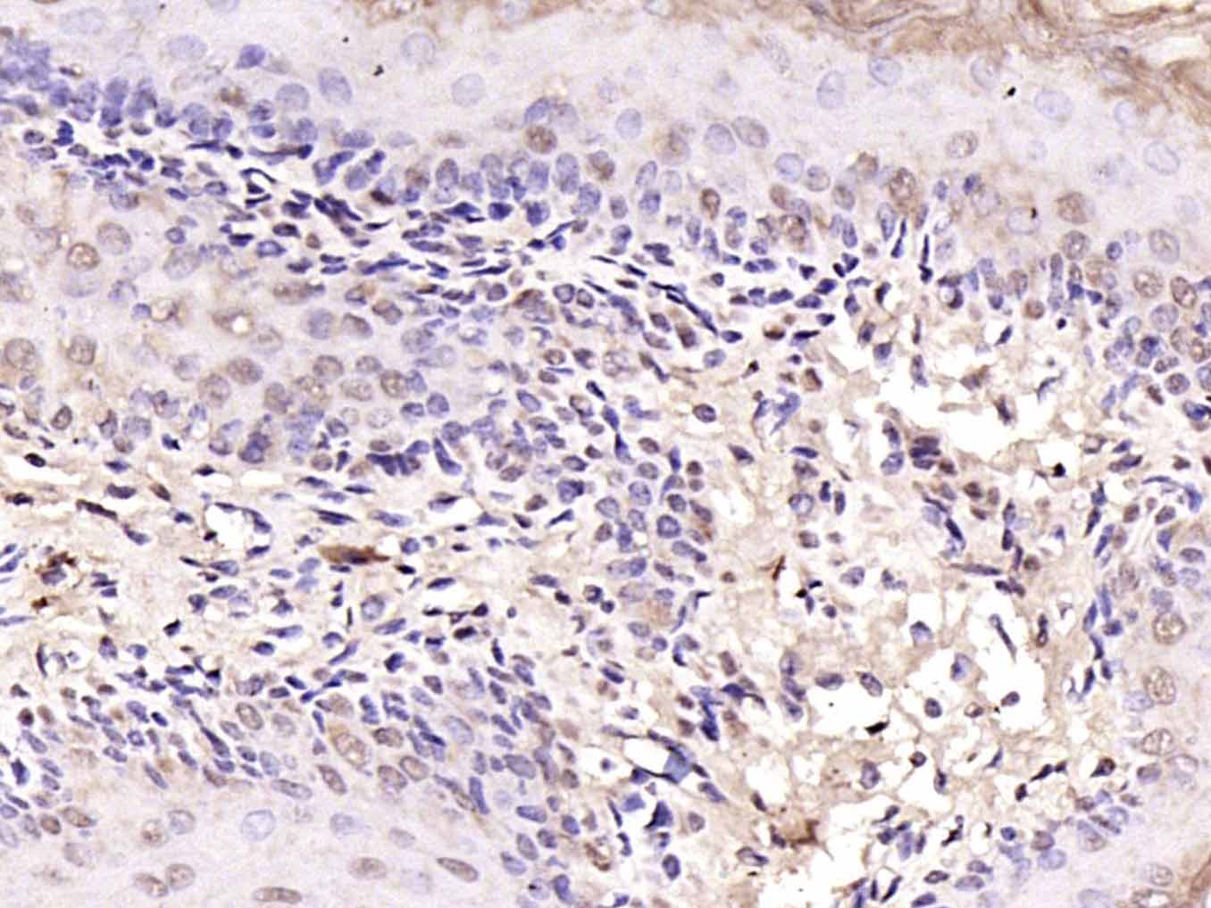
Tissue/cell: Human endometrium carcinoma; 4% Paraformaldehyde-fixed and paraffin-embedded;
Antigen retrieval: citrate buffer ( 0.01M, pH 6.0 ), Boiling bathing for 15min; Block endogenous peroxidase by 3% Hydrogen peroxide for 30min; Blocking buffer (normal goat serum,C-0005) at 37℃ for 20 min;
Incubation: Anti-Notch1 Polyclonal Antibody, Unconjugated(bs-1335R) 1:200, overnight at 4°C, followed by conjugation to the secondary antibody(SP-0023) and DAB(C-0010) staining
-
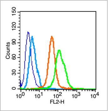
Blank control (blue line): Hela (blue).
Primary Antibody (green line): Rabbit Anti-Notch1 antibody (bs-1335R)
Dilution: 1μg /10^6 cells;
Isotype Control Antibody (orange line): Rabbit IgG .
Secondary Antibody (white blue line): Goat anti-rabbit IgG-PE
Dilution: 1μg /test.
Protocol
The cells were fixed with 70% ethanol overnight at 4℃ and then permeabilized with 90% ice-cold methanol for 30 min on ice. Cells stained with Primary Antibody for 30 min at room temperature. The cells were then incubated in 1 X PBS/2%BSA/10% goat serum to block non-specific protein-protein interactions followed by the antibody for 15 min at room temperature. The secondary antibody used for 40 min at room temperature. Acquisition of 20,000 events was performed.
-
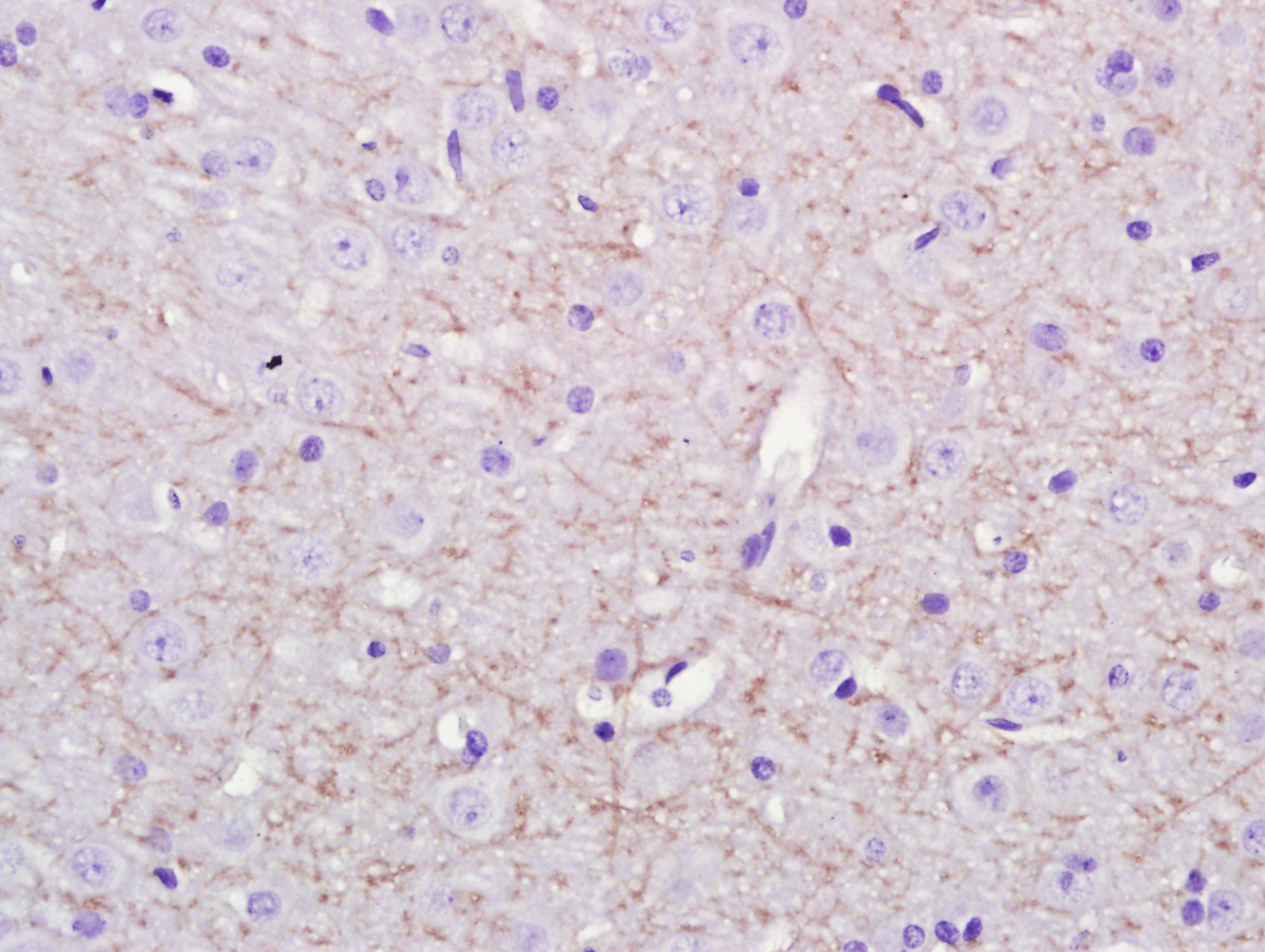
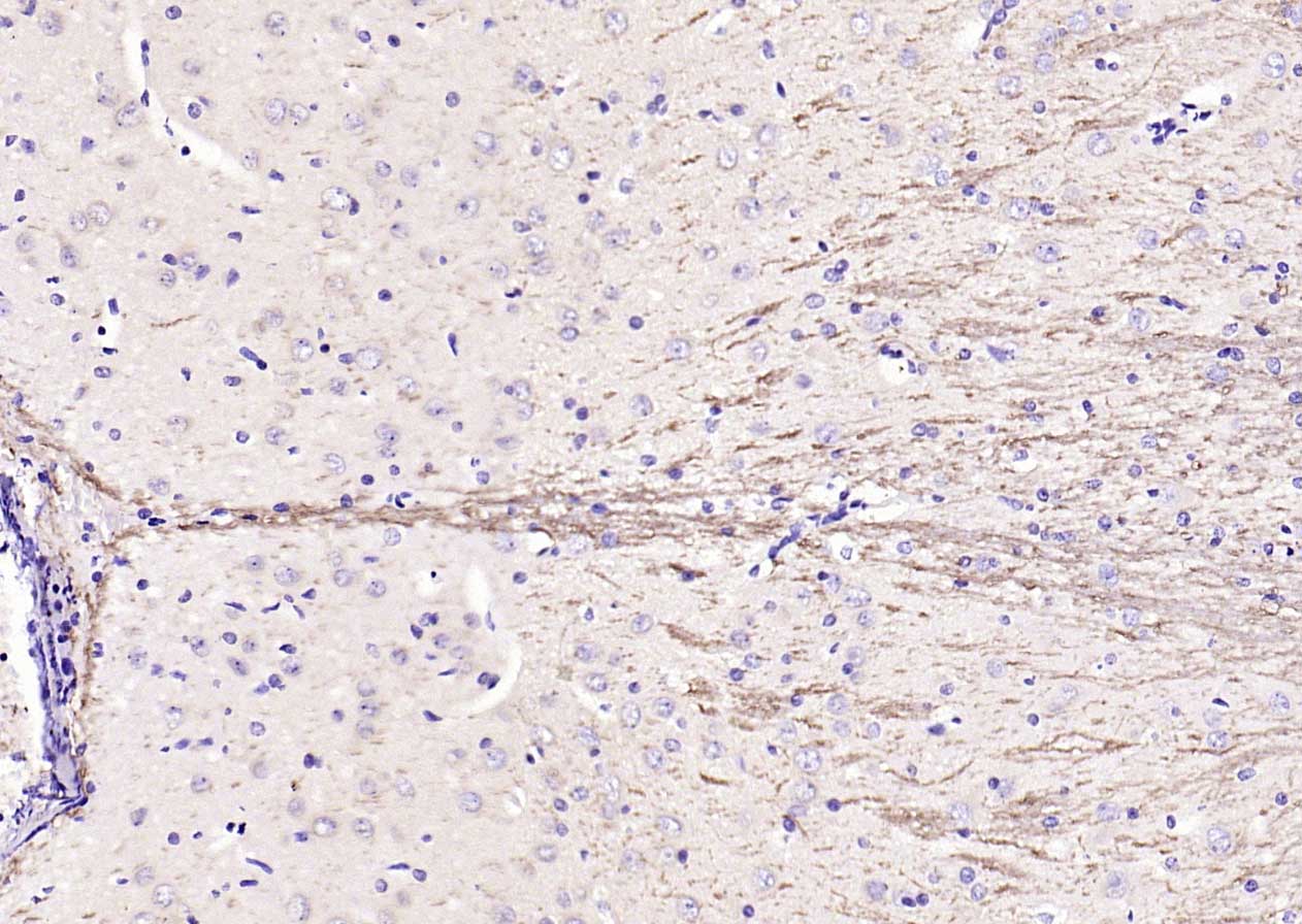
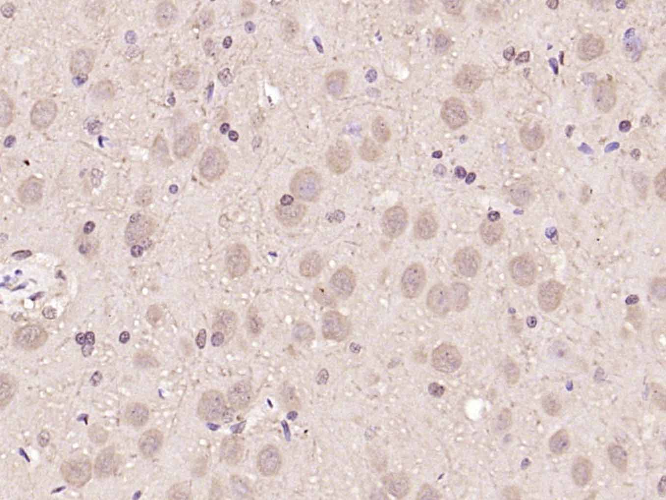
Paraformaldehyde-fixed, paraffin embedded (Rat brain); Antigen retrieval by boiling in sodium citrate buffer (pH6.0) for 15min; Block endogenous peroxidase by 3% hydrogen peroxide for 20 minutes; Blocking buffer (normal goat serum) at 37°C for 30min; Antibody incubation with (Notch1) Polyclonal Antibody, Unconjugated (bs-1335R) at 1:400 overnight at 4°C, followed by a conjugated secondary antibody (sp-0023) for 20 minutes and DAB staining.
-
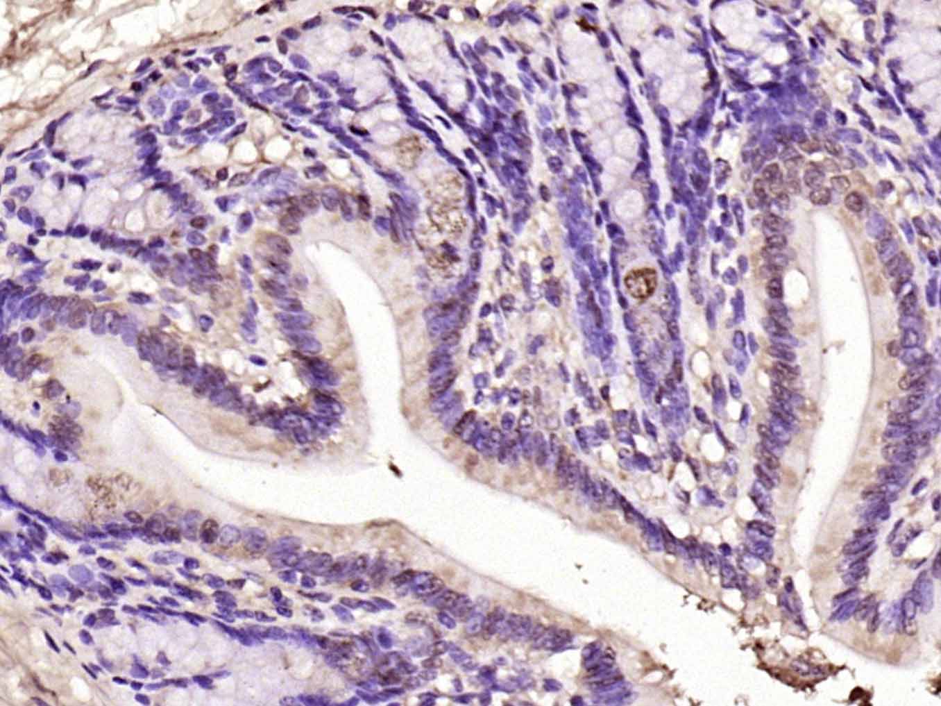
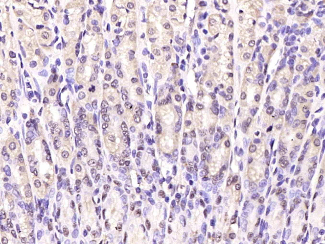
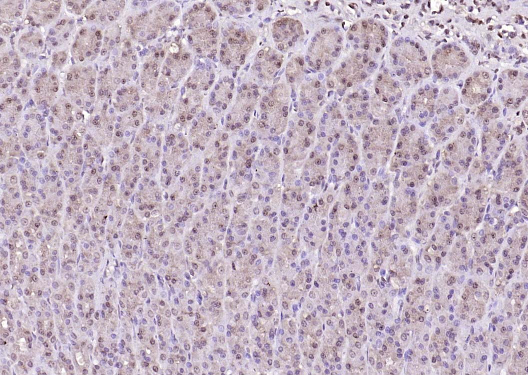
Tissue/cell: human stomach tissue; 4% Paraformaldehyde-fixed and paraffin-embedded;
Antigen retrieval: citrate buffer ( 0.01M, pH 6.0 ), Boiling bathing for 15min; Block endogenous peroxidase by 3% Hydrogen peroxide for 30min; Blocking buffer (normal goat serum,C-0005) at 37℃ for 20 min;
Incubation: Anti-Notch1 Polyclonal Antibody, Unconjugated(bs-1335R) 1:500, overnight at 4°C, followed by conjugation to the secondary antibody(SP-0023) and DAB(C-0010) staining
-
Paraformaldehyde-fixed, paraffin embedded (rat colon); Antigen retrieval by boiling in sodium citrate buffer (pH6.0) for 15min; Block endogenous peroxidase by 3% hydrogen peroxide for 20 minutes; Blocking buffer (normal goat serum) at 37°C for 30min; Antibody incubation with (Notch1) Polyclonal Antibody, Unconjugated (bs-1335R) at 1:200 overnight at 4°C, followed by operating according to SP Kit(Rabbit) (sp-0023) instructionsand DAB staining.
-
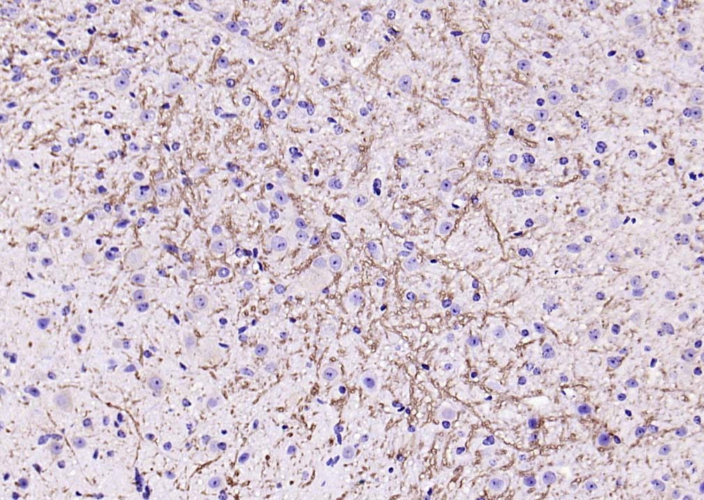
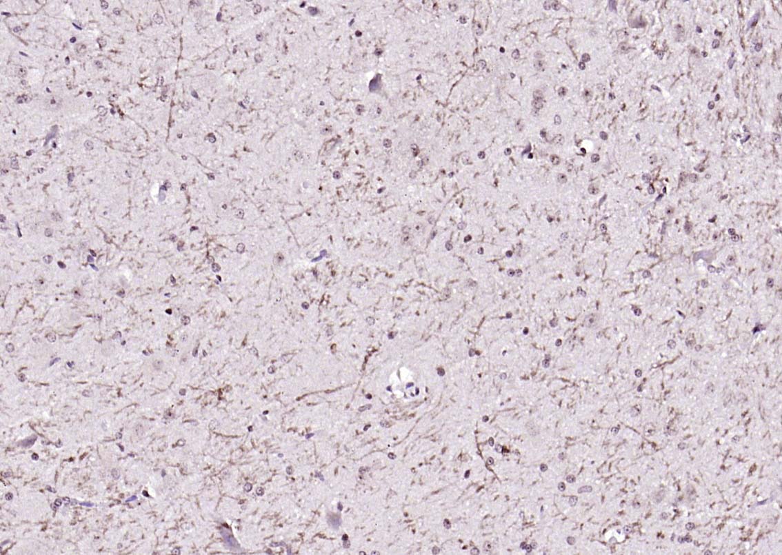
Paraformaldehyde-fixed, paraffin embedded (rat brain); Antigen retrieval by boiling in sodium citrate buffer (pH6.0) for 15min; Block endogenous peroxidase by 3% hydrogen peroxide for 20 minutes; Blocking buffer (normal goat serum) at 37°C for 30min; Antibody incubation with (Notch1) Polyclonal Antibody, Unconjugated (bs-1335R) at 1:200 overnight at 4°C, followed by operating according to SP Kit(Rabbit) (sp-0023) instructionsand DAB staining.
-
Paraformaldehyde-fixed, paraffin embedded (mouse brain); Antigen retrieval by boiling in sodium citrate buffer (pH6.0) for 15min; Block endogenous peroxidase by 3% hydrogen peroxide for 20 minutes; Blocking buffer (normal goat serum) at 37°C for 30min; Antibody incubation with (Notch1) Polyclonal Antibody, Unconjugated (bs-1335R) at 1:200 overnight at 4°C, followed by operating according to SP Kit(Rabbit) (sp-0023) instructionsand DAB staining.
-
Paraformaldehyde-fixed, paraffin embedded (rat brain); Antigen retrieval by boiling in sodium citrate buffer (pH6.0) for 15min; Block endogenous peroxidase by 3% hydrogen peroxide for 20 minutes; Blocking buffer (normal goat serum) at 37°C for 30min; Antibody incubation with (Notch1) Polyclonal Antibody, Unconjugated (bs-1335R) at 1:200 overnight at 4°C, followed by operating according to SP Kit(Rabbit) (sp-0023) instructionsand DAB staining.
-
A549 cell; 4% Paraformaldehyde-fixed; Triton X-100 at room temperature for 20 min; Blocking buffer (normal goat serum, C-0005) at 37°C for 20 min; Antibody incubation with (Notch1) polyclonal Antibody, Unconjugated (bs-1335R) 1:100, 90 minutes at 37°C; followed by a conjugated Goat Anti-Rabbit IgG antibody at 37°C for 90 minutes, DAPI (blue, C02-04002) was used to stain the cell nuclei.
-
Paraformaldehyde-fixed, paraffin embedded (rat stomach); Antigen retrieval by boiling in sodium citrate buffer (pH6.0) for 15min; Block endogenous peroxidase by 3% hydrogen peroxide for 20 minutes; Blocking buffer (normal goat serum) at 37°C for 30min; Antibody incubation with (Notch1) Polyclonal Antibody, Unconjugated (bs-1335R) at 1:200 overnight at 4°C, followed by operating according to SP Kit(Rabbit) (sp-0023) instructionsand DAB staining.
-
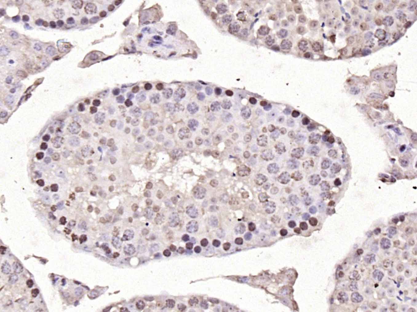
Paraformaldehyde-fixed, paraffin embedded (mouse testis); Antigen retrieval by boiling in sodium citrate buffer (pH6.0) for 15min; Block endogenous peroxidase by 3% hydrogen peroxide for 20 minutes; Blocking buffer (normal goat serum) at 37°C for 30min; Antibody incubation with (Notch1) Polyclonal Antibody, Unconjugated (bs-1335R) at 1:200 overnight at 4°C, followed by operating according to SP Kit(Rabbit) (sp-0023) instructionsand DAB staining.
-
Paraformaldehyde-fixed, paraffin embedded (mouse stomach); Antigen retrieval by boiling in sodium citrate buffer (pH6.0) for 15min; Block endogenous peroxidase by 3% hydrogen peroxide for 20 minutes; Blocking buffer (normal goat serum) at 37°C for 30min; Antibody incubation with (Notch1) Polyclonal Antibody, Unconjugated (bs-1335R) at 1:200 overnight at 4°C, followed by operating according to SP Kit(Rabbit) (sp-0023) instructionsand DAB staining.
-
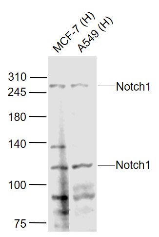
Sample:
Lane 1: MCF-7 (Human) Cell Lysate at 30 ug
Lane 2: A549 (Human) Cell Lysate at 30 ug
Primary: Anti-Notch1 (bs-1335R) at 1/1000 dilution
Secondary: IRDye800CW Goat Anti-Rabbit IgG at 1/20000 dilution
Predicted band size: 270/120/110 kD
Observed band size: 270/110 kD
-
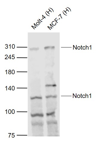
Sample:
Lane 1: Molt-4 (Human) Cell Lysate at 30 ug
Lane 2: MCF-7 (Human) Cell Lysate at 30 ug
Primary: Anti-Notch1 (bs-1335R) at 1/1000 dilution
Secondary: IRDye800CW Goat Anti-Rabbit IgG at 1/20000 dilution
Predicted band size: 270/110/120 kD
Observed band size: 270/110 kD
-
Paraformaldehyde-fixed, paraffin embedded (rat stomach); Antigen retrieval by boiling in sodium citrate buffer (pH6.0) for 15min; Block endogenous peroxidase by 3% hydrogen peroxide for 20 minutes; Blocking buffer (normal goat serum) at 37°C for 30min; Antibody incubation with (Notch1) Polyclonal Antibody, Unconjugated (bs-1335R) at 1:200 overnight at 4°C, followed by operating according to SP Kit(Rabbit) (sp-0023) instructionsand DAB staining.
-
Paraformaldehyde-fixed, paraffin embedded (rat brain); Antigen retrieval by boiling in sodium citrate buffer (pH6.0) for 15min; Block endogenous peroxidase by 3% hydrogen peroxide for 20 minutes; Blocking buffer (normal goat serum) at 37°C for 30min; Antibody incubation with (Notch1) Polyclonal Antibody, Unconjugated (bs-1335R) at 1:200 overnight at 4°C, followed by operating according to SP Kit(Rabbit) (sp-0023) instructionsand DAB staining.
RRID:AB_10854452
产品名称:Rabbit Anti-Notch1 antibody
别名: Neurogenic locus notch homolog protein 1; Notch 1 extracellular truncation; Notch 1 intracellular domain; hN1; Lin-12; LIN12; MIS6; Motch A; mT14; Neurogenic locus notch homolog protein 1; Neurogenic locus notch protein homolog; NICD; Notch1 intracellular
中文名称:跨膜受体蛋白Notch-1抗体
英文名称:Rabbit Anti-Notch1 antibody
抗体来源: Rabbit
克隆类型:多克隆
细胞定位:细胞核,细胞膜
性 状:Liquid
亚 型:IgG
纯化方法:affinity purified by Protein A
保存条件:Shipped at 4℃. Store at -20 °C for one year. Avoid repeated freeze/thaw cycles.
免 疫 原:KLH conjugated synthetic peptide derived from human C-terminal sequence of Notch 1 extracellular truncation and Notch 1 intracellular domain
抗原表位:2101-2300/2555
SWISS:P46531
Gene ID :4851
Human Gene ID:4851
This gene encodes a member of the Notch family. Members of this Type 1 transmembrane protein family share structural characteristics including an extracellular domain consisting of multiple epidermal growth factor-like (EGF) repeats, and an intracellular domain consisting of multiple, different domain types. Notch family members play a role in a variety of developmental processes by controlling cell fate decisions. The Notch signaling network is an evolutionarily conserved intercellular signaling pathway which regulates interactions between physically adjacent cells. In Drosophilia, notch interaction with its cell-bound ligands (delta, serrate) establishes an intercellular signaling pathway that plays a key role in development. Homologues of the notch-ligands have also been identified in human, but precise interactions between these ligands and the human notch homologues remain to be determined. This protein is cleaved in the trans-Golgi network, and presented on the cell surface as a heterodimer. This protein functions as a receptor for membrane bound ligands, and may play multiple roles during development. [provided by RefSeq, Jul 2008].
Function:Functions as a receptor for membrane-bound ligands Jagged1, Jagged2 and Delta1 to regulate cell-fate determination. Upon ligand activation through the released notch intracellular domain (NICD) it forms a transcriptional activator complex with RBPJ/RBPSUH
Subunit:Heterodimer of a C-terminal fragment N(TM) and an N-terminal fragment N(EC) which are probably linked by disulfide bonds. Interacts with DNER, DTX1, DTX2 and RBPJ/RBPSUH. Also interacts with MAML1, MAML2 and MAML3 which act as transcriptional coactivators
Subcellular Location:Cell membrane and Nucleus. Following proteolytical processing NICD is translocated to the nucleus.
Tissue Specificity:In fetal tissues most abundant in spleen, brain stem and lung. Also present in most adult tissues where it is found mainly in lymphoid tissues.
Post-translational modifications:Synthesized in the endoplasmic reticulum as an inactive form which is proteolytically cleaved by a furin-like convertase in the trans-Golgi network before it reaches the plasma membrane to yield an active, ligand-accessible form. Cleavage results in a C-t
DISEASE:Defects in NOTCH1 are a cause of bicuspid aortic valve (BAV) [MIM:109730]. A common defect in the aortic valve in which two rather than three leaflets are present. It is often associated with aortic valve calcification and insufficiency. In extreme cases,
Similarity:Belongs to the NOTCH family.
Contains 5 ANK repeats.
Contains 36 EGF-like domains.
Contains 3 LNR (Lin/Notch) repeats.
Important Note:This product as supplied is intended for research use only, not for use in human, therapeutic or diagnostic applications.
 400-901-9800


 说明书
说明书 联系我们
联系我们 打印此页面
打印此页面 收藏
收藏