| Rabbit Anti-RAGE antibody |
| 产品应用(已验证) |
WB,IHC,FCM |
| 产品应用(可尝试) |
IF,ELISA |
| 推荐稀释比例 |
WB=1:500-2000,Elisa=1:5000-10000,IHC-P=1:100-500,IHC-F=1:100-500,Flow Cyt=1μg /test,IF=1:100-500, |
| 研究领域 |
肿瘤,心血管,免疫学,生长因子和激素,糖尿病,内分泌病, |
| 标签 |
Array |
-
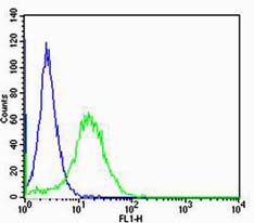
Cell: NIH/3T3
Concentration:1:100
Host/Isotype:Rabbit/IgG
Flow cytometric analysis of Rabbit IgG isotype control (Cat#: bs-0177R) on NIH/3T3(green) compared with control in the absence of primary antibody (blue) followed by Alexa Fluor 488-conjugated goat anti-rabbit IgG(H+L) secondary antibody .
-
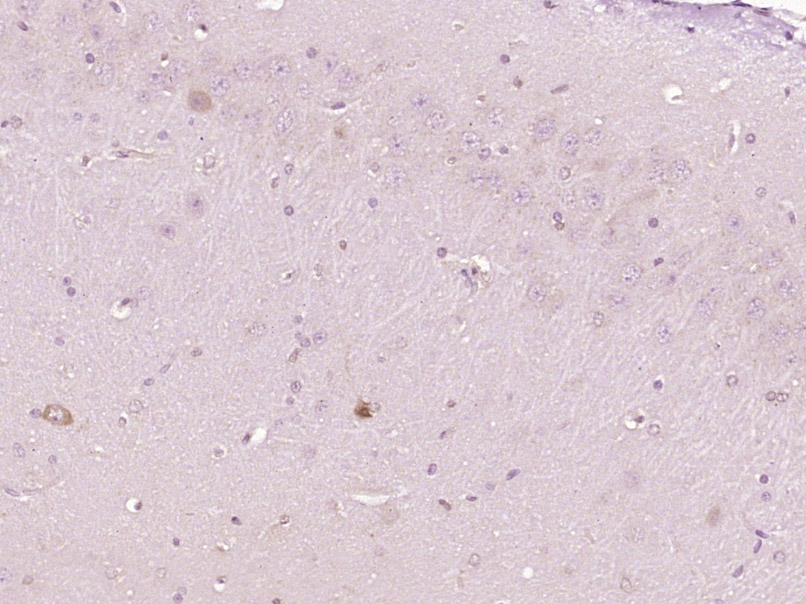
Paraformaldehyde-fixed, paraffin embedded (Rat brain); Antigen retrieval by boiling in sodium citrate buffer (pH6.0) for 15min; Block endogenous peroxidase by 3% hydrogen peroxide for 20 minutes; Blocking buffer (normal goat serum) at 37°C for 30min; Antibody incubation with (RAGE) Polyclonal Antibody, Unconjugated (bs-0177R) at 1:400 overnight at 4°C, followed by operating according to SP Kit(Rabbit) (sp-0023) instructionsand DAB staining.
-
Blank control: MCF7.
Primary Antibody (green line): Rabbit Anti-RAGE antibody (bs-0177R)
Dilution: 1μg /10^6 cells;
Isotype Control Antibody (orange line): Rabbit IgG .
Secondary Antibody : Goat anti-rabbit IgG-AF647
Dilution: 1μg /test.
Protocol
The cells were incubated in 5%BSA to block non-specific protein-protein interactions for 30 min at room temperature .Cells stained with Primary Antibody for 30 min at room temperature. The secondary antibody used for 40 min at room temperature. Acquisition of 20,000 events was performed.
-
Sample:
Lane 1: Kidney (Mouse) Lysate at 40 ug
Lane 2: Adrenal gland (Mouse) Lysate at 40 ug
Lane 3: Kidney (Rat) Lysate at 40 ug
Lane 4: Adrenal gland (Rat) Lysate at 40 ug
Primary: Anti-RAGE (bs-0177R) at 1/1000 dilution
Secondary: IRDye800CW Goat Anti-Rabbit IgG at 1/20000 dilution
Predicted band size: 50/43/55 kD
Observed band size: 58/50 kD
RRID:AB_10857130
产品名称:Rabbit Anti-RAGE antibody
别名: Advanced glycosylation end product specific receptor; Advanced glycosylation end product-specific receptor; AGER; EC 2.7.11.22; LE 9211 A antigen;LE-9211-A antigen; MGC22357; MOK; RAGE 1; RAGE1; MOK protein kinase; Receptor for advanced glycation endprodu
中文名称:晚期糖基化终末产物特异性受体抗体
英文名称:Rabbit Anti-RAGE antibody
抗体来源: Rabbit
克隆类型:多克隆
细胞定位:细胞膜,分泌型蛋白
性 状:Liquid
亚 型:IgG
纯化方法:affinity purified by Protein A
保存条件:Shipped at 4℃. Store at -20 °C for one year. Avoid repeated freeze/thaw cycles.
免 疫 原:KLH conjugated synthetic peptide derived from rat AGER
抗原表位:151-250/403
抗原细胞定位:Extracellular
SWISS:Q63495
Gene ID :81722
Human Gene ID:177
CAS:4
Advanced glycosylation end product-specific receptor (AGER; RAGE) is a member of the immunoglobulin superfamily of cell surface molecules that binds molecules that have been irreversibly modified by non-enzymatic glycation and oxidation, and are know as advanced glycation end products (AGEs). It is expressed by endothelium, mononuclear phagocytes, neurons and smooth muscle cells. Whereas RAGE is present at high levels during development, especially in the central nervous system, its levels decline during maturity.The increased expression of RAGE is associated with several pathological states, such as diabetic vasculopathy, neuropathy, retinopathy and other disorders, including Alzheimer's disease and immune/inflammatory reactions of the vessel walls. In diabetic tissues, the production of RAGE is due to the overproduction of AGEs that eventually overwhelm the protective properties of RAGE. This results in oxidative stress and endothelial cell dysfunction that leads to vascular disease in diabetics. In the brain, RAGE also binds amyloid beta (Ab). Because Ab is overproduced in neurons and vessels in the brains of Alzheimer disease, this leads to the hyperstimulation of RAGE. The RAGE-Ab interaction is thought to result in oxidative stress leading to neuronal degeneration.
Function:Mediates interactions of advanced glycosylation end products (AGE). These are nonenzymatically glycosylated proteins which accumulate in vascular tissue in aging and at an accelerated rate in diabetes. Acts as a mediator of both acute and chronic vascular
Subunit:Interacts with S100B, S100A1 and APP. Interacts with S100A12.
Subcellular Location:Isoform 1: Cell membrane; Single-pass type I membrane protein.
Isoform 2: Secreted.
Tissue Specificity:Endothelial cells and cardiomyocytes.
Similarity:Contains 2 Ig-like C2-type (immunoglobulin-like) domains.
Contains 1 Ig-like V-type (immunoglobulin-like) domain.
Important Note:This product as supplied is intended for research use only, not for use in human, therapeutic or diagnostic applications.
 400-901-9800


 说明书
说明书 联系我们
联系我们 打印此页面
打印此页面 收藏
收藏