| Rabbit Anti-HSP27 antibody |
| 反应物种(预测) |
Dog,Pig,Cow |
| 产品应用(已验证) |
WB,IHC,FCM |
| 产品应用(可尝试) |
ICC,IF,ELISA |
| 推荐稀释比例 |
WB=1:500-2000,Elisa=1:5000-10000,IHC-P=1:100-500,IHC-F=1:100-500,Flow Cyt=2μg/Test,IF=1:100-500,ICC=1:100, |
| 研究领域 |
肿瘤,免疫学,信号转导 |
| 标签 |
Array |
-
-
Blank control (blue line): A431 cells (blue).
Primary Antibody (green line): Rabbit Anti-HSP27 antibody (bs-0730R)
Dilution: 2μg /10^6 cells;
Isotype Control Antibody (orange line): Rabbit IgG .
Secondary Antibody (white blue line): Goat anti-rabbit IgG-FITC
Dilution: 1μg /test.
Protocol
The cells were fixed with 70% methanol (Overnight at 4℃) and then permeabilized with 90% ice-cold methanol for 20 min at -20℃. Cells stained with Primary Antibody for 30 min at room temperature. The cells were then incubated in 1 X PBS/2%BSA/10% goat serum to block non-specific protein-protein interactions followed by the antibody for 15 min at room temperature. The secondary antibody used for 40 min at room temperature. Acquisition of 20,000 events was performed.
-
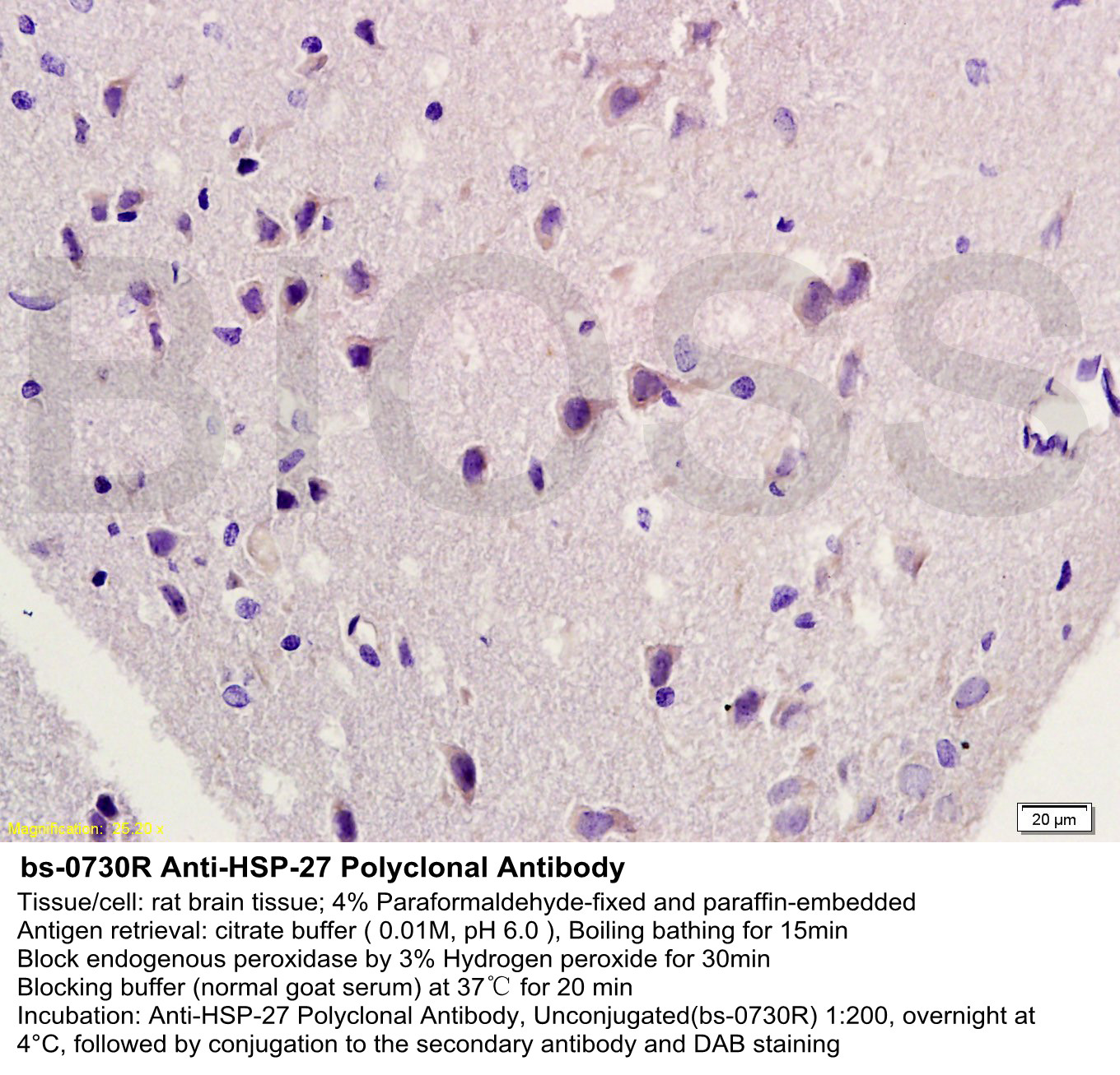
Tissue/cell: rat brain tissue; 4% Paraformaldehyde-fixed and paraffin-embedded;
Antigen retrieval: citrate buffer ( 0.01M, pH 6.0 ), Boiling bathing for 15min; Block endogenous peroxidase by 3% Hydrogen peroxide for 30min; Blocking buffer (normal goat serum,C-0005) at 37℃ for 20 min;
Incubation: Anti-HSP-27 Polyclonal Antibody, Unconjugated(bs-0730R) 1:200, overnight at 4°C, followed by conjugation to the secondary antibody(SP-0023) and DAB(C-0010) staining
-
Tissue/cell: rat brain tissue;4% Paraformaldehyde-fixed and paraffin-embedded;
Antigen retrieval: citrate buffer ( 0.01M, pH 6.0 ), Boiling bathing for 15min; Blocking buffer (normal goat serum,C-0005) at 37℃ for 20 min;
Incubation: Anti-HSP-27 Polyclonal Antibody, Unconjugated(bs-0730R) 1:200, overnight at 4°C; The secondary antibody was Goat Anti-Rabbit IgG, Cy3 conjugated(bs-0295G-Cy3)used at 1:200 dilution for 40 minutes at 37°C. DAPI(5ug/ml,blue,C-0033) was used to stain the cell nuclei
-
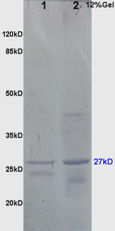
Sample:
Brain(Mouse) lysate at 30ug;
Liver(Mouse) lysate at 30ug;
Primary: Anti-HSP-27 (bs-0730R) at 1:200;
Secondary: AP conjugated Goat Anti-Rabbit IgG(bs-0295G-AP) at 1: 3000 dilutionNBT/BCIP staining;
Predicted band size : 27kD
Observed band size : 27kD
-
Paraformaldehyde-fixed, paraffin embedded (rat skeletal muscle); Antigen retrieval by boiling in sodium citrate buffer (pH6.0) for 15min; Block endogenous peroxidase by 3% hydrogen peroxide for 20 minutes; Blocking buffer (normal goat serum) at 37°C for 30min; Antibody incubation with (HSP27) Polyclonal Antibody, Unconjugated (bs-0730R ) at 1:200 overnight at 4°C, followed by operating according to SP Kit(Rabbit) (sp-0023) instructionsand DAB staining.
-
Paraformaldehyde-fixed, paraffin embedded (rat stomach tissue); Antigen retrieval by boiling in sodium citrate buffer (pH6.0) for 15min; Block endogenous peroxidase by 3% hydrogen peroxide for 20 minutes; Blocking buffer (normal goat serum) at 37°C for 30min; Antibody incubation with (HSP27) Polyclonal Antibody, Unconjugated (bs-0730R) at 1:400 overnight at 4°C, followed by operating according to SP Kit(Rabbit) (sp-0023) instructionsand DAB staining.
-
Paraformaldehyde-fixed, paraffin embedded (Rat uterus); Antigen retrieval by boiling in sodium citrate buffer (pH6.0) for 15min; Block endogenous peroxidase by 3% hydrogen peroxide for 20 minutes; Blocking buffer (normal goat serum) at 37°C for 30min; Antibody incubation with (HSP27) Polyclonal Antibody, Unconjugated (bs-0730R) at 1:400 overnight at 4°C, followed by operating according to SP Kit(Rabbit) (sp-0023) instructionsand DAB staining.
-
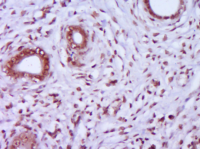
Paraformaldehyde-fixed, paraffin embedded (Human breast cancer); Antigen retrieval by boiling in sodium citrate buffer (pH6.0) for 15min; Block endogenous peroxidase by 3% hydrogen peroxide for 20 minutes; Blocking buffer (normal goat serum) at 37°C for 30min; Antibody incubation with (HSP27) Polyclonal Antibody, Unconjugated (bs-0730R) at 1:400 overnight at 4°C, followed by operating according to SP Kit(Rabbit) (sp-0023) instructions and DAB staining.
-
Sample:
Lane 1: Hela (Human) Cell Lysate at 30 ug
Lane 2: MCF-7 (Human) Cell Lysate at 30 ug
Lane 3: A431 (Human) Cell Lysate at 30 ug
Lane 4: Huvec (Human) Cell Lysate at 30 ug
Lane 5: HepG2 (Human) Cell Lysate at 30 ug
Primary:
Anti-HSP27 (bs-0730R) at 1/1000 dilution
Secondary: IRDye800CW Goat Anti-Rabbit IgG at 1/20000 dilution
Predicted band size: 27-30 kD
Observed band size: 27 kD
RRID:AB_10857781
产品名称:Rabbit Anti-HSP27 antibody
别名: Heat shock 27kDa protein; 28 kDa heat shock protein; CMT2F; DKFZp586P1322; Estrogen regulated 24 kDa protein; Estrogen-regulated 24 kDa protein; Heat shock 25kDa protein 1; Heat shock 25kDa protein 1; Heat shock 27 kDa protein; Heat shock 27kD protein 1;
中文名称:热休克蛋白27/HSP25抗体
英文名称:Rabbit Anti-HSP27 antibody
抗体来源: Rabbit
克隆类型:多克隆
细胞定位:细胞核,细胞浆
性 状:Liquid
亚 型:IgG
纯化方法:affinity purified by Protein A
保存条件:Shipped at 4℃. Store at -20 °C for one year. Avoid repeated freeze/thaw cycles.
免 疫 原:KLH conjugated synthetic peptide derived from human HSP27
抗原表位:101-205/205
SWISS:P04792
Gene ID :3315
Human Gene ID:3315
The protein encoded by this gene is induced by environmental stress and developmental changes. The encoded protein is involved in stress resistance and actin organization and translocates from the cytoplasm to the nucleus upon stress induction. Defects in this gene are a cause of Charcot-Marie-Tooth disease type 2F (CMT2F) and distal hereditary motor neuropathy (dHMN). [provided by RefSeq, Oct 2008]
Function:Involved in stress resistance and actin organization.
Subunit:Interacts with TGFB1I1. Associates with alpha- and beta-tubulin, microtubules and CRYAB. Interacts with HSPB8 and HSPBAP1.
Subcellular Location:Cytoplasm. Nucleus. Cytoplasm, cytoskeleton, spindle. Note=Cytoplasmic in interphase cells. Colocalizes with mitotic spindles in mitotic cells. Translocates to the nucleus during heat shock and resides in sub-nuclear structures known as SC35 speckles or n
Tissue Specificity:Detected in all tissues tested: skeletal muscle, heart, aorta, large intestine, small intestine, stomach, esophagus, bladder, adrenal gland, thyroid, pancreas, testis, adipose tissue, kidney, liver, spleen, cerebral cortex, blood serum and cerebrospinal
Post-translational modifications:Phosphorylated in MCF-7 cells on exposure to protein kinase C activators and heat shock.
Phosphorylation by MAPKAPK2 and MAPKAPK3 in response to stress leads to dissociate HSP27/HSPB1 from large small heat-shock protein (sHsps) oligomers and impair its
DISEASE:Defects in HSPB1 are the cause of Charcot-Marie-Tooth disease type 2F (CMT2F) [MIM:606595]. CMT2F is a form of Charcot-Marie-Tooth disease, the most common inherited disorder of the peripheral nervous system. Charcot-Marie-Tooth disease is classified in t
Similarity:Belongs to the small heat shock protein (HSP20) family.
Important Note:This product as supplied is intended for research use only, not for use in human, therapeutic or diagnostic applications.
 400-901-9800


 说明书
说明书 联系我们
联系我们 打印此页面
打印此页面 收藏
收藏