| Rabbit Anti-XIAP/BIRC4 antibody |
| 产品应用(已验证) |
WB,ICC,FCM |
| 产品应用(可尝试) |
IHC,IF,ELISA |
| 推荐稀释比例 |
WB=1:500-2000,Elisa=1:5000-10000,IHC-P=1:100-500,IHC-F=1:100-500,Flow Cyt=3ug/Test,IF=1:100-500,ICC=1:100, |
| 研究领域 |
肿瘤,细胞生物,细胞凋亡,新陈代谢, |
| 标签 |
Array |
-
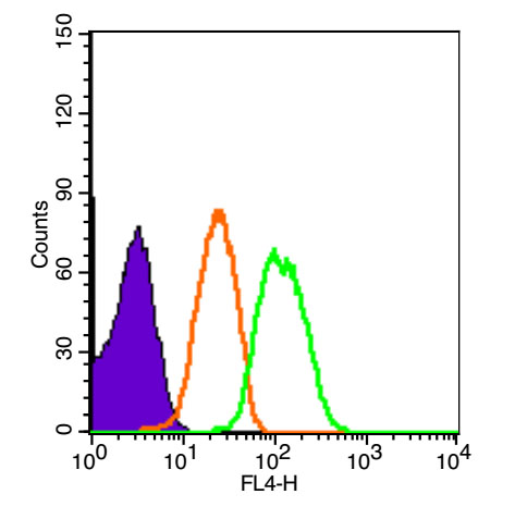
Blank control (Black line): HUVEC(Black).
Primary Antibody (green line): Rabbit Anti-XIAP antibody (bs-1281R)
Dilution: 1μg /10^6 cells;
Isotype Control Antibody (orange line): Rabbit IgG .
Secondary Antibody (white blue line): Goat anti-rabbit IgG-PE
Dilution: 1μg /test.
Protocol
The cells were fixed with 4% PFA (10min at room temperature)and then permeabilized with 90% ice-cold methanol for 20 min at room temperature. The cells were then incubated in 5%BSA to block non-specific protein-protein interactions for 30 min at room temperature .Cells stained with Primary Antibody for 30 min at room temperature. The secondary antibody used for 40 min at room temperature. Acquisition of 20,000 events was performed.
-
Tissue/cell: rat brain tissue;4% Paraformaldehyde-fixed and paraffin-embedded;
Antigen retrieval: citrate buffer ( 0.01M, pH 6.0 ), Boiling bathing for 15min; Blocking buffer (normal goat serum,C-0005) at 37℃ for 20 min;
Incubation: Anti-XIAP/BIRC4 Polyclonal Antibody, Unconjugated(bs-1281R) 1:200, overnight at 4°C; The secondary antibody was Goat Anti-Rabbit IgG, PE conjugated(bs-0295G-PE)used at 1:200 dilution for 40 minutes at 37°C. DAPI(5ug/ml,blue,C-0033) was used to stain the cell nuclei
-
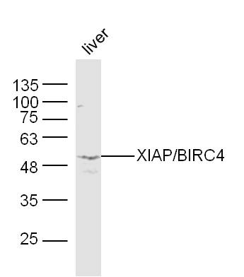
Sample:Liver(Mouse) Lysate at 40 ug
Primary: Anti-XIAP/BIRC4(bs-1281R) at 1/300 dilution
Secondary: IRDye800CW Goat Anti-Rabbit IgG at 1/20000 dilution
Predicted band size: 55 kD
Observed band size: 53 kD
-
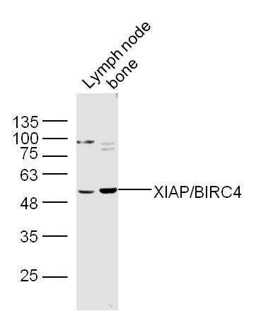
Sample:
Lymph node(Mouse) Lysate at 40 ug
Bone(Mouse) Lysate at 40 ug
Primary: Anti-XIAP/BIRC4(bs-1281R) at 1/300 dilution
Secondary: IRDye800CW Goat Anti-Rabbit IgG at 1/20000 dilution
Predicted band size: 55 kD
Observed band size: 53 kD
-
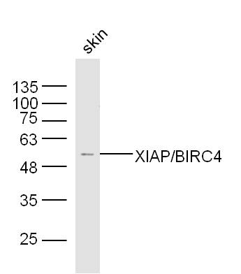
Sample:Skin(Mouse) Lysate at 40 ug
Primary: Anti-XIAP/BIRC4(bs-1281R) at 1/300 dilution
Secondary: IRDye800CW Goat Anti-Rabbit IgG at 1/20000 dilution
Predicted band size: 55 kD
Observed band size: 53 kD
-
-
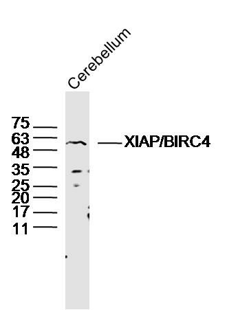
Sample: Cerebellum (Mouse) Lysate at 40 ug
Primary: Anti-XIAP/BIRC4 (bs-1281R) at 1/300 dilution
Secondary: IRDye800CW Goat Anti-Rabbit IgG at 1/20000 dilution
Predicted band size: 55 kD
Observed band size: 55 kD
-
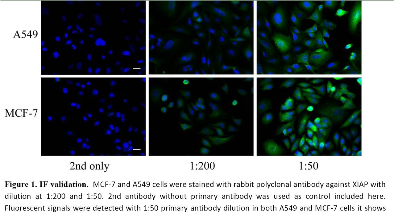
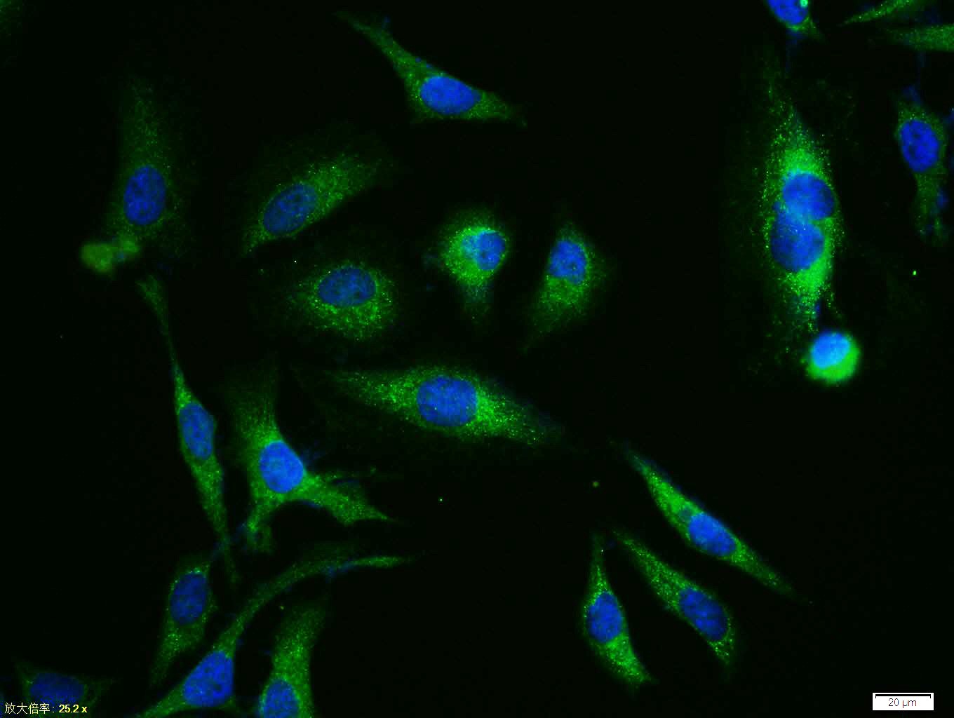
Tissue/cell:Hela cell; 4% Paraformaldehyde-fixed; Triton X-100 at room temperature for 20 min; Blocking buffer (normal goat serum,C-0005) at 37°C for 20 min; Antibody incubation with (XIAP/BIRC4) polyclonal Antibody, Unconjugated (bs-1281R) 1:100, 90 minutes at 37°C; followed by a FITC conjugated Goat Anti-Rabbit IgG antibody at 37°C for 90 minutes, DAPI (blue, C02-04002) was used to stain the cell nuclei.
RRID:AB_10856518
产品名称:Rabbit Anti-XIAP/BIRC4 antibody
别名: X-linked Inhibitor of Apoptosis Protein; RIAP-3; Baculoviral IAP repeat-containing protein 4; E3 ubiquitin-protein ligase XIAP; Inhibitor of apoptosis protein 3; X-linked IAP; IAP-like protein; HILP; BIRC4; HILP; XLP2; ILP1; Xiap; MIHA; hILP; IAP3; API3;
中文名称:X-连锁凋亡蛋白/性连锁凋亡抑制蛋白抗体
英文名称:Rabbit Anti-XIAP/BIRC4 antibody
抗体来源: Rabbit
克隆类型:多克隆
细胞定位:细胞浆
性 状:Liquid
亚 型:IgG
纯化方法:affinity purified by Protein A
保存条件:Shipped at 4℃. Store at -20 °C for one year. Avoid repeated freeze/thaw cycles.
免 疫 原:KLH conjugated synthetic peptide derived from human XIAP
抗原表位:201-330/496
SWISS:Q60989
Gene ID :331
Human Gene ID:331
This gene encodes a protein that belongs to a family of apoptotic suppressor proteins. Members of this family share a conserved motif termed, baculovirus IAP repeat, which is necessary for their anti-apoptotic function. This protein functions through binding to tumor necrosis factor receptor-associated factors TRAF1 and TRAF2 and inhibits apoptosis induced by menadione, a potent inducer of free radicals, and interleukin 1-beta converting enzyme. This protein also inhibits at least two members of the caspase family of cell-death proteases, caspase-3 and caspase-7. Mutations in this gene are the cause of X-linked lymphoproliferative syndrome. Alternate splicing results in multiple transcript variants. Pseudogenes of this gene are found on chromosomes 2 and 11.[provided by RefSeq, Feb 2011]
Function:Apoptotic suppressor. Has E3 ubiquitin-protein ligase activity. Mediates the proteasomal degradation of target proteins, such as caspase-3, SMAC or AIFM1. Inhibitor of caspase-3, -7 and -9. Mediates activation of MAP3K7/TAK1, leading to the activation of
Subunit:Monomer, and homodimer. Interacts with SMAC and with PRSS25; these interactions inhibit apoptotic suppressor activity. Interacts with MAP3K7IP1 and AIFM1. Interaction with SMAC hinders binding of MAP3K7IP1 and AIFM1. Interacts with TCF25.
Subcellular Location:Cytoplasm.
Tissue Specificity:Ubiquitous, except peripheral blood leukocytes.
Post-translational modifications:Ubiquitinated and degraded by the proteasome in apoptotic cells.
Phosphorylation by PKB/AKT protects XIAP against ubiquitination and protects the protein against proteasomal degradation.
DISEASE:Defects in XIAP are the cause of lymphoproliferative syndrome X-linked type 2 (XLP2) [MIM:300635]. XLP is a rare immunodeficiency characterized by extreme susceptibility to infection with Epstein-Barr virus (EBV). Symptoms include severe or fatal mononucl
Similarity:Belongs to the IAP family.
Contains 3 BIR repeats.
Contains 1 RING-type zinc finger.
Important Note:This product as supplied is intended for research use only, not for use in human, therapeutic or diagnostic applications.
 400-901-9800


 说明书
说明书 联系我们
联系我们 打印此页面
打印此页面 收藏
收藏