| Rabbit Anti-PTEN antibody |
| 反应物种(预测) |
Rat |
| 产品应用(已验证) |
WB,IHC,ICC,FCM |
| 产品应用(可尝试) |
IF,ELISA |
| 推荐稀释比例 |
WB=1:500-2000,Elisa=1:5000-10000,IHC-P=1:100-500,IHC-F=1:100-500,Flow Cyt=1μg/Test,IF=1:100-500,ICC=1:100, |
| 研究领域 |
肿瘤,细胞生物,细胞周期蛋白,激酶和磷酸酶,表观遗传学, |
| 标签 |
Array |
-
Blank control: Hela.
Primary Antibody (green line): Rabbit Anti-PTEN antibody (bs-0748R)
Dilution: 1μg /10^6 cells;
Isotype Control Antibody (orange line): Rabbit IgG .
Secondary Antibody : Goat anti-rabbit IgG-AF647
Dilution: 1μg /test.
Protocol
The cells were fixed with 4% PFA (10min at room temperature)and then permeabilized with 90% ice-cold methanol for 20 min at -20℃. The cells were then incubated in 5%BSA to block non-specific protein-protein interactions for 30 min at room temperature .Cells stained with Primary Antibody for 30 min at room temperature. The secondary antibody used for 40 min at room temperature. Acquisition of 20,000 events was performed.
-
Tissue/cell: Human nasopharyngeal carcinoma; 4% Paraformaldehyde-fixed and paraffin-embedded;
Antigen retrieval: citrate buffer ( 0.01M, pH 6.0 ), Boiling bathing for 15min; Block endogenous peroxidase by 3% Hydrogen peroxide for 30min; Blocking buffer (normal goat serum,C-0005) at 37∩ for 20 min;
Incubation: Anti-PTEN Polyclonal Antibody, Unconjugated(bs-0748R) 1:200, overnight at 4∑C, followed by conjugation to the secondary antibody(SP-0023) and DAB(C-0010) staining
-
Sample: Lung (Mouse) Lysate at 30 ug
Primary: Anti- PTEN (bs-0748R) at 1/300 dilution
Secondary: IRDye800CW Goat Anti-Mouse IgG at 1/20000 dilution
Predicted band size: 44 kD
Observed band size: 51 kD
-
Sample: Brain(Mouse) Lysate at 30 ug
Primary: Anti- PTEN (bs-0748R) at 1/300 dilution
Secondary: IRDye800CW Goat Anti-Mouse IgG at 1/20000 dilution
Predicted band size: 44 kD
Observed band size: 51 kD
-
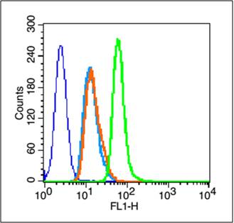
Blank control (blue line): A431 cells (blue).
Primary Antibody (green line): Rabbit Anti-PTEN antibody (bs-0748R)
Dilution: 1μg /10^6 cells;
Isotype Control Antibody (orange line): Rabbit IgG .
Secondary Antibody (white blue line): Goat anti-rabbit IgG-FITC
Dilution: 1μg /test.
Protocol
The cells were fixed with 70% methanol (Overnight at 4℃) and then permeabilized with 90% ice-cold methanol for 20 min at -20℃. Cells stained with Primary Antibody for 30 min at room temperature. The cells were then incubated in 1 X PBS/2%BSA/10% goat serum to block non-specific protein-protein interactions followed by the antibody for 15 min at room temperature. The secondary antibody used for 40 min at room temperature. Acquisition of 20,000 events was performed.
-
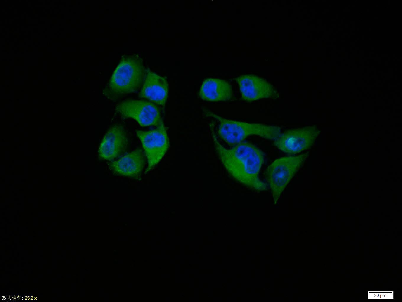
Hela cell; 4% Paraformaldehyde-fixed; Triton X-100 at room temperature for 20 min; Blocking buffer (normal goat serum, C-0005) at 37°C for 20 min; Antibody incubation with (PTEN) polyclonal Antibody, Unconjugated (bs-0748R) 1:100, 90 minutes at 37°C; followed by a conjugated Goat Anti-Rabbit IgG antibody at 37°C for 90 minutes, DAPI (blue, C02-04002) was used to stain the cell nuclei.
RRID:AB_10856502
产品名称:Rabbit Anti-PTEN antibody
别名: ITGA 2; MGC11227; MHAM; MMAC 1; MMAC1; Bannayan Zonana; BZS a; Multiple hamartoma (Cowden syndrome); Mutated in Mutiple Advanced Cancers 1; Phosphatase and Tensin Homolog; Phosphatidylinositol 345 trisphosphate 3 phosphatase and dual specificity protein p
中文名称:磷酸酶和张力蛋白同系物抗体
英文名称:Rabbit Anti-PTEN antibody
抗体来源: Rabbit
克隆类型:多克隆
细胞定位:细胞核,细胞浆
性 状:Liquid
亚 型:IgG
纯化方法:affinity purified by Protein A
保存条件:Shipped at 4℃. Store at -20 °C for one year. Avoid repeated freeze/thaw cycles.
免 疫 原:KLH conjugated synthetic peptide derived from human PTEN
抗原表位:101-200/403
SWISS:P60484
Gene ID :5728
Human Gene ID:5728
Potential tumor suppressor. Acts as a phosphoinositide3-phosphatase by regulating PtdIns (3,4,5)P3 levels. Involved in regulation of the AKT1 signaling pathway. The unphosphorylated form cooperates with AIP1 to suppress AKT1 activation.The PTEN/MMAC1 discovers the first to have the suppress of the phosphoric acid enzyme activity cancer gene currently.The gene of PTEN locates the chromosome10q23 area, sending forth sex tumor and a few households cancers with the variety to suffer from the comprehensive disease easilyrelevant.The activity that passes to repress the Akt regulates the cell period, the cell ground rule decease and glues to connect.This text discussed PTEN structure, function and its correlationses, the PTEN is in tumor repress function mechanism.
Function:Tumor suppressor. Acts as a dual-specificity protein phosphatase, dephosphorylating tyrosine-, serine- and threonine-phosphorylated proteins. Also acts as a lipid phosphatase, removing the phosphate in the D3 position of the inositol ring from phosphatidy
Subunit:Monomer. The unphosphorylated form interacts with the second PDZ domain of AIP1 and with DLG1 and MAST2 in vitro. Interacts with MAGI2, MAGI3, MAST1 and MAST3, but neither with MAST4 nor with DLG5. Interaction with MAGI2 increases protein stability. Inter
Subcellular Location:Cytoplasm. Nucleus. Nucleus, PML body. Note=Monoubiquitinated form is nuclear. Nonubiquitinated form is cytoplasmic. Colocalized with PML and USP7 in PML nuclear bodies. XIAP/BIRC4 promotes its nuclear localization.
Tissue Specificity:Expressed at a relatively high level in all adult tissues, including heart, brain, placenta, lung, liver, muscle, kidney and pancreas.
Post-translational modifications:Constitutively phosphorylated by CK2 under normal conditions. Phosphorylated in vitro by MAST1, MAST2 and MAST3. Phosphorylation results in an inhibited activity towards PIP3. Phosphorylation can both inhibit or promote PDZ-binding. Phosphorylation at Tyr
DISEASE:Defects in PTEN are a cause of Cowden disease (CD) [MIM:158350]; also known as Cowden syndrome (CS). CD is an autosomal dominant cancer predisposition syndrome associated with elevated risk for tumors of the breast, thyroid and skin. The predominant pheno
Similarity:Contains 1 C2 tensin-type domain.
Contains 1 phosphatase tensin-type domain.
Important Note:This product as supplied is intended for research use only, not for use in human, therapeutic or diagnostic applications.
 400-901-9800


 说明书
说明书 联系我们
联系我们 打印此页面
打印此页面 收藏
收藏