| Rabbit Anti-Runx3 antibody |
| 反应物种(预测) |
Rat,Chicken,Dog,Cow |
| 产品应用(已验证) |
WB,FCM |
| 产品应用(可尝试) |
IHC,IF,ELISA |
| 推荐稀释比例 |
WB=1:500-2000,Elisa=1:5000-10000,IHC-P=1:100-500,IHC-F=1:100-500,Flow Cyt=1ug/test,IF=1:100-500, |
| 研究领域 |
肿瘤,细胞生物 |
| 标签 |
Array |
-
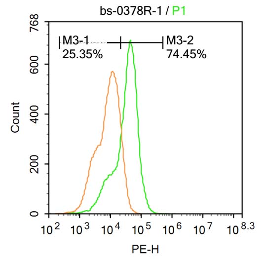
U-937 cells were fixed with 4% PFA for 10min at room temperature,permeabilized with 90% ice-cold methanol for 20 min at room temperature,and incubated in 5% BSA blocking buffer for 30 min at room temperature. Cells were then stained with Runx3 Antibody(bs-0378R) at 1:500 dilution in blocking buffer and incubated for 30 min at room temperature, washed twice with 2%BSA in PBS, followed by secondary antibody incubation for 40 min at room temperature. Acquisitions of 20,000 events were performed.Cells stained with primary antibody (green), and isotype control (orange).
-
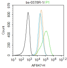
Blank control: Jurkat.
Primary Antibody (green line): Rabbit Anti-Runx3 antibody (bs-0378R)
Dilution: 1μg /10^6 cells;
Isotype Control Antibody (orange line): Rabbit IgG .
Secondary Antibody : Goat anti-rabbit IgG-AF647
Dilution: 1μg /test.
Protocol
The cells were fixed with 4% PFA (10min at room temperature)and then permeabilized with 90% ice-cold methanol for 20 min at-20℃. The cells were then incubated in 5%BSA to block non-specific protein-protein interactions for 30 min at room temperature .Cells stained with Primary Antibody for 30 min at room temperature. The secondary antibody used for 40 min at room temperature. Acquisition of 20,000 events was performed.
-
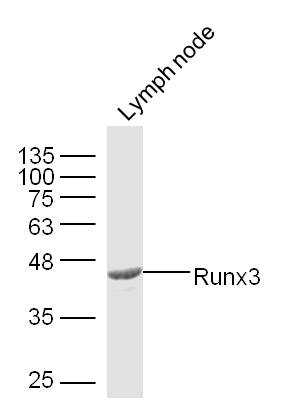
Sample: Lymph node (Mouse) Lysate at 30 ug
Primary: Anti- Runx3 (bs-0378R) at 1/300 dilution
Secondary: IRDye800CW Goat Anti-Mouse IgG at 1/20000 dilution
Predicted band size: 44 kD
Observed band size: 44 kD
-
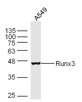
Sample: A549 Cell Lysate at 30 ug
Primary: Anti- Runx3 (bs-0378R) at 1/300 dilution
Secondary: IRDye800CW Goat Anti-Mouse IgG at 1/20000 dilution
Predicted band size: 44 kD
Observed band size: 44 kD
-
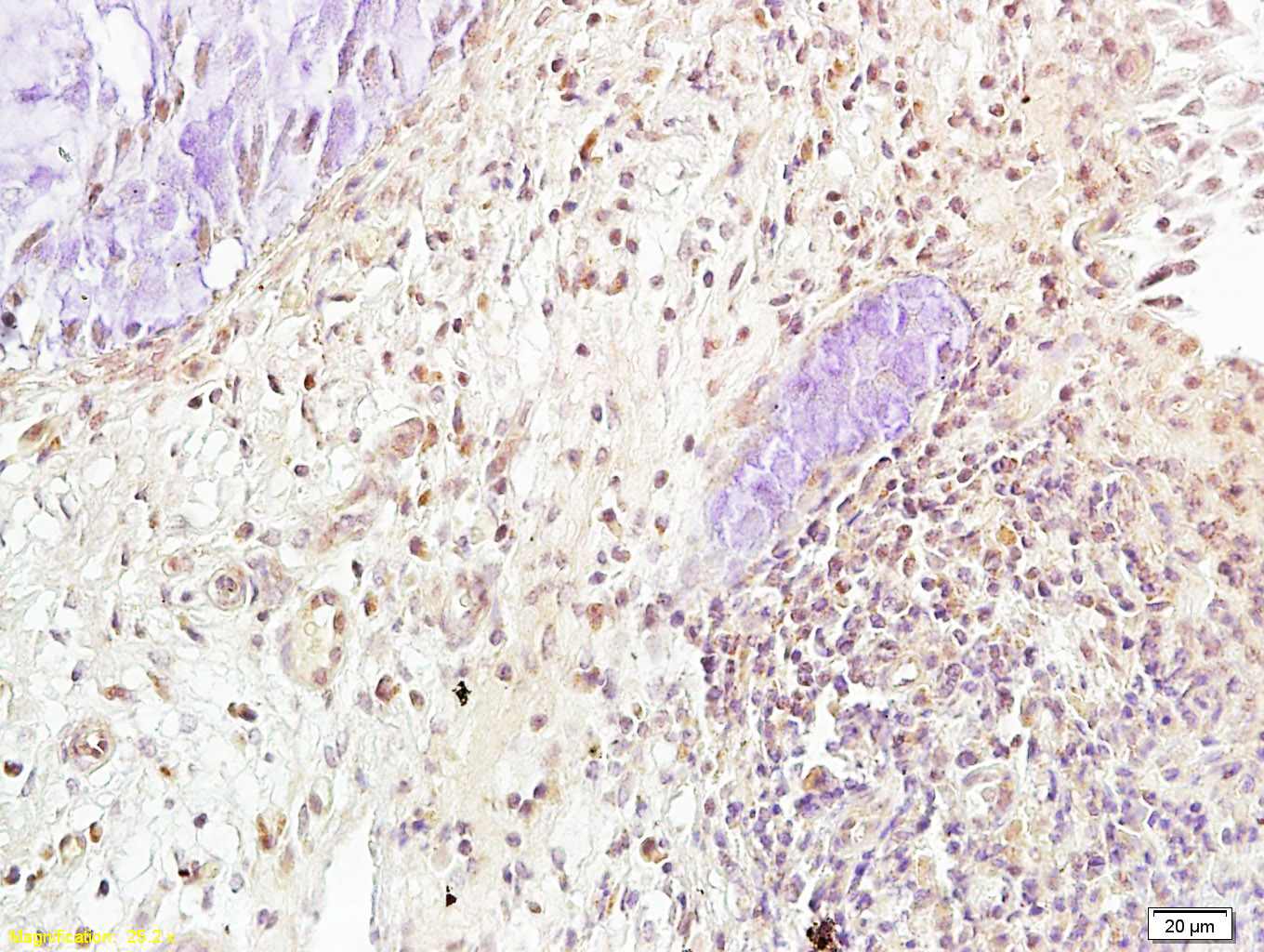
Tissue/cell: human cervical carcinoma; 4% Paraformaldehyde-fixed and paraffin-embedded;
Antigen retrieval: citrate buffer ( 0.01M, pH 6.0 ), Boiling bathing for 15min; Block endogenous peroxidase by 3% Hydrogen peroxide for 30min; Blocking buffer (normal goat serum,C-0005) at 37℃ for 20 min;
Incubation: Anti-Runx3 Polyclonal Antibody, Unconjugated(bs-0378R) 1:300, overnight at 4°C, followed by conjugation to the secondary antibody(SP-0023) and DAB(C-0010) staining
RRID:AB_10855649
产品名称:Rabbit Anti-Runx3 antibody
别名: RUNX3_HUMAN; Runt-related transcription factor 3; AML2; CBFA3; PEBP2A3; Acute myeloid leukemia 2 protein; Core-binding factor subunit alpha-3 (CBF-alpha-3); Oncogene AML-2; Polyomavirus enhancer-binding protein 2 alpha C subunit (PEA2-alpha C; PEBP2-alpha
中文名称:Runx3抗体
英文名称:Rabbit Anti-Runx3 antibody
抗体来源: Rabbit
克隆类型:多克隆
细胞定位:细胞核,细胞浆
性 状:Liquid
亚 型:IgG
纯化方法:affinity purified by Protein A
保存条件:Shipped at 4℃. Store at -20 °C for one year. Avoid repeated freeze/thaw cycles.
免 疫 原:KLH conjugated synthetic peptide derived from human Runx3
抗原表位:151-250/415
SWISS:Q13761
Gene ID :864
Human Gene ID:864
This gene encodes a member of the runt domain-containing family of transcription factors. A heterodimer of this protein and a beta subunit forms a complex that binds to the core DNA sequence 5'-PYGPYGGT-3' found in a number of enhancers and promoters, and can either activate or suppress transcription. It also interacts with other transcription factors. It functions as a tumor suppressor, and the gene is frequently deleted or transcriptionally silenced in cancer. Multiple transcript variants encoding different isoforms have been found for this gene. [provided by RefSeq, Jul 2008]
Function:CBF binds to the core site, 5'-PYGPYGGT-3', of a number of enhancers and promoters, including murine leukemia virus, polyomavirus enhancer, T-cell receptor enhancers, lck, IL-3 and GM-CSF promoters.
Subunit:Heterodimer of an alpha and a beta subunit. The alpha subunit binds DNA as a monomer and through the Runt domain. DNA-binding is increased by heterodimerization. Interacts with TLE1 and SUV39H1. The tyrosine phosphorylated form (via runt domain) interacts
Subcellular Location:Nucleus. Cytoplasm. Note=The tyrosine phosphorylated form localizes to the cytoplasm.
Post-translational modifications:Phosphorylated on tyrosine residues by SRC. Phosphorylated by LCK and FYN.
Similarity:Contains 1 Runt domain.
Important Note:This product as supplied is intended for research use only, not for use in human, therapeutic or diagnostic applications.
 400-901-9800


 说明书
说明书 联系我们
联系我们 打印此页面
打印此页面 收藏
收藏