| Rabbit Anti-PPARGC1A antibody |
| 反应物种(预测) |
Rat,Chicken,Dog,Pig,Cow,Horse,Rabbit |
| 产品应用(已验证) |
WB,IHC,FCM |
| 产品应用(可尝试) |
IF,ELISA |
| 推荐稀释比例 |
WB=1:500-2000,Elisa=1:5000-10000,IHC-P=1:100-500,IHC-F=1:100-500,Flow Cyt=1μg/Test,IF=1:100-500, |
| 研究领域 |
免疫学,转录调节因子,激酶和磷酸酶 |
| 标签 |
Array |
-
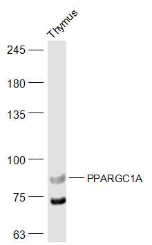
Sample:
Thymus (Mouse) Lysate at 40 ug
Primary: Anti-PPARGC1A (bs-1832R) at 1/500 dilution
Secondary: IRDye800CW Goat Anti-Rabbit IgG at 1/20000 dilution
Predicted band size: 88 kD
Observed band size: 88 kD
-
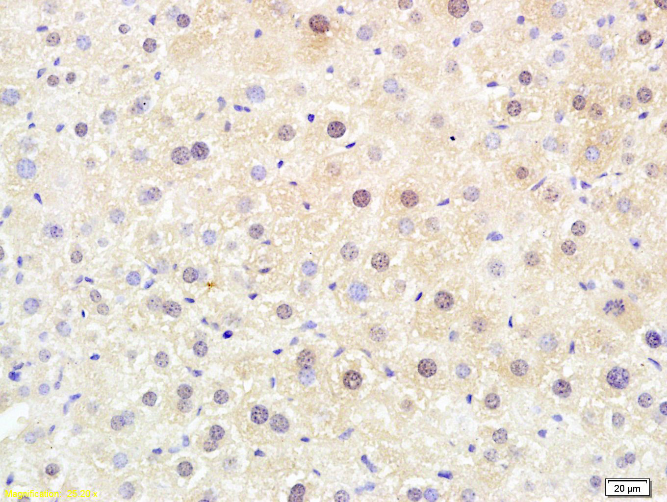
Tissue/cell: mouse liver tissue; 4% Paraformaldehyde-fixed and paraffin-embedded;
Antigen retrieval: citrate buffer ( 0.01M, pH 6.0 ), Boiling bathing for 15min; Block endogenous peroxidase by 3% Hydrogen peroxide for 30min; Blocking buffer (normal goat serum,C-0005) at 37℃ for 20 min;
Incubation: Anti-PPARGC1A/PGC1 alpha Polyclonal Antibody, Unconjugated(bs-1832R) 1:200, overnight at 4°C, followed by conjugation to the secondary antibody(SP-0023) and DAB(C-0010) staining
-
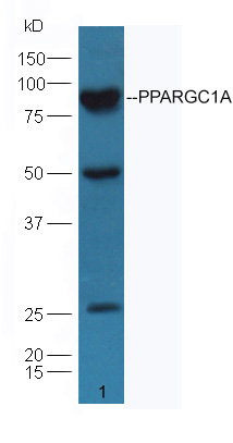
Sample: Heart (Mouse) Lysate at 30 ug
Primary: Anti- PPARGC1A (bs-1832R) at 1/300 dilution
Secondary: IRDye800CW Goat Anti-Rabbit IgG at 1/10000 dilution
Predicted band size: 88 kD
Observed band size: 88 kD
-
Blank control: HepG2.
Primary Antibody (green line): Rabbit Anti-PPARGC1A antibody (bs-1832R)
Dilution: 1μg /10^6 cells;
Isotype Control Antibody (orange line): Rabbit IgG .
Secondary Antibody : Goat anti-rabbit IgG-AF647
Dilution: 1μg /test.
Protocol
The cells were fixed with 4% PFA (10min at room temperature)and then permeabilized with 90% ice-cold methanol for 20 min at -20℃. The cells were then incubated in 5%BSA to block non-specific protein-protein interactions for 30 min at room temperature .Cells stained with Primary Antibody for 30 min at room temperature. The secondary antibody used for 40 min at room temperature. Acquisition of 20,000 events was performed.
-
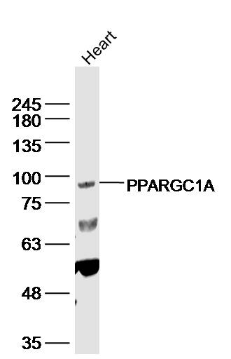
Sample: Heart (Mouse) Lysate at 40 ug
Primary: Anti-PPARGC1A (bs-1832R) at 1/300 dilution
Secondary: IRDye800CW Goat Anti-Rabbit IgG at 1/20000 dilution
Predicted band size: 88 kD
Observed band size: 90 kD
RRID:AB_10853206
产品名称:Rabbit Anti-PPARGC1A antibody
别名: LEM6; PGC-1 Alpha; PGC1 Alpha; Ligand effect modulator 6; Peroxisome proliferative activated receptor, gamma, coactivator 1 alpha; Peroxisome proliferative activated receptor, gamma, coactivator 1 ; Peroxisome proliferator activated receptor gamma coactiv
中文名称:过氧化物酶体增殖物激活受体γ辅激活子1α抗体
英文名称:Rabbit Anti-PPARGC1A antibody
抗体来源: Rabbit
克隆类型:多克隆
细胞定位:细胞核
性 状:Liquid
亚 型:IgG
纯化方法:affinity purified by Protein A
保存条件:Shipped at 4℃. Store at -20 °C for one year. Avoid repeated freeze/thaw cycles.
免 疫 原:KLH conjugated synthetic peptide derived from human PGC-1
抗原表位:601-700/798
SWISS:Q9UBK2
Gene ID :10891
Human Gene ID:10891
The protein encoded by this gene is a transcriptional coactivator that regulates the genes involved in energy metabolism. This protein interacts with PPARgamma, which permits the interaction of this protein with multiple transcription factors. This protein can interact with, and regulate the activities of cAMP response element binding protein (CREB) and nuclear respiratory factors (NRFs). It provides a direct link between external physiological stimuli and the regulation of mitochondrial biogenesis, and is a major factor that regulates muscle fiber type determination. This protein may be also involved in controlling blood pressure, regulating cellular cholesterol homoeostasis, and the development of obesity (referenced from entrez gene.
Function:Transcriptional coactivator for steroid receptors and nuclear receptors. Greatly increases the transcriptional activity of PPARG and thyroid hormone receptor on the uncoupling protein promoter. Can regulate key mitochondrial genes that contribute to the p
Subunit:Binds MYBBP1A, which inhibits transcriptional activation by this protein. Interacts with PRDM16. Interacts with LRPPRC. Homooligomer. Interacts with LPIN1.
Subcellular Location:Nucleus.
Tissue Specificity:Heart, skeletal muscle, liver and kidney. Expressed at lower levels in brain and pancreas and at very low levels in the intestine and white adipose tissue. In skeletal muscle, levels were lower in obese than in lean subjects and fasting induced a 2-fold i
Post-translational modifications:hosphorylation by AMPK in skeletal muscle increases activation of its own promoter. Phosphorylated by CLK2.
Heavily acetylated by GCN5 and biologically inactive under conditions of high nutrients. Deacetylated by SIRT1 in low nutrients/high NAD condit
Similarity:Contains 1 RRM (RNA recognition motif) domain.
Important Note:This product as supplied is intended for research use only, not for use in human, therapeutic or diagnostic applications.
 400-901-9800


 说明书
说明书 联系我们
联系我们 打印此页面
打印此页面 收藏
收藏