| Rabbit Anti-CD86 antibody |
| 反应物种(预测) |
Dog,Pig,Cow,Sheep |
| 产品应用(已验证) |
WB,IHC,IF |
| 产品应用(可尝试) |
ELISA |
| 推荐稀释比例 |
WB=1:500-2000,Elisa=1:5000-10000,IHC-P=1:100-500,IHC-F=1:100-500,IF=1:100-500, |
| 研究领域 |
免疫学,微生物学,干细胞,细胞表面分子,糖蛋白,淋巴细胞,T-淋巴细胞,B-淋巴细胞, |
| 标签 |
Array |
-
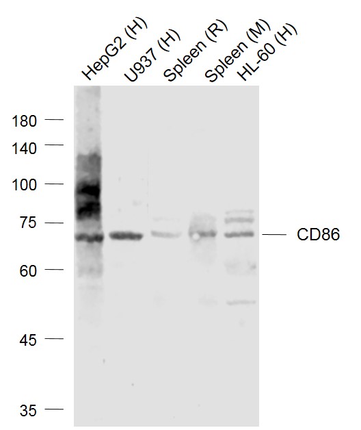
Sample:
Lane 1: HepG2 (Human) Cell Lysate at 30 ug
Lane 2: U937 (Human) Cell Lysate at 30 ug
Lane 3: Spleen (Rat) Lysate at 40 ug
Lane 4: Spleen (Mouse) Lysate at 40 ug
Lane 5: HL-60 (Human) Cell Lysate at 30 ug
Primary: Anti-CD86 (bs-1035R) at 1/1000 dilution
Secondary: IRDye800CW Goat Anti-Rabbit IgG at 1/20000 dilution
Predicted band size: 72-74 kD
Observed band size: 72 kD
-
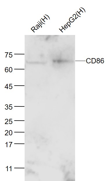
Sample:
Raji(Human) Cell Lysate at 30 ug
HepG2(Human) Cell Lysate at 30 ug
Primary: Anti- CD86 (bs-1035R) at 1/300 dilution
Secondary: IRDye800CW Goat Anti-Rabbit IgG at 1/20000 dilution
Predicted band size: 70/80 kD
Observed band size: 70 kD
-
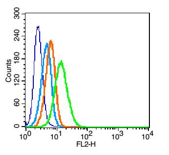
Blank control: U937(blue).
Primary Antibody: Rabbit Anti-CD86 antibody(bs-1035R), Dilution: 1μg in 100 μL 1X PBS containing 0.5% BSA;
Isotype Control Antibody: Rabbit IgG (orange) ,used under the same conditions.
Secondary Antibody: Goat anti-rabbit IgG-PE(white blue), Dilution: 1:200 in 1 X PBS containing 0.5% BSA.
Protocol
The cells were fixed with 2% paraformaldehyde (10 min).Primary antibody (bs-1035R, 1μg /1x10^6 cells) were incubated for 30 min on the ice, followed by 1 X PBS containing 0.5% BSA + 10% goat serum (15 min) to block non-specific protein-protein interactions. Then the Goat Anti-rabbit IgG/PE antibody was added into the blocking buffer mentioned above to react with the primary antibody at 1/200 dilution for 30 min on ice. Acquisition of 20,000 events was performed.
-
Tissue/cell: rat lung tissue; 4% Paraformaldehyde-fixed and paraffin-embedded;
Antigen retrieval: citrate buffer ( 0.01M, pH 6.0 ), Boiling bathing for 15min; Block endogenous peroxidase by 3% Hydrogen peroxide for 30min; Blocking buffer (normal goat serum,C-0005) at 37℃ for 20 min;
Incubation: Anti-CD86/B7-2 Polyclonal Antibody, Unconjugated(bs-1035R) 1:200, overnight at 4°C, followed by conjugation to the secondary antibody(SP-0023) and DAB(C-0010) staining
-
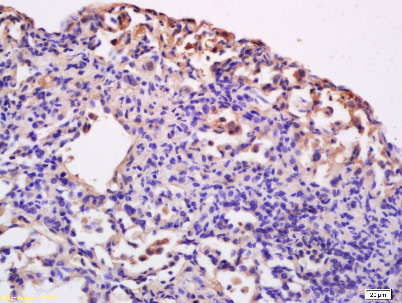
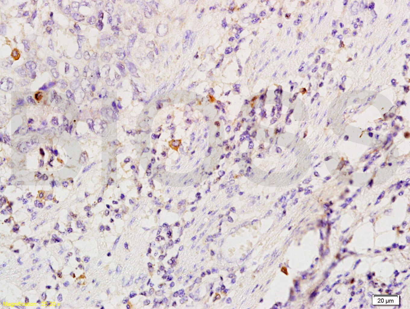
Tissue/cell: Human esophageal carcinoma; 4% Paraformaldehyde-fixed and paraffin-embedded;
Antigen retrieval: citrate buffer ( 0.01M, pH 6.0 ), Boiling bathing for 15min; Block endogenous peroxidase by 3% Hydrogen peroxide for 30min; Blocking buffer (normal goat serum,C-0005) at 37℃ for 20 min;
Incubation: Anti-CD86/B7-2 Polyclonal Antibody, Unconjugated(bs-1035R) 1:200, overnight at 4°C, followed by conjugation to the secondary antibody(SP-0023) and DAB(C-0010) staining
RRID:AB_10856252
产品名称:Rabbit Anti-CD86 antibody
别名: CD28LG2; FUN1; LAB72; Activation B7 2 antigen; Activation B7-2 antigen 3; CD86_HUMAN; Activation B7-2 antigen; Activation B72 antigen; B lymphocyte activation antigen B7 2; B lymphocyte activation antigen B72; B-lymphocyte activation antigen B7-2 2; B-lym
中文名称:CD86抗体
英文名称:Rabbit Anti-CD86 antibody
抗体来源: Rabbit
克隆类型:多克隆
细胞定位:细胞膜
性 状:Liquid
亚 型:IgG
纯化方法:affinity purified by Protein A
保存条件:Shipped at 4℃. Store at -20 °C for one year. Avoid repeated freeze/thaw cycles.
免 疫 原:KLH conjugated synthetic peptide derived from the middle of rat CD86
抗原表位:140-175/313
抗原细胞定位:Extracellular
SWISS:O35531
Gene ID :56822
Human Gene ID:942
This gene encodes a type I membrane protein that is a member of the immunoglobulin superfamily. This protein is expressed by antigen-presenting cells, and it is the ligand for two proteins at the cell surface of T cells, CD28 antigen and cytotoxic T-lymphocyte-associated protein 4. Binding of this protein with CD28 antigen is a costimulatory signal for activation of the T-cell. Binding of this protein with cytotoxic T-lymphocyte-associated protein 4 negatively regulates T-cell activation and diminishes the immune response. Alternative splicing results in several transcript variants encoding different isoforms.[provided by RefSeq, May 2011].
Function:Receptor involved in the costimulatory signal essential for T-lymphocyte proliferation and interleukin-2 production, by binding CD28 or CTLA-4. May play a critical role in the early events of T-cell activation and costimulation of naive T-cells, such as d
Subunit:Homodimer. Interacts with MARCH8. Interacts with human herpesvirus 8 MIR2 protein (Probable). Interacts with adenovirus subgroup B fiber proteins and acts as a receptor for these viruses.
Subcellular Location:Cell membrane; Single-pass type I membrane protein.
Tissue Specificity:Expressed by activated B-lymphocytes and monocytes.
Post-translational modifications:Polyubiquitinated; which is promoted by MARCH8 and results in endocytosis and lysosomal degradation.
Similarity:Contains 1 Ig-like C2-type (immunoglobulin-like) domain.
Contains 1 Ig-like V-type (immunoglobulin-like) domain.
Important Note:This product as supplied is intended for research use only, not for use in human, therapeutic or diagnostic applications.
 400-901-9800


 说明书
说明书 联系我们
联系我们 打印此页面
打印此页面 收藏
收藏