| Rabbit Anti-FUT4 antibody |
| 反应物种(预测) |
Rat |
| 产品应用(已验证) |
WB,IHC,FCM |
| 产品应用(可尝试) |
IF,ELISA |
| 推荐稀释比例 |
WB=1:500-2000,Elisa=1:5000-10000,IHC-P=1:100-500,IHC-F=1:100-500,Flow Cyt=1μg/Test,IF=1:100-500, |
| 研究领域 |
肿瘤,细胞生物,免疫学,细胞膜受体 |
| 标签 |
Array |
-
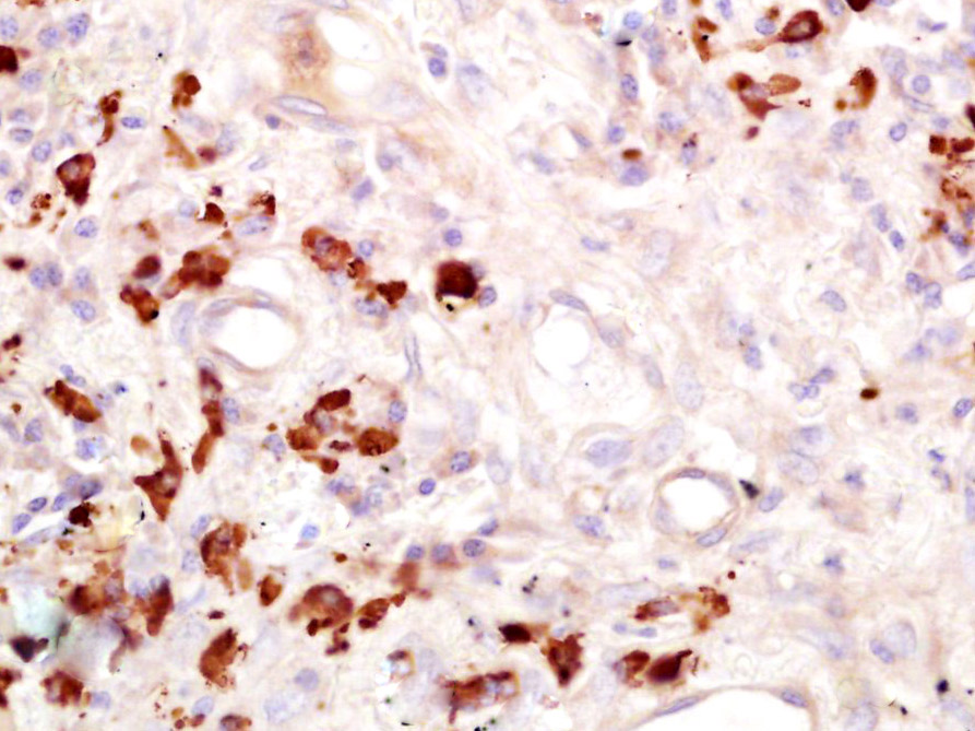
Paraformaldehyde-fixed, paraffin embedded (Human lung cancer); Antigen retrieval by boiling in sodium citrate buffer (pH6.0) for 15min; Block endogenous peroxidase by 3% hydrogen peroxide for 20 minutes; Blocking buffer (normal goat serum) at 37°C for 30min; Antibody incubation with (FUT4) Polyclonal Antibody, Unconjugated (bs-1702R) at 1:400 overnight at 4°C, followed by operating according to SP Kit(Rabbit) (sp-0023) instructionsand DAB staining.
-
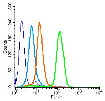
Overlay histogram showing HL 60 cells stained with bs-1702R (Green line).
The cells were fixed with 90% methanol (5 min) and then permeabilized with 0.01M PBS-Tween for 20 min. The cells were then incubated in 1x PBS / 10% normal goat serum to block non-specific protein-protein interactions followed by the antibody (bs-1702R,1μg/1x10^6 cells) for 30 min at 22℃. The secondary antibody used was fluorescein isothiocyanate goat anti-rabbit IgG (H+L) (bs- 0295G-FITC , Brillant blue line) at 1/200 dilution for 30 min at 22℃. Isotype control antibody was rabbit IgG (polyclonal,bs-0295P,Orange line) (1μg/1x10^6 cells) used under the same conditions. Unlabelled sample (blue line) was also used as a control. Acquisition of 20,000 events were collected using a 20mW Argon ion laser (488nm) and 525/30 bandpass filter.
-
Blank control: Mouse spleen.
Primary Antibody (green line): Rabbit Anti-FUT4/FITC Conjugated antibody (bs-1702R-FITC)
Dilution: 1μg /10^6 cells;
Isotype Control Antibody (orange line): Rabbit IgG-FITC .
Protocol
The cells were fixed with 4% PFA (10min at room temperature)and then permeabilized with 0.1% PBST for 20 min at-20℃. The cells were then incubated in 5% BSA to block non-specific protein-protein interactions for 30 min at room temperature. The cells were stained with Primary Antibody for 30 min at room temperature. Acquisition of 20,000 events was performed.
-
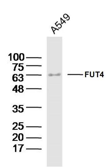
Sample:A549 Cell (Human) Lysate at 40 ug
Primary: Anti-FUT4(bs-1702R)at 1/300 dilution
Secondary: IRDye800CW Goat Anti-Rabbit IgG at 1/20000 dilution
Predicted band size: 58kD
Observed band size: 63kD
-
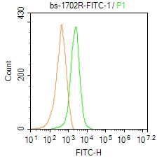
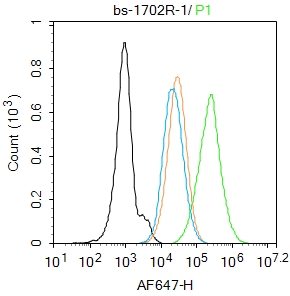
Blank control:HL-60.
Primary Antibody (green line): Rabbit Anti-FUT4 (bs-1702R)
Dilution: 1μg /10^6 cells;
Isotype Control Antibody (orange line): Rabbit IgG .
Secondary Antibody : Goat anti-rabbit IgG-AF647
Dilution: 1μg /test.
Protocol
The cells were fixed with 4% PFA (10min at room temperature)and then permeabilized with 0.1% PBST for 20 min at room temperature. The cells were then incubated in 5%BSA to block non-specific protein-protein interactions for 30 min at room temperature.Cells stained with Primary Antibody for 30 min at room temperature. The secondary antibody used for 40 min at room temperature. Acquisition of 20,000 events was performed.
RRID:AB_10856399
产品名称:Rabbit Anti-FUT4 antibody
别名: SSEA-1; 3-FAL; Alpha (1,3) fucosyltransferase; Alpha 13 fucosyltransferase FucT; EC 2.4.1.; ELAM 1 ligand fucosyltransferase; ELAM ligand fucosyltransferase; ELAM1 ligand fucosyltransferase; ELFT; FCT3A; Fuc TIV; Fucosyltransferase 4 alpha 1 3 fucosyltran
中文名称:α-(1,3)-岩藻糖基转移酶 4抗体
英文名称:Rabbit Anti-FUT4 antibody
抗体来源: Rabbit
克隆类型:多克隆
细胞定位:细胞浆,细胞膜
性 状:Liquid
亚 型:IgG
纯化方法:affinity purified by Protein A
保存条件:Shipped at 4℃. Store at -20 °C for one year. Avoid repeated freeze/thaw cycles.
免 疫 原:KLH conjugated synthetic peptide derived from human FUT4
抗原表位:251-295/433
SWISS:P22083
Gene ID :2526
Human Gene ID:2526
The Lewis histo-blood group system comprises a set of fucosylated glycosphingolipids that are synthesized by exocrine epithelial cells and circulate in body fluids. The glycosphingolipids function in embryogenesis, tissue differentiation, tumor metastasis, inflammation, and bacterial adhesion. They are secondarily absorbed to red blood cells giving rise to their Lewis phenotype. This gene is a member of the fucosyltransferase family, which catalyzes the addition of fucose to precursor polysaccharides in the last step of Lewis antigen biosynthesis. It encodes an enzyme with alpha(1,3)-fucosyltransferase and alpha(1,4)-fucosyltransferase activities. Mutations in this gene are responsible for the majority of Lewis antigen-negative phenotypes. Multiple alternatively spliced variants, encoding the same protein, have been found for this gene. [provided by RefSeq].
Function:May catalyze alpha-1,3 glycosidic linkages involved in the expression of Lewis X/SSEA-1 and VIM-2 antigens.
Subcellular Location:Golgi apparatus, Golgi stack membrane; Single-pass type II membrane protein. Note=Membrane-bound form in trans cisternae of Golgi.
Tissue Specificity:Highest expression in stomach and colon. It is also expressed in the lung, testis, uterus, small intestine and to a lesser extent in spleen, and ovary. Present in trace amounts in brain, thymus, heart, smooth muscle, kidney and bone marrow. Not found in l
Similarity:Belongs to the glycosyltransferase 10 family.
Important Note:This product as supplied is intended for research use only, not for use in human, therapeutic or diagnostic applications.
 400-901-9800


 说明书
说明书 联系我们
联系我们 打印此页面
打印此页面 收藏
收藏